Instructions for Side by Side Printing
- Print the notecards
- Fold each page in half along the solid vertical line
- Cut out the notecards by cutting along each horizontal dotted line
- Optional: Glue, tape or staple the ends of each notecard together
Lower Extremity Muscles
front 1 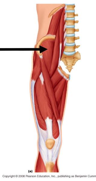 | back 1 Iliacus O: Iliac Fossa I: Lesser trochanter of femur A: Flexion of Hip A: Assists with adduction & lateral rotation of hip A: Anterior rotation of pelvis with femur stabilized N: Femoral nerve (L2-L4) |
front 2 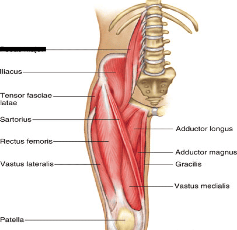 | back 2 Psoas Major O: L1-L4 - bodies and transverse process, IV discs at same levels I: Lesser trochanter A: Flexion of hip A: Assists with adduction & lateral rotation of hip; some rotation of the lumbar spine A: Flexion of lumbar spine with femur stabilized N: Femoral Nerve (L2-L4) |
front 3 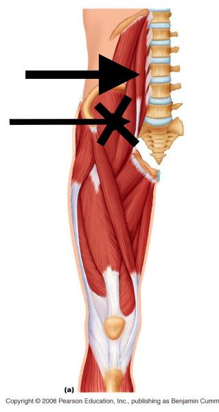 | back 3 Psoas Minor A: Assists psoas major with flexion of lumbar spine *Not a true muscle of the hip joint (coxofemoral joint) *Not present in some people |
front 4 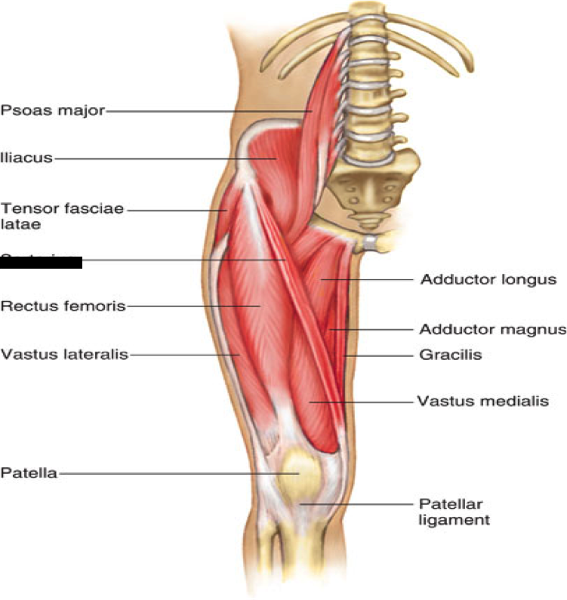 | back 4 Sartorius "Tailor's Muscle" O: Anterior superior iliac spine (ASIS) I: Medial surface of tibia (pes anserine) A: Flexes hip & knee: crosses both joints A: Also acts to laterally rotate the knee at the tibiofemoral joint N: Femoral Nerve (L2-L4) |
front 5 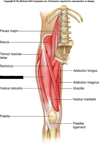 | back 5 Rectus Femoris O: Anterior inferior iliac spine I: Tibial tuberosity A: Assists iliopsoas with flexion of hip A: Knee extension and stabilization N: Femoral Nerve (L2-L4) |
front 6 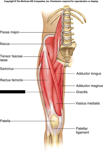 | back 6 Vastus Lateralis O: Lateral lip of linea aspera & greater trochanter I: Tibial tuberosity A: Knee extension N: Femoral nerve (L2-L4) *Largest muscle of the quads |
front 7 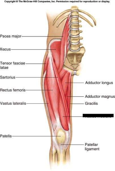 | back 7 Vastus Medialis O: Medial lip of linea aspera I: Tibial tuberosity A: Knee extension N: Femoral Nerve (L2-L4) *Vastus medialis obliquus |
front 8 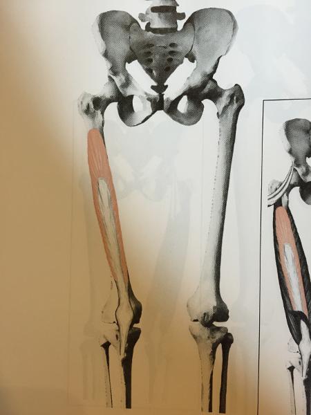 | back 8 Vastus Intermedius O: Proximal, anterolateral shaft of femur I: Tibial Tuberosity A: Knee extension N: Femoral Nerve (L2-L4) |
front 9 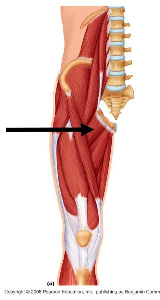 | back 9 Pectineus O: Superior Ramus of Pubis I: Intertrochanteric line to line aspera A: Adduction & Flexion of hip N: Femoral Nerve |
front 10 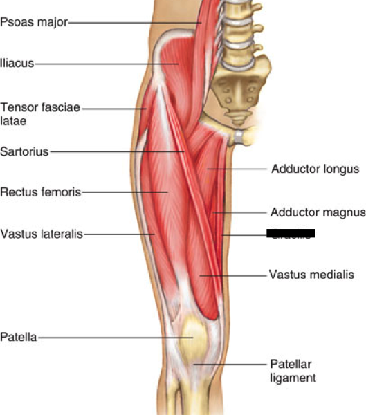 | back 10 Gracilis O: Inferior ramus & body of pubis I: Medial surface of tibia (pes anserine) A: Adduction & flexion of femur A: Medial rotation of tibia N: Obturator nerve (L2-L4) |
front 11 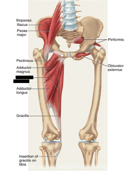 | back 11 Adductor Brevis O: Inferior ramus & body of pubis I: Upper 1/3 of linea aspera A: Adduct & laterally of the hip N: Obturator (L2-L4) |
front 12 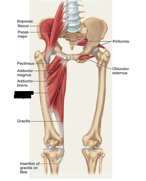 | back 12 Adductor Longus O: Inferior ramus & body of pubis I: Middle 1/3 of linea aspera A: Adducts & laterally rotates femur N: Obturator Nerve (L2-L4) |
front 13 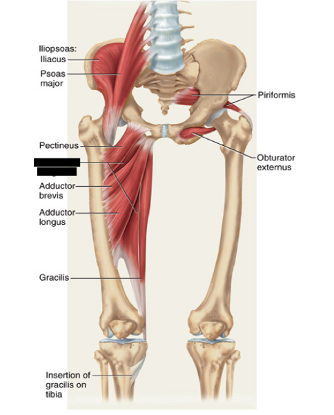 | back 13 Adductor Magnus O: Ischial tuberosity & Pubis I: Linea aspera & adductor tubercle - anterior & posteior portions (BIG!) A: Adduction of femur Anterior = flex & lateral rotation Posterior = extend & medial rotation N: Obturator (L2-L4) N: Sciatic nerve (L4-S3) |
front 14 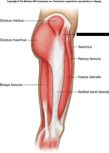 | back 14 Tensor Fascia Latae (TFL) O: Anterior iliac crest I: Fascia of the thigh-Iliotibial (IT) Band A: Abduct femur A: Flex hip joint A: Helps depress femoral head into acetabulum N: Superior gluteal nerve (L4-S1) |
front 15 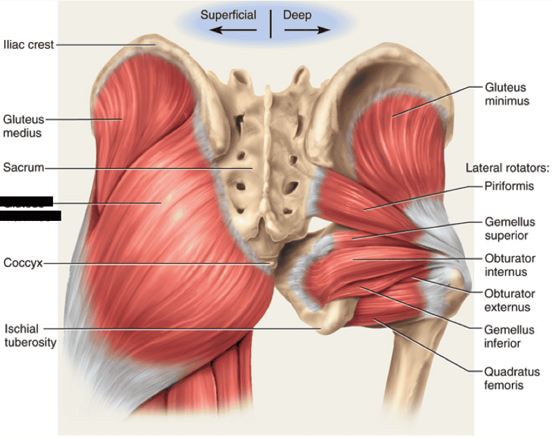 | back 15 Gluteus Maximus O: Iliac crest, Sacrum, Coccyx I: Posterior portion of femur - blends with IT band A: Hip extension (prime mover) A: Also laterally rotates A: Also abducts & adducts N: Inferior Gluteal Nerve (L5-S1) |
front 16 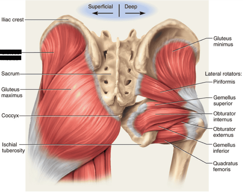 | back 16 Gluteus Medius O: Posterior aspect of ilium I: Greater trochanter of femus -portion of gluteus medium is deep to the gluteus maximus A: Abduct & medially rotate femur N: Superior gluteal nerve (L4-S1) |
front 17 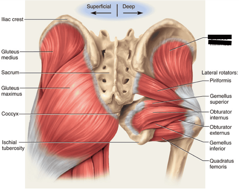 | back 17 Gluteus Minimus O: Posterior aspect of ilium I: Greater trochanter of femus -portion of gluteus medium is deep to the gluteus maximus A: Abduct & medially rotate femur N: Superior gluteal nerve (L4-S1) |
front 18 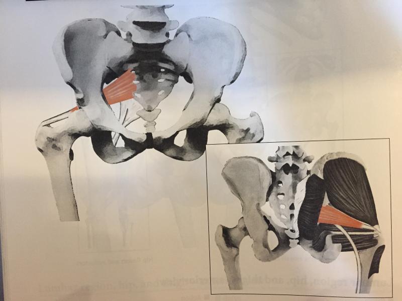 | back 18 Piriformis O: Upper portion of sacrum I: Greater trochanter - crosses greater sciatic notch A: Lateral rotation of femur N: Anterior rami of first and second sacral nerves |
front 19 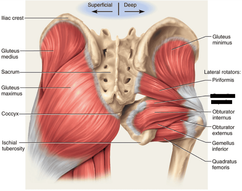 | back 19 Gemellus superior |
front 20 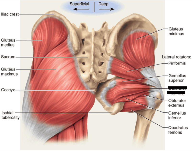 | back 20 Obturator internus |
front 21  | back 21 Obturator externus |
front 22 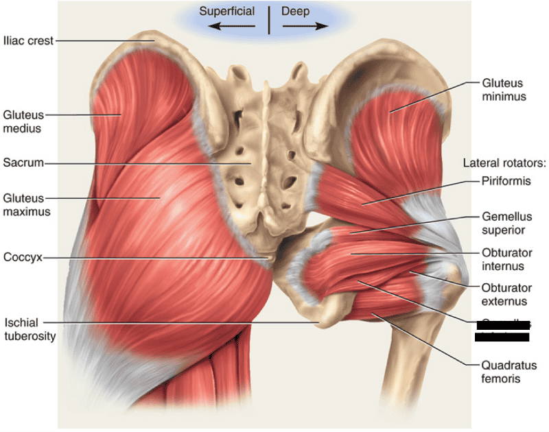 | back 22 Gemellus inferior |
front 23 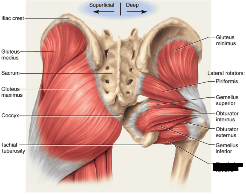 | back 23 Quadratus femoris |
front 24 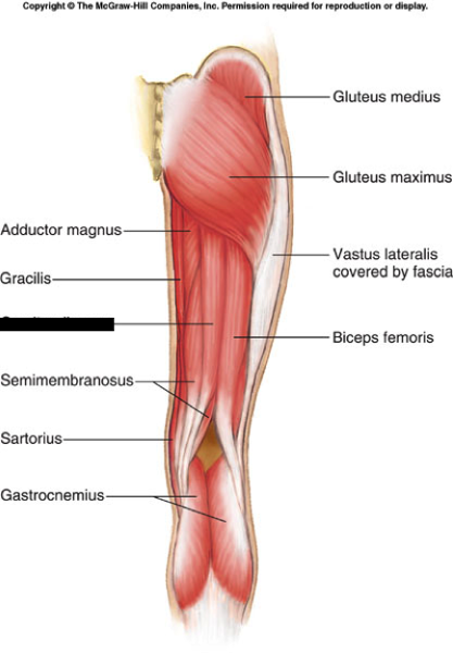 | back 24 Semitendinosus O: Ischial tuberosity I: Medial surface of tibia (pes anserine) A: Knee flexion A: Medial rotation of the tibia A: Hip extension N: Sciatic Nerve (L5, S1, S2) |
front 25 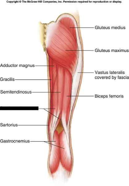 | back 25 Semimembranosus O: Ischial tuberosity I: Posterior, medial condyle of tibia A: Flex knee & medially rotate femur N: Sciatic Nerve (L5, S1, S2) |
front 26 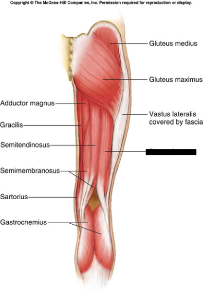 | back 26 Biceps Femoris O: Long head - ischial tuberosity O: Short head - posterior femur (line aspera) - not a hip extender I: Fibular head, lateral condyle of tibia A: Flex knee & laterally rotate femur N: LH - tibial part of sciatic nerve (S1-S3) N: SH - common perennial part of sciatic nerve (L5, S1, S2) |
front 27 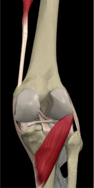 | back 27 Popliteus O: Lateral epicondyle of femur I: Shaft of tibia A: Unlocks knee to allow flexion to occur Open kinetic chain (OKC) position: Medial rotation of the tibia Closed kinetic chain (CKC) position: Lateral rotation of the femur N: Tibial Nerve |
front 28 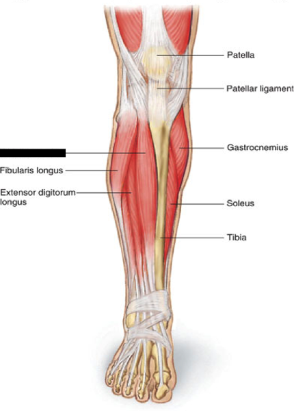 | back 28 Tibialis Anterior O: Shaft of the tibia I: Medial cuneiform & 1st metatarsal A: Dorsiflexion & inversion N: Deep peroneal nerve (L4-S1) *Most medial of the anterior leg muscles |
front 29 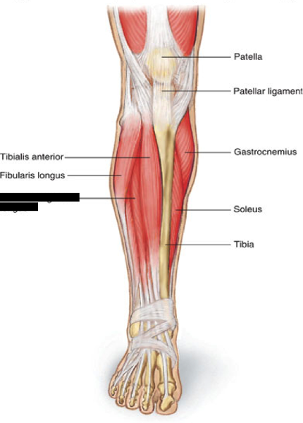 | back 29 Extensor Digitorum Longus O: Lateral condyle of tibia I: Distal phalanx; digits 2-5 A: Extend digits 2-5 A: Dorsiflex foot N: Deep peroneal nerve (L4-S1) |
front 30 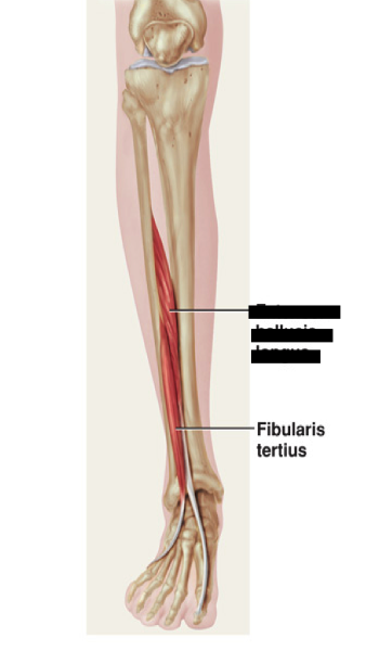 | back 30 Extensor Hallucis Longus O: Fibula I: Distal phalanx - digit 1 A: Extends digit 1 A: Inversion N: Deep peroneal nerve (L4-S1) |
front 31 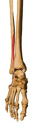 | back 31 Peroneus Tertius O: Anterior shaft of fibula I: Base of 5th metatarsal A: Everts & dorsiflexes foot *Tendon crosses anterior to lateral malleolus *Causes dorsiflexion N: Deep peroneal nerve (L4-S1) |
front 32 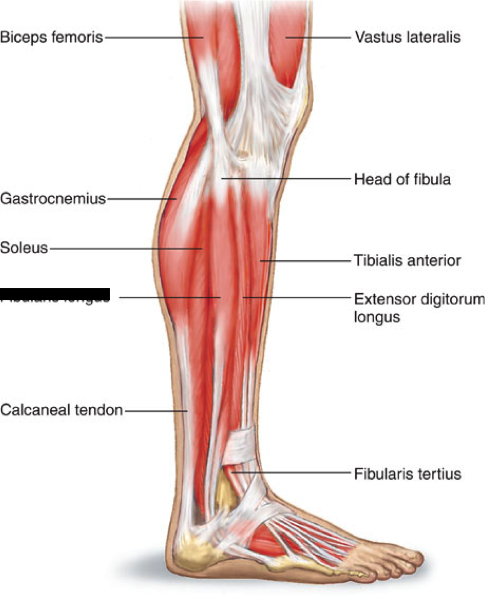 | back 32 Peroneus Longus O: Head & shaft of fibula I: First metatarsal & medial cuneiform A: Evert foot (strongest pronator) A: Plantar flexion of the ankle A: Tendon travels behind lateral malleolus, then under foot to medial side N: Superficial peroneal |
front 33 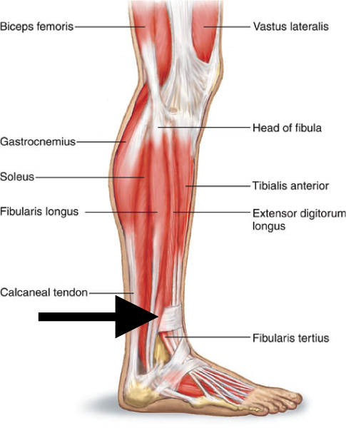 | back 33 Peroneus Brevis O: Lateral aspect of shaft of fibula I: Base of 5th metatarsal A: Eversion & plantar flexion of ankle *tendon travels behind lateral malleolus N: Superficial peroneal |
front 34 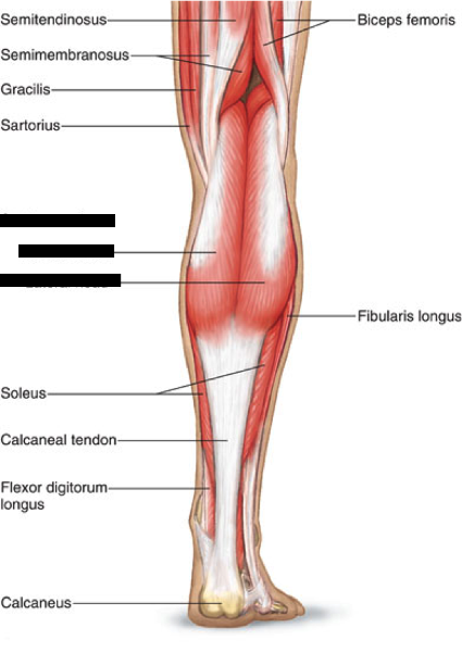 | back 34 Gastrocnemius O: Lateral & medial epicondyles of femur I: Calcaneus via calcaneal (Achilles') tendon A: Plantar flexion & assists knee flexion *Important in walking/running activities N: Tibial Nerve |
front 35 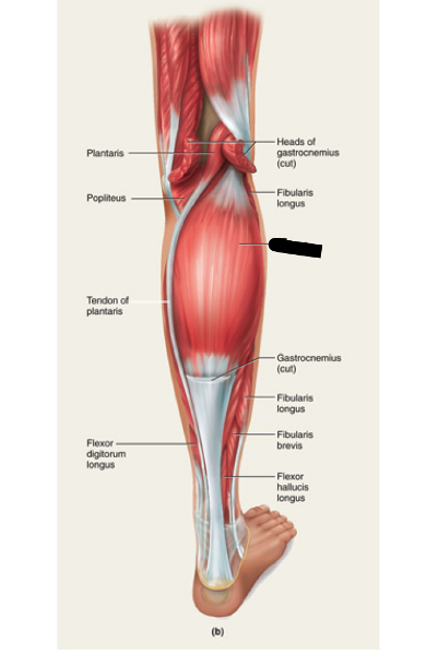 | back 35 Soleus O: Head/shaft of fibular & posterior surface of tibia I: Calcaneus via calcaneal (Achilles') tendon A: Plantar flex foot N: Tibial Nerve *Mostly lies deep to the gastrocnemius |
front 36 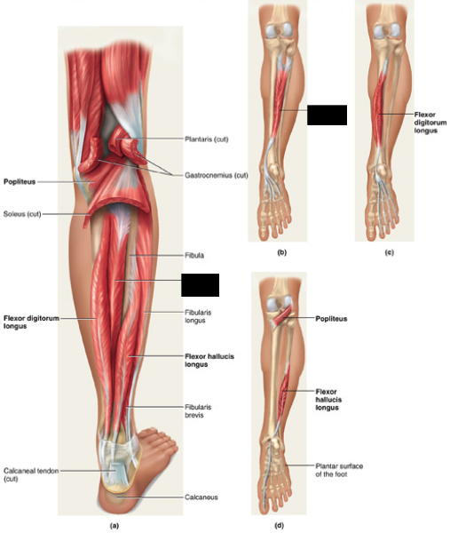 | back 36 Tibialis Posterior O: Posterior upper half of tibia & fibular I: Lower surfaces of navicular, cuneiform, & bases of metatarsals 2-5 A: Plantar flex and invert foot N: Tibial nerve |
front 37 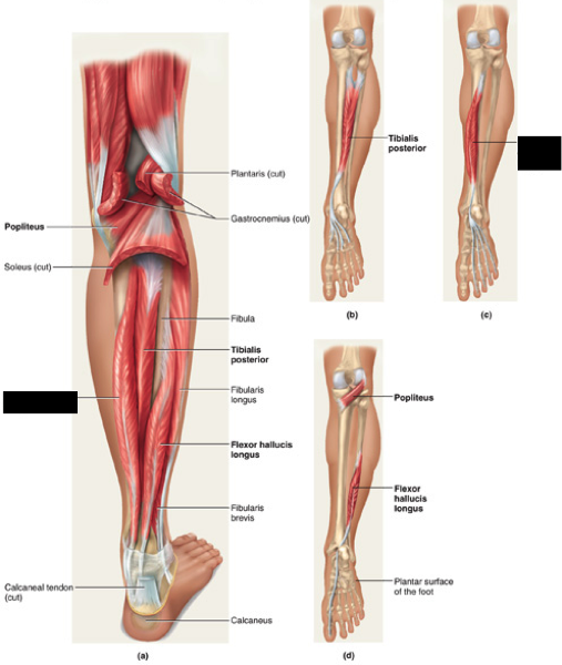 | back 37 Flexor Digitorum Longus O: Posterior surface of tibia I: Distal phalanges of 2-5 A: Flex digits 2-5 & plantar flexion N: Tibial Nerve *Helps support the arch |
front 38 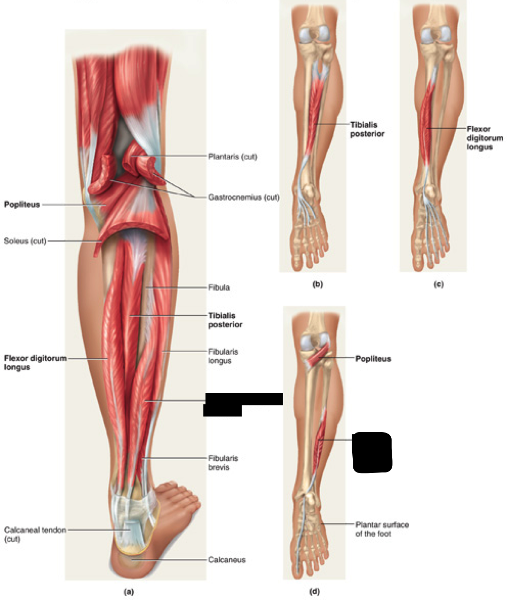 | back 38 Flexor Hallucis Longus O: Middle 2/3 of posterior fibular I: Base of distal phalanx of big toe A: Flex digit 1, inversion, plantar flexion N: Tibial Nerve |
front 39 What is the name for the iliacus & psoas major together? | back 39 Iliopsoas |
front 40 What is the "Groin"? | back 40 Anterior aspect of the hip joint, but commonly accepted as adductor group |
front 41 What is the Femoral Nerve? | back 41 Innervates the quadriceps & hip flexors |
front 42 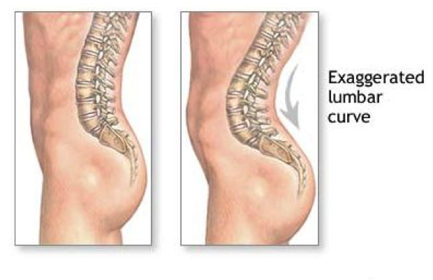 What causes this? | back 42 Over activation or tightness |
front 43 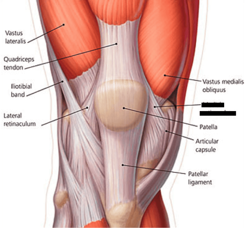 | back 43 Medial retinaculum |
front 44 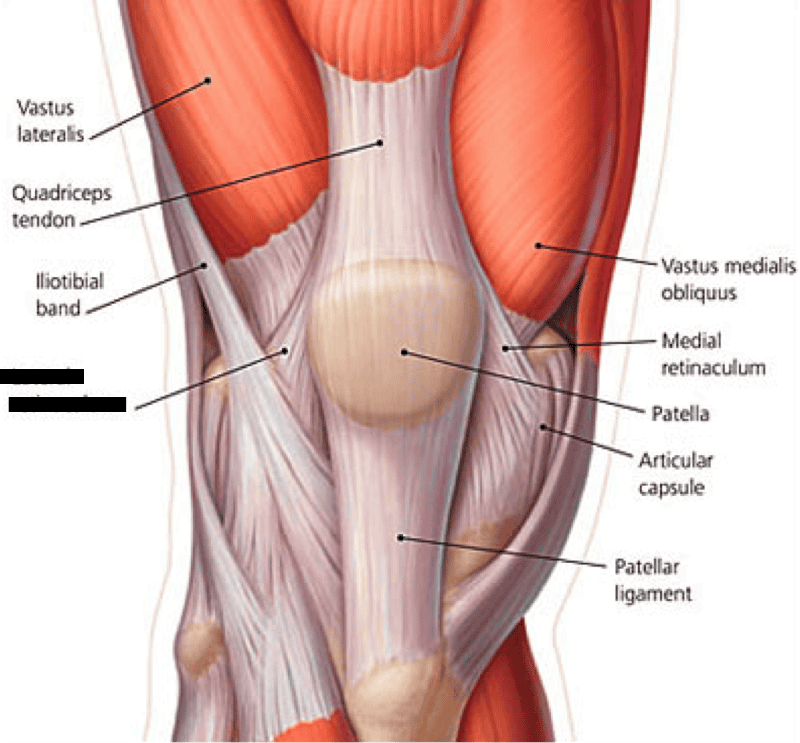 | back 44 Lateral retinaculum |
front 45 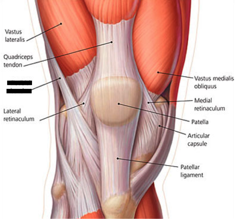 | back 45 Iliotibial band |
front 46 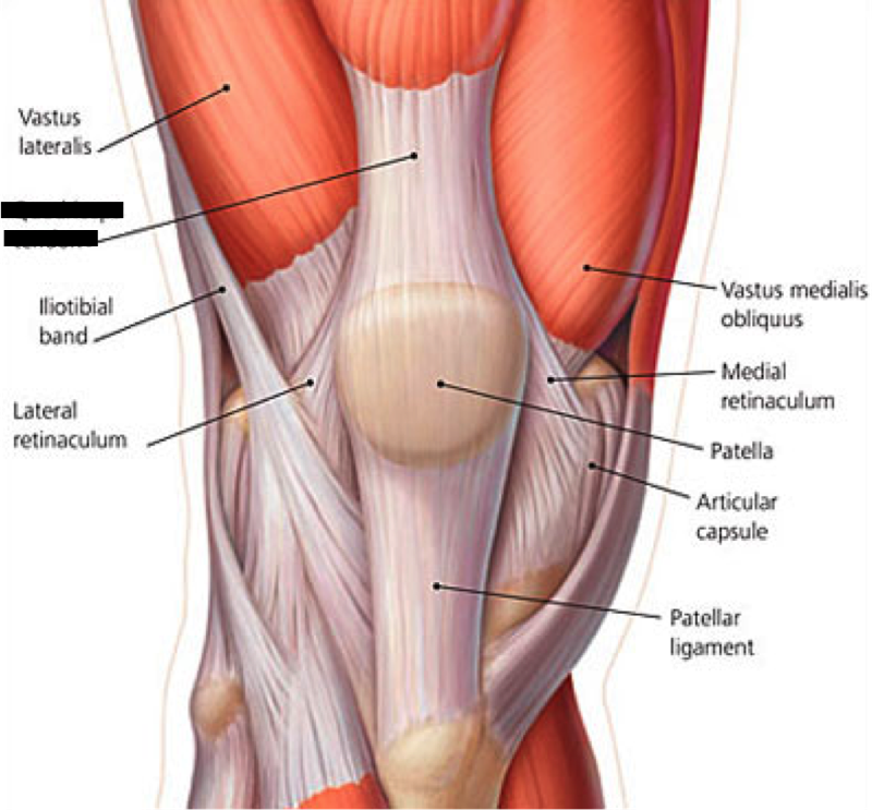 | back 46 Quadriceps Tendon |
front 47 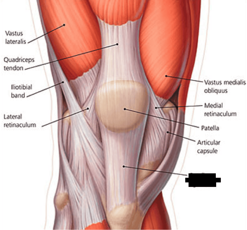 | back 47 Patellar Tendon |
front 48 What is the quadriceps group? | back 48 Rectus Femoris Vastus Medialis Vastus Intermedius Vastus Lateralis |
front 49 What muscles are involved in a Groin strain? | back 49 Pectineus Gracilis Adductor Brevis Adductor Longus Adductor Magnus |
front 50 What are the hip adductors? | back 50 Adductor Magnus Adductor Brevis Adductor Longus Gracilis Pectineus |
front 51 What do the gluteus minimum & medium do together? | back 51 Stabilize pelvis when: -Foot is on the ground -They prevent the pelvis from dropping toward opposite side during walking --Swinging leg --Trendelenburg's Sign |
front 52 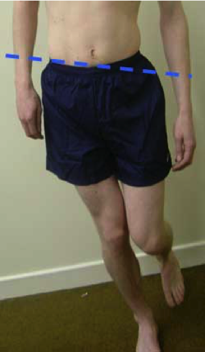 | back 52 Trendelenburg’s Sign |
front 53 What is sciatica? | back 53 Compression of the sciatic nerve Radiating or shooting pain down the leg |
front 54 What are the "Deep 6" External rotators? | back 54 Piriformis Gemellus superior Obturator internus Obturator externus Gemellus inferior Quadratus femoris |
front 55 What are the hamstring muscles? | back 55 Biceps Femoris (long & short head) Semimembranosus Semitendinosus |
front 56 Innervation of anterior compartment | back 56 Deep peroneal nerve (L4-S1) |
front 57 Innervation of lateral compartment | back 57 Superficial peroneal nerve (L4-S1) |
front 58 Innervation of deep posterior compartment | back 58 Tibial nerve (L4-S3) |
front 59 Innervation of superficial posterior | back 59 Tibial nerve (L4-S3) |
front 60 Anterior compartment muscles | back 60 Tibialis anterior Extensor digitorum longus Extensor hallucis longus Peroneus tertius |
front 61 Lateral compartment muscles | back 61 Peroneus longus Peroneus brevis |
front 62 Superficial Posterior compartments | back 62 Gastrocnemius Soleus |
front 63 Deep Posterior compartments | back 63 Tibialis posterior Flexor digitorum longus Flexor hallicis longus |
front 64 What encloses a compartment? | back 64 Fascia |
front 65 When tendons pass behind lateral malleolus what does it make them? | back 65 Plantar flexors |
front 66 Another name for the calf muscle | back 66 Triceps surae |
front 67 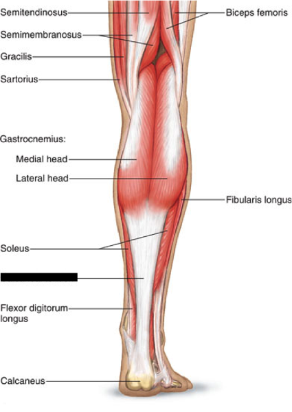 | back 67 Calcaneal tendon |
front 68 Strengthening the Triceps surae in the standing position | back 68 Work primarily gastrocnemius because it is stretched (lengthened position) & in direct line of pull |
front 69 Strengthening the Triceps surae in the seated position | back 69 Work primarily soles because gastrocnemius is relaxed (shortened position) across knee joint |
front 70 Hip abductors | back 70 Gluteus Maximus Gluteus medius Gluteus minimus TFL Sartorius Piriformis (when hip is flexed) |
front 71 Hip adductors | back 71 Adductor Magnus Adductor Longus Adductor Brevis Pectineus Gracilis Psoas major Iliacus Gluteus Maximus (lower fibers) |
front 72 Internal (Medial) Rotation | back 72 Semimembranosus Semitendinosus Gluteus Medius (anterior fibers) Gluteus Minimus |
front 73 External (Lateral) Rotation | back 73 Biceps Femoris Gluteus Maximus (all fibers) Gluteus Medius (posterior fibers) Sartorius Piriformis Quadratus Femoris Obturator Internus/Externus Gemellus Superior/Inferior Iliopsoas |
front 74 Hip Flexion | back 74 Rectus Femoris Gluteus medius (anterior fibers) Gluteus minimus Add. magnus/longus/brevis (assists) Pectineus TFL Sartorius Psoas major/Iliacus |
front 75 Hip Extension | back 75 Biceps femoris Semitendinosus Semimebranosus Gluteus Maximus (all fibers) Gluteus Medius (posterior fibers) Adductor Magnus (posterior fibers) |
front 76 Internal (Medial) Rotation | back 76 Semitendinosus Semimembranosus Gracilis Sartorius Popliteus |
front 77 External (Lateral) Rotation | back 77 Biceps Femoris |
front 78 Knee Flexion | back 78 Biceps Femoris Semitendinosus Semimembranosus Gracilis Sartorius Gastrocnemius Popliteus Plantaris (weak) |
front 79 Knee Extension | back 79 Rectus Femoris Vastus Lateralis Vastus Medialis Vastus Intermedius |
front 80 Plantar Flexion | back 80 Gastrocnemius Soleus Tibialis Posterior Peroneus Longus Peroneus Brevis Flexor Digitorum Longus (weak) Flexor Hallucis longus (weak) Plantaris (weak) |
front 81 Dorsiflexion | back 81 Tibialis Anterior Extensor Digitorum Longus Extensor Hallucis Longus |
front 82 Inversion | back 82 Tibialis Anterior Tibialis Posterior Flexor Digitorum Longus Flexor Hallucis Longus Extensor Hallucis Longus |
front 83 Eversion | back 83 Peroneus Brevis Peroneus Longus Extensor Digitorum Longus |