Print Options

Card layout: ?

← Back to notecard set|Easy Notecards home page

Instructions for Side by Side Printing
  1. Print the notecards
  2. Fold each page in half along the solid vertical line
  3. Cut out the notecards by cutting along each horizontal dotted line
  4. Optional: Glue, tape or staple the ends of each notecard together
  1. Verify Front of pages is selected for Viewing and print the front of the notecards
  2. Select Back of pages for Viewing and print the back of the notecards
    NOTE: Since the back of the pages are printed in reverse order (last page is printed first), keep the pages in the same order as they were after Step 1. Also, be sure to feed the pages in the same direction as you did in Step 1.
  3. Cut out the notecards by cutting along each horizontal and vertical dotted line
To print: Ctrl+PPrint as a list

7 notecards = 2 pages (4 cards per page)

Viewing:

Anatomy of the veins of the trunk - Lecture 6

front 1

Describe the features and tributaries of the Superior Vena Cava (SVC)

back 1

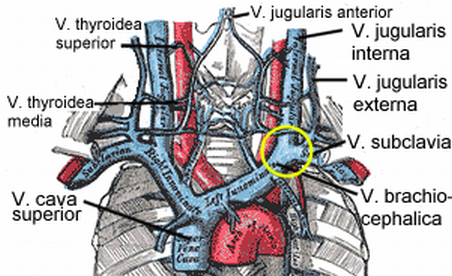
Tributaries from R. and L.Brachiocephalic veins

  • Brachiocephalic formed by tributaries of subclavian, internal jugular and internal thoracic veins
    • Subclavian tributary are external jugular veins

Single Azygous vein tributary

Begins in lower border of 1st costal cartilage and descends behind 2nd-3rd intercostal spaces into RA

L.Lateral border is aortic arch and trachea

R.Lateral border is pleura and right upper lobe of lung

Anterior border is thymus and manubrium

front 2

Describe the features and tributaries of the Inferior Vena Cava (IVC)

back 2

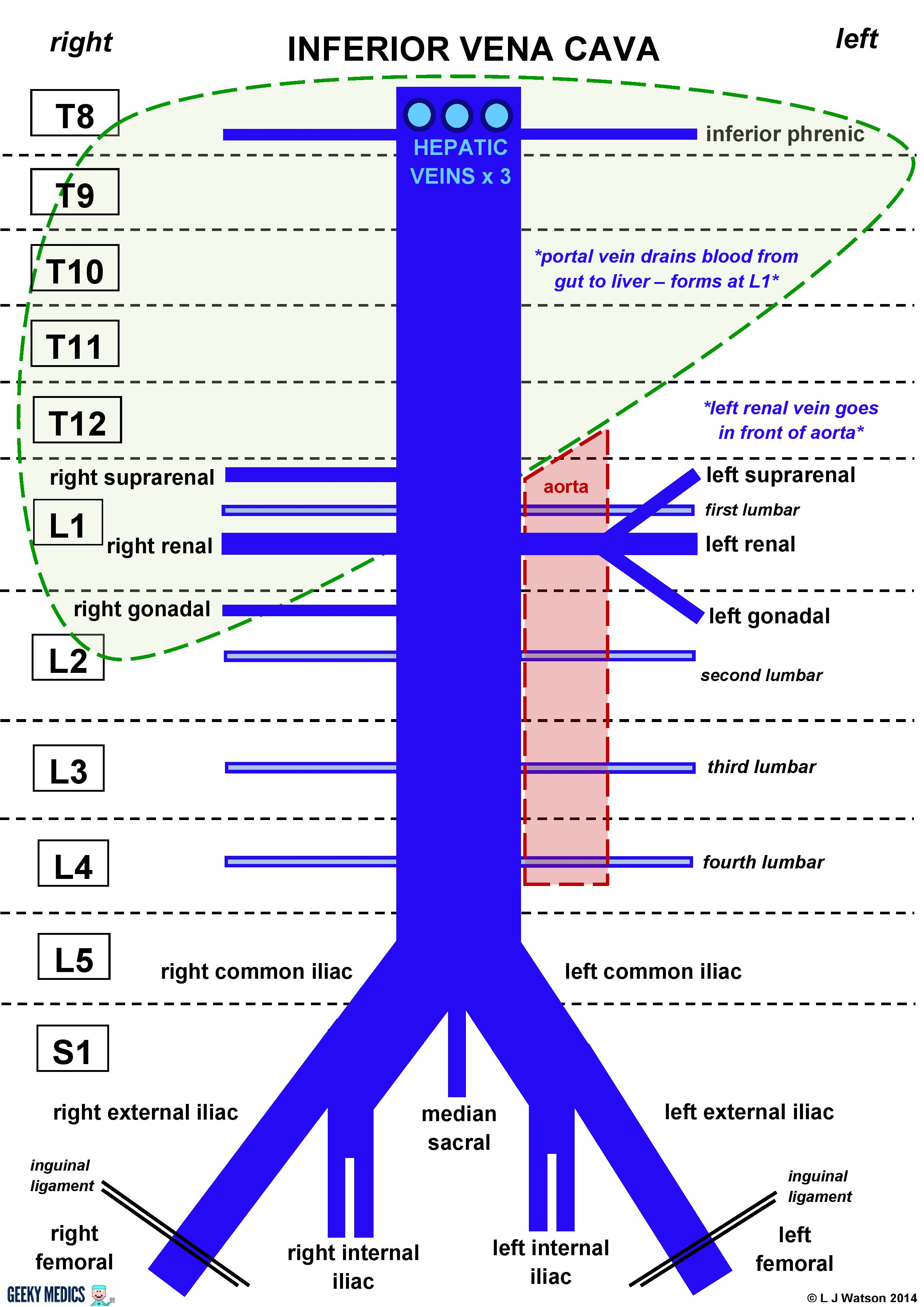
Formed by the joining of the common iliac veins at L5

Is a retroperitoneal structure - Posterior to abdominal cavity next to vertebral colum

Anastomoses the azygous system on the Right side of the vertebral column

Caval opening is at T8

Right side

  • Gonadal vein and suprarenal vein drain into IVC

Left side

  • Gonadal vein and suprarenal vein drain into renal vein then into IVC
  • L.Inferior phrenic veins drain into L.Renal vein

All lumbar and hepatic veins drain into IVC

Tributaries from superior to inferior

  • Hepatic - T8
  • Inferior phrenic - T8
    • 1-2 per side from diaphragm
  • R.Suprarenal - L1
  • Renal - L1
  • R.Gonadal - L2
  • Lumbar - L1-5
  • Common iliac - L5
    • Tributaries of internal and external iliac veins

front 3

Describe the epigastric veins, their tributaries and their relationship to the anterior abdominal wall

back 3

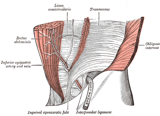
The veins accompany the arteries in the abdominal wall

Superior epigastric

  • Drains into internal thoracic vein - inferior to external iliac vein

Both the superior and inferior anastomose with each other at the level of the umbilicus, and with paraumbilical veins

Inferior epigastric vein

  • Arises from the superior epigastric vein
  • Drains into external iliac vein

front 4

Describe the azygous system, its function and the relation to the SVC

back 4

The azygous or hemiazygous vein arise from the ascending lumbar vein from the lumbar veins and lateral sacral vein that come from the common iliac vein

  • When these ascending lumbar veins cross the subcostal vein (vein along the bottom of the 12th rib) it becomes the azygous or hemiazygous

Comprises the azygous, hemiazygous, accessory hemiazygous and the L.superior intercostal vein

Function

  • Drain the posterior abdominal wall, thoracic wall and the upper lumbar region via the lumbar and posterior intercostal veins

Azygous

  • Arises from the union of the R.ascending lumbar and the R.subcostal vein ~T12
  • Drains the R.lower 8 posterior intercostal veins and the R.bronchial veins
  • Enters thorax via aortic hiatus
  • Ascends along vertebral column on the RHS within the posterior mediastinum
  • Arches over R.main bronchus T5-6
  • Enters SVC at T4 or can enter into R.Brachiocephalic or R.subclavian
  • Tributaries
    • Hemiazygous joins at T8 as it crosses from L. to R.
    • R.superior intercostal vein joins superiorly

Hemiazygous

  • May or may not be present
  • Arises from the L.ascending lumbar vein
  • Passes through L.crus of diaphragm
  • Drains L.9-11 posterior intercostal veins and L.subcostal vein
  • Crosses vertebral column from L. to R. at T8 where it anastomoses with the azygous

Accessory hemiazygous

  • Not always present
  • On the LHS
  • Arises from the 4th-8th L.posterior intercostal veins
  • Drains L.bronchial vein
  • Crosses behind the oesophagus at T8 to anastomose with the azygous or can anastomose superiorly to the L.brachiocephalic vein to join into the SVC

front 5

List all of the veins and their tributaries of the trunk (abdomen, thorax and neck region)

back 5

  1. SVC
  2. L. and R. Brachiocephalic
  3. Internal Jugular
  4. External Jugular
  5. Subclavian
  6. IVC
  7. Hepatic veins
  8. R. Suprarenal
  9. R. gonadal
  10. R. Renal
  11. L. Suprarenal
  12. L. Renal
  13. L. Gonadal
  14. Common iliac
  15. Internal iliac
  16. External iliac
  17. Femoral
  18. R.Ascending lumbar vein
  19. Azygous
  20. Hemiazygous
  21. Accessory Hemiazygous
  22. L.Ascending lumbar

front 6

Describe in brief the major drainage vessels of the 3 section of the gut (fore, mid and hind) via the hepatic portal system

back 6

Foregut ~ oesophagus, duodenum, stomach

  • Drained by splenic vein
  • Supplied by coeliac trunk

Midgut ~ Small intestine

  • Drained by superior mesenteric vein
  • Supplied by superior mesenteric artery

Hindgut ~ Large intestine

  • Drained by inferior mesenteric vein
  • Supplied by inferior mesenteric artery

front 7

Describe the relationship between surface/cross sectional anatomy and that truncal venous system

back 7

R.1st costal cartilage = Brachiocephalic veins become SVC

Manubriosternal joint = Azygous terminates at SVC

Transpyloric plane = Confluence of superior mesenteric and splenic veins

L5 = Common iliac veins joint o form IVC