Instructions for Side by Side Printing
- Print the notecards
- Fold each page in half along the solid vertical line
- Cut out the notecards by cutting along each horizontal dotted line
- Optional: Glue, tape or staple the ends of each notecard together
Human Anatomy and Physiology: Chapter 3 Cells The Living Unit
front 1 Cell | back 1 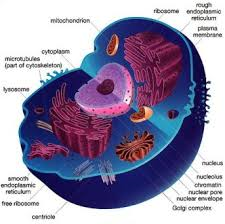 The basic structural and functional unit of a living organisms. |
front 2 Plasma Membrane | back 2 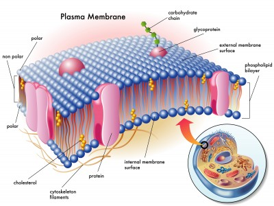 Defines the extent of the cell and is the flexible outer boundary. The lipid bilayer and proteins constantly changing in fluid mosaic. Plays a dynamic role in cellular activity. Separates intracellular fluid from extracellular fluid. Contains thousands of substances (amino acids, sugars, fatty acids, vitamins, hormones, salts, waste products) Allows cell to take in what it needs, when it needs it, and keep out what it does not |
front 3 What are the 3 basic parts of a human cell? | back 3 Plasma membrane Cytoplasm Nuclues |
front 4 Cytoplasm | back 4 Intracellular fluid |
front 5 Nucleus | back 5 Control Center |
front 6 Phospholipid (lipid bilayer) | back 6  75% of membrane lipids Phosphate heads are polar and hydrophilic Fatty acid tails are non-polar and hydrophobic |
front 7 Glycolipids | back 7  5% of membrane lipids Lipids with polar sugar groups on outer membrane surface Fatty acid tails are non-polar |
front 8 Cholesterol | back 8  20% of membrane lipids Increases membrane stability Hydroxyl group is polar fused ring system is non-polar |
front 9 Membrane Proteins | back 9  Allow communication with environment 1/2 of the mass of plasma membrane Most specialized membrane functions Some float freely, others are "tethered" to intracellular structures that make up the cytoskeleton and are restricted in their movement. 2 types - Integral and peripheral |
front 10 Integral Proteins | back 10 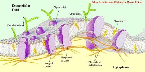 Firmly inserted into the lipid bilayer Some protrude from one membrane face only, but most are transmembrane that span the entire membrane and protrude on both sides Have both hydrophobic and hydrophilic regions Can interact with lipid tails and water Function as transport proteins (channels and carriers), enzymes, or receptors |
front 11 Peripheral Proteins | back 11  Loosely attached to integral proteins Include filaments on intracellular surface for membrane support Function as enzymes; motor proteins for shape change during cell division and muscle contraction; cell-to-cell connections |
front 12 Six Functions of Membrane Proteins | back 12
|
front 13 Lipid Rafts | back 13 ~ 20% of outer membrane surface Contain phospholipids, sphingolipids and cholesterol More stable; less fluid that rest of membrane May function as stable platforms for cell-signaling molecules, membrane invagination, and other functions |
front 14 The Glycocalyx | back 14 -"Sugar covering" at cell surface Lipids and proteins with attached carbohydrates (sugar groups) - Every cell has different patterns of sugars Specific biological markers for cell to cell recognition Allows immune system to recognize "self" and "non-self" Cancerous cells change it continuously |
front 15 Cell Junctions - Tight Junctions | back 15  Adjacent integral proteins fuse to form impermeable junction encircling cell Prevents fluids and most molecules from moving between cells Tight junctions can be found between epithelial cells in the GI tract |
front 16 Cell Junctions - Desmosomes | back 16  "Rivets" or "spot-welds" that anchor cells together at plaques (thickening on plasma membrane) Linker proteins between cells contain plaques Keratin filaments extend through cytosol to opposite plaque giving stability to cell Reduces possibility of tearing They are abundant in tissues subjected to great mechanical stress |
front 17 Cell Junctions - Gap Junctions | back 17 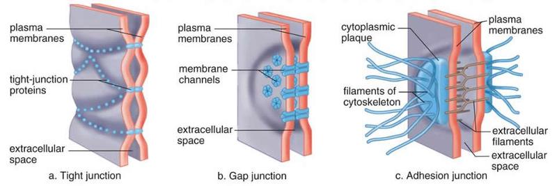 Communication between adjacent cells Transmembrane proteins form pores (connexons) that allow small molecules to pass cell to cell For spread of ions, simple sugars, and other small molecules between cardiac or smooth muscle cells Found in excitable tissues, such as the heart and smooth muscle to synchronize electrical activity and contraction |
front 18 Passive Process Membrane Transport | back 18 Diffusion and filtration No ATP required Substances move down its concentration gradient |
front 19 Diffusion | back 19 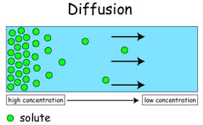 The tendency of molecules or ions to move from an area where they are in higher concentration to an area where they are in lower concentration. Three forms - simple diffusion carrier and channel mediated facilitated diffusion Osmossis |
front 20 Simple Diffusion | back 20 Non-polar and lipid soluble (hydrophobic) substances diffuse directly through the lipid bilayer. E.g. oxygen, carbon dioxide, fat-soluble vitamins |
front 21 Facilitated Diffusion | back 21 Certain molecules and ions are transported passively even though they are unable to pass through the lipid bilayer. They move through the membrane by binding to protein carriers in the membrane and is ferried across or by moving through water filled protein canals. E.g. glucose, amino acids, and ions |
front 22 Channel Mediated Facilitated Diffusion | back 22 Aqueous channels formed by transmembrane proteins Selectively transport ions or water Two Types - Leakage Channels - always open Gated Channels - Controlled by chemical or electrical signals |
front 23 Osmosis (Passive Process) | back 23 Movement of solvent across selectively permeable membrane (water) Water diffuses through plasma membranes though lipid bilayer/through specific water channels called aquaporins Occurs when water concentration different on the two sides of a membrane |
front 24 1. Osmolarity 2. Hydrostatic Pressure 3. Osmotic Pressure | back 24 1. Measure of total concentration of solute particles 2. The back pressure exerted by water against the membrane 3. The tendency of water to move into the cell by osmossis |
front 25 Does osmosis cause the cell to shrink? | back 25 Yes, because change in cell volume disrupts cell function, especially in neurons. |
front 26 Isotonic solutions | back 26  Have the same concentrations of non-penetrating solutes as those found in cells. Cells will retain their normal size and shape. |
front 27 Hypertonic Solutions | back 27 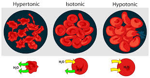 Have a higher concentration of non-penetrating solutes as those found in cells. Cells lose water by osmosis and shrink. the solution contains a higher concentration of solutes that are present inside the cell. |
front 28 Hypotonic Solutions | back 28  Solutions are more dilute (contain a lower concentration of non-penetrating solutes) than cells. Cells take on water by osmosis until they become bloated and burst (lyse). Lower concentration of solutes that are present inside cells. |
front 29 Membrane Transport - Active Processes | back 29 Two types - Active Transport Vesicular Transport Both require ATP to move solutes across a living plasma membrane because -Solute is too large for chemicals -Solute not lipid soluble -Solute not able to move down concentration gradient |
front 30 Active Transport- Primary Active Transport | back 30 Requires carrier proteins (solute pumps) Bind specifically and reversibly with substance Moves against concentration gradient Requires energy directly from ATP hydrolysis |
front 31 Active Transport- Secondary Active Transport | back 31 Requires carrier proteins (solute pumps) Bind specifically and reversibly with substance Moves against concentration gradient Requires energy indirectly from ionic gradients created by primary active transport |
front 32 Sodium - Potassium Pump | back 32 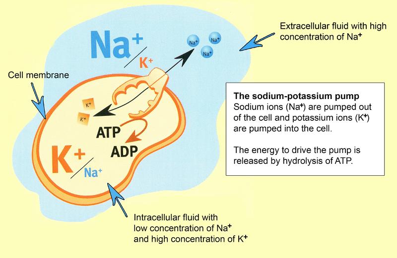 Located in all plasma membranes Involved in primary and secondary active transport of nutrients and ions Pumps against Na+ and K+ gradients to maintain high intracellular K+ concentration and high extracellular Na+ concentration Maintains electrochemical gradients essential for functions of muscle and nerve tissues It drives Na+ out of the cell against a steep concentration gradient and pumps K+ back into the cell. It is crucial for cardiac, skeletal muscle, and neuron function. |
front 33 Secondary Active Transport Cotransport | back 33 Always transports more that one substance at a time |
front 34 1 - Symport 2 - Antiport | back 34 1 - Substances transported in the same direction 2 - Substances transported in the opposite directions |
front 35 Vesicular Transport | back 35 Transport of large particles, macromolecules, and fluids across membrane in membranous sacs called vesicles Requires cellular energy (ATP) |
front 36 Vesicular Transport Functions 1) Exocytosis 2) Endocytosis 3) Transcytosis 4) Vesicular Trafficking | back 36 1) transport out of cell 2) transport into cell (phagocytosis, pinocytosis, receptor-mediated endocytosis 3) Transcytosis - transport into, across, then out of the cell 4) transport from one area or organelle in cell one cell to another |
front 37 Phagocytosis (Endocytosis) | back 37 Pseudopods engulf solids (often large or solid material, such as bacteria, cell debris, or inanimate particles) and bring them into the cell's interior. These cells (phagocytes) are experts at ingesting and disposing of bacteria, other foreign substances, and dead tissue cells. |
front 38 Pinocytosis (Endocytosis) | back 38 Fluid phase endocytosis (cell drinking) Plasma membrane unfolds, bringing extracellular fluid and dissolved solutes inside of cell, then fuses with an endosome. A routine activity in most cells Most cells utilize to "sample" environment Nutrient absorption in the small intestine |
front 39 Receptor-mediated endocytosis | back 39 Allows specific endocytosis and transcytosis cells use to concentrate materials in limited supply Clathrin-coated pits provide main route for endocytosis and transcytosis Uptake of enzymes, low-density lipoproteins, iron, insulin, and viruses, diphtheria, and cholera toxins |
front 40 Exocytosis | back 40 Usually activated by cell surface signal or change in membrane voltage Substances enclosed in secretory vesicle v-snares - on vesicle t-snares - on membrane and bind Functions - hormone secretion, neurotransmitter release, mucus secretion, ejection of wastes |
front 41 Generation of Resting Membrane Potential (RMP) | back 41 Produced by separation of oppositely charged particles (voltage) across me membrane in all cells Cells described as polarized Typically ranges from -50 to -100 millivolts depending on cell type |
front 42 Selective Diffusion Establishes RMP | back 42 In many cells Na+ affects RMP Attracted into cell due to negative charge |
front 43 Potassium's role in Resting Membrane Potential | back 43 The resting membrane potential is largely determined by K+ because at rest the membrane is much more permeable to K+ than Na+. |
front 44 Sodium's role in Resting Membrane Potential | back 44 Sodium also contributes to a resting membrane potential because sodium is strongly attracted to the cell interior by its concentration gradient. |
front 45 Ligands (1st messenger) | back 45 Binds to the receptor, changes shape and activates. |
front 46 G Protein | back 46 A regulatory molecule that acts as a middleman or relay to activate (or inactivate) a membrane bound enzyme or ion channel. |
front 47 Second Messengers | back 47 An intracellular chemical signal which connects plasma membrane events to the internal metabolic machinery of the cell. |
front 48 Cyclic AMP and Ion Calcium | back 48 Typically activate protein kinase enzymes, which transfer phosphate group from ATP to other proteins. |
front 49 Cytoplasm | back 49 "Cell forming material" The cellular material between the plasma membrane and the nucleus. Made up of 3 elements - the cytosol, organelles, and inclusions |
front 50 Cytosol | back 50 The viscous, semi-transparent fluid in which the other cytoplasmic elements are suspended. |
front 51 Organelles | back 51 The metabolic machinery of the cell. Each carries out a specific function for the cell. Some synthesize proteins, others package those proteins, etc. |
front 52 Inclusions | back 52 Chemical substances that may or may not be present depending on the cell type. Such as stored nutrients, glycogen granules in the liver and muscle cells, lipid droplets in fat cells, melanin in skin and hair cells. |
front 53 Mitochondria | back 53 Threadlike or lozenge-shaped membraneous organelles. They squirm, elongate, and change shape almost continuously. They are the power plants of the cells, providing most of its ATP supply.and ribosomes Enclosed by 2 membranes. The outer membrane is smooth and featureless and the inner membrane folds inward, forming shelf like cristae (crests) that protrude into the matrix, the gel-like substance. They contain their own DNA, RNA, and ribosomes that can reproduce themselves. |
front 54 Ribosomes | back 54 Small, dark staining granules composed of proteins and a variety of RNAs. Each has 2 globular subunits that fit together like the body and cap of an acorn. The are the site of protein synthesis. Some float freely, some are attached to membranes. |
front 55 Free Ribosomes | back 55 Float freely in the cytoplasm. They make soluble proteins that function in the cytosol, as well as those imported into mitochondria and some other organelles |
front 56 Membrane Bound Ribosomes | back 56 Attached to membranes, forming a complex. They synthesize proteins destined either for incorporation into cell membranes or lysosomes, or for export from cell. |
front 57 Endoplasmic Reticulum | back 57 An extensive system of interconnected tubes and parallel membranes enclosing fluid filled cavities. It coils and twists through the cytosol, with continuous outer nuclear membrane and accounts for about half of the cell's membrane. |
front 58 Rough Endoplasmic Reticulum | back 58 The external surface is studded with ribosomes. Proteins assembled on these ribosomes thread their way into the fluid-filled interior of the ER cisterns. When complete, the newly made proteins are enclosed in vesicles for their journey to the Golgi apparatus where they undergo further processing. |
front 59 Smooth Endoplasmic Reticulum | back 59 Consists of tubules arranged in a looping network. Its network does not play a role in protein synthesis. Enzyme catalyze reactions involved with the following: metabolize lipids synthesize steroid-based hormones absorb, synthesize, and transport fats detoxify drugs breakdown stored glycogen to form free glucose (in liver cells especially) Skeletal and cardiac muscle cells have an elaborate amount that plays an important role in storing and releasing calcium ions during muscle contraction. |
front 60 Golgi Apparatus | back 60 Consists of stacked and flattened membranous sacs, shaped like hollow dinner plates, associated with timey membranous vesicles. Acts as the "traffic director" for cellular proteins Major function is to modify, concentrate, and package the proteins and lipids made at the rough ER and destined for export from the cell. The "finishing touches" The membranes are shaped like flattened rubber bands. |
front 61 Peroxisomes | back 61 Membranous sacs containing powerful oxidases and catalases Detoxify harmful or toxic substances Catalysis and synthesis of fatty acids Neutralize dangerous free radicals -Oxidases convert to H2O (also toxic) -Catalases convert H2O2 to water and oxygen |
front 62 Lysosomes | back 62 Spherical membranous bags containing digestive enzymes "Safe" site for intracellular digestion Digest ingested bacteria, viruses, and toxins Degrade nonfunctional organelles Destroy cells in injured or non-useful tissue Break down bone to release Ca2+ |
front 63 Endomembrane System | back 63 Overall function -produce, degrade, store, and export biological molecules -degrade potentially harmful substances Includes ER, Golgi apparatus, secretory vesicles, lysosomes, nuclear and plasma membranes |
front 64 Cytoskeleton | back 64 Elaborate series of rods throughout cytosol; proteins to other cell structures, non-membranous, gives it the shape of the cell 3 types
|
front 65 Microfilaments | back 65 Thinnest of cytoskeletal elements Dynamic stands of protein actin Each cell unique arrangement of strands Dense web attached to cytoplasmic side of plasma membrane-terminal web that gives strength, compression resistance Involved in cell motility, change in shape, endocytosis and exocytosis |
front 66 Intermediate Filaments | back 66 Tough, insoluble, ropelike protein fibers Composed of tetramer fibrils Resist pulling forces on cell; attach to desmosomes (Neurofilaments in nerve cells; keratin filaments in epithelial cells) |
front 67 Microtubules | back 67 Largest of cytoskeletal elements; dynamic hollow tubes; most radiate from centrosome Composed of tetramer fibrils Determine overall shape of cell and distribution of organelles Mitochondria, lysosomes, secretory vesicles attach to microtubules, moved throughout cell by motor proteins |