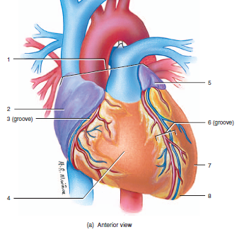
front 1 label the anterior surface features of the heart | back 1 1. base of the heart
2. auricle of right atrium
3. coronary sulcus
4. right ventricle
5. auricle of the left atrium
6. anterior interventricular sulcus
7. left ventricle
8. apex of heart |
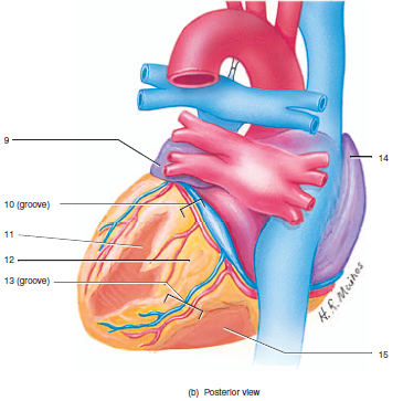
front 2 label the posterior surface features of the heart | back 2 9. left auricle
10. coronary sulcus
11. left ventricle
12. adipose tissue
13. posterior interventricular sulcus
14. right atrium
15. right ventricle |
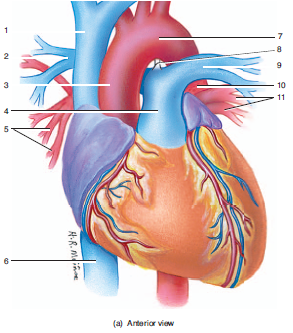
front 3 label the anterior great vessels of the heart | back 3 1. superior vena cava
2. right pulmonary artery
3. ascending aorta
4. pulmonary trunk
5. right puqlmonary veins
6. inferior vena cava
7. aortic arch
8. ligamentum arteriosum
9. left pulmonary artery
10. descending aorta
11. left pulmonary veins |
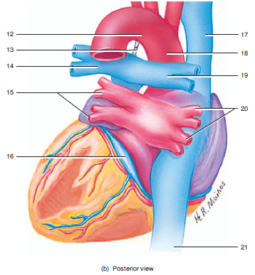
front 4 label the posterior great vessels of the heart | back 4 12. aortic arch
13. ligamentum arteriosum
14. left pulmonary artery
15. left pulmonary veins
16. coronary sinus
17. superior vena cava
18. ascending aorta
19. right pulmonary artery
20. right pulmonary veins
21. inferior vena cava |
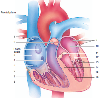
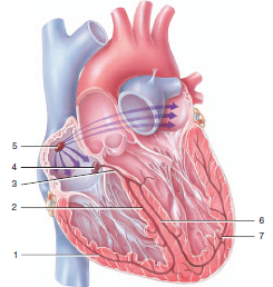
front 5 label the internal features of the heart | back 5 1. superior vena cava opening
2. pulmonary (semilunar) valve
3. right atrium
4. coronary sinus opening
5. inferior vena cava opening
6. tricuspid valve
7. right ventricle
8.trabeculae carneae
9. pulmonary vein openings
10. left atrium
11. aortic (semilunar) valve
12. bicuspid valve (mitral)
13. chordae tendineae
14. interventricular septum
15. papillary muscle
16. left ventricle |
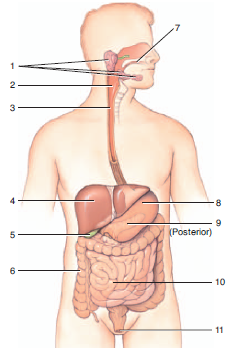
front 6 label the the organs of the digestive system | back 6 1. salivary glands
2. pharynx
3. esophagus
4. liver
5. gall bladder
6. large intestine
7. mouth
8. stomach
9. pancreas
10. small intestine
11. anus |
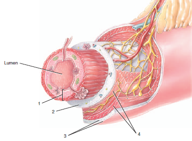
front 7 label the layers of the gastrointestinal tract | back 7 1. mucosa
2. submucosa
3. serosa
4. muscularis |
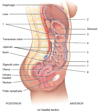
front 8 label the peritoneal folds | back 8 1. retroperitoneal organs
2. lesser omentum
3. Mesentery
4. greater omentum
5. mesocolon
6. parietal peritoneum
7. visceral peritoneum
8. peritoneal cavity |
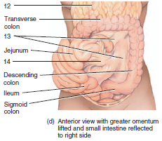
front 9 label the inner parts of the peritoneal folds | back 9 12. greater omentum (reflected)
13. Mesocolon
14. Mesentery |
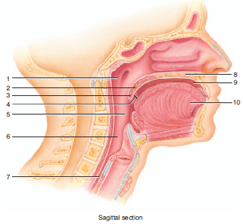
front 10 label the oral cavities and components | back 10 1. nasopharynx
2.soft palate
3. uvula
4. fauces
5. oropharynx
6. laryngopharynx
7. esophagus
8. hard palate
9. oral cavity
10. tongue |
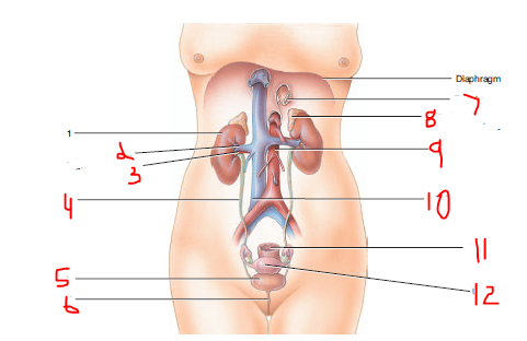
front 11 label the female urinary organs (specify right or left) | back 11 1. right kidney
2. right renal vein
3. right renal artery
4. right ureter
5. urinary bladder
6. urethra
7. esophagus
8. left adrenal (suprarenal) gland
9. abdominal aorta
10. interior vena cava
11. rectum
12. uterus |
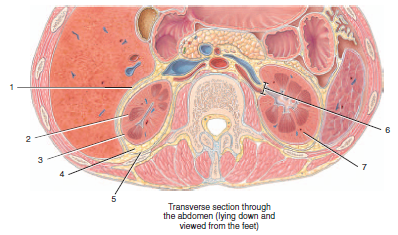
front 12 label the coverings of the kidney | back 12 1. peritoneum
2. right kidney
3. renal capsule
4. adipose capsule
5. renal fascia
6. renal hilum
7. left kidney |
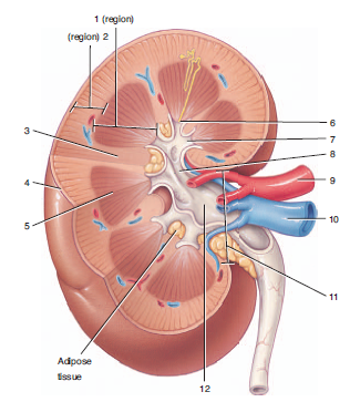
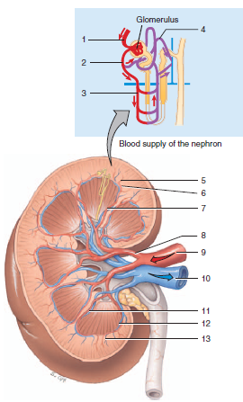
front 13 label the internal structures of the kidney | back 13 1. renal medulla
2. renal cortex
3. renal column
4. renal capsule
5. renal pyramid
6. renal papilla
7. minor calyx
8. major calyx
9. renal artery
10. renal vein
11. renal hilum
12, renal pelvis in renal sinus |
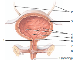
front 14 label the parts of the urinary organs | back 14 1. internal urethral sphincter
2. ureters
3. detrusor muscle of urinary bladder
4. ureteral openings
5. trigone
6. internal urethral orifice
7. urethra
8. external urethral sphincter
9. external urethral oriface |
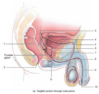
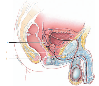
front 15 label the sagittal section of the male pelvis | back 15 1. rectum
2. anus
3. ureter
4. urinary bladder
5. ureteral opening
6. internal urethral orifice
7. prostatic urethra
8. membranous urethra
9. spongy urethra
10. external urethral orifice |
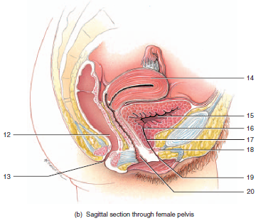
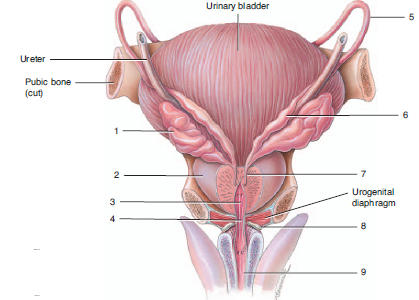
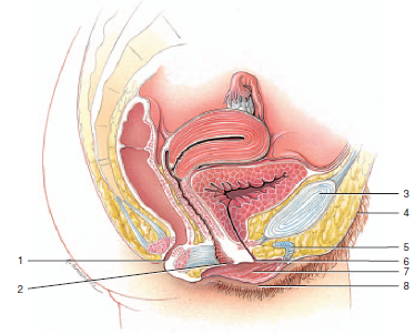
front 16 label the sagittal section of the female pelvis | back 16 11. urogenital diaphragm
12. rectum
13. anus
14. uterus
15. urinary bladder
16. internal urethral orifice
17. urethra
18. urogenitial diaphragm
19. external urethral orifice
20. vagina |
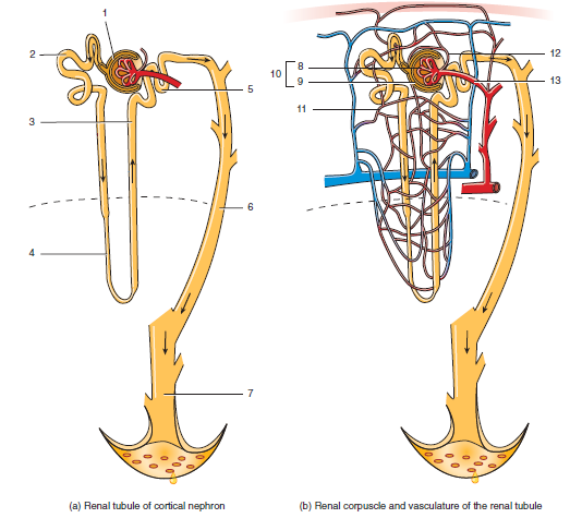
front 17 label the parts of the nephron | back 17 1. glomerular capsule
2. proximal convoluted tubule
3. ascending limb of loop of henle
4. descending limb of loop of henle
5. distal convoluted tubule
6. collecting duct
7. papillary duct
8. glomerulus
9. glomerular capsule
10. renal corpuscle
11. peritubular capillary
12. efferent arteriole
13. afferent arteriole |
front 18 label the blood supply of the kidney | back 18 1. afferent arteriole
2. efferent arteriole
3. vasa recta
4. peritubular capillaries
5. interlobular artery
6. arcuate artery
7. interlobar artery
8. segmental artery
9. renal artery
10. renal vein
11. interlobar vein
12. arcuate vein
13. interlobular vein |
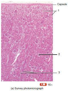
front 19 label the structure and parts | back 19 1. proximal and distal convoluted tubules (outer cortex)
2. glomerulus
3. loop of henle and collecting ducts |
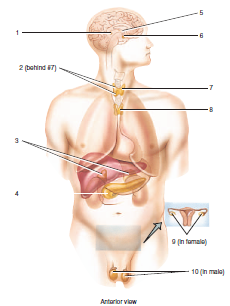
front 20 label the major endocrine glands | back 20 1. pineal gland
2. parathyroid gland
3. adrenal glands
4. pancreas
5. hypothalamus
6. pituitary gland
7. thyroid gland
8. thymus gland
9. ovaries
10. testes |
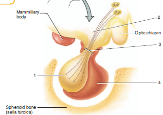
front 21 name the structure and label its parts | back 21 pituitary gland
1. posterior pituitary
2. hypothalamus
3. infundibulum
4. anterior pituitary |
front 22 name the structure and label its parts | back 22 pituitary gland
5. hypothalamus
6. infundibulum
7. anterior pituitary
8. posterior pituitary
9. posterior pituitary
10. anterior pituitary |
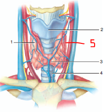
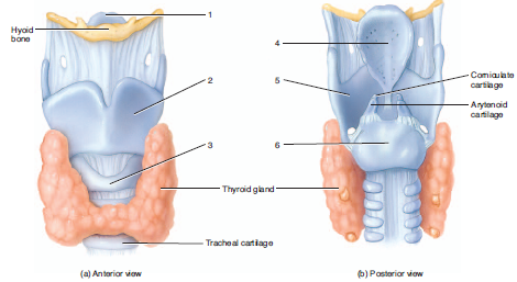
front 23 Label the parts of the anterior thyroid and parathyroid glands | back 23 1. right lobe of thyroid gland
2. thyroid cartilage of larynx
3. isthmus of thyroid gland
4. trachea
5. left lobe of thyroid gland |
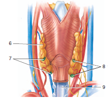
front 24 label the parts of the posterior thyroid and parathyroid glands | back 24 6. left lobe of thyroid gland
7. left parathyroid gland
8. right parathyroid glands
9. trachea |
| back 25 1. parathyroid gland
2. thyroid gland |
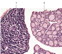
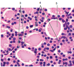
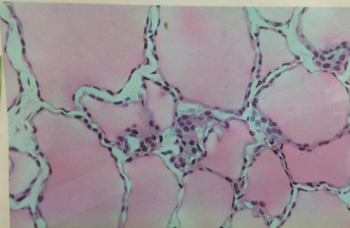
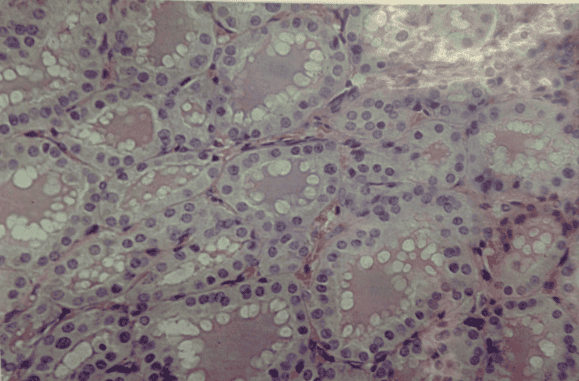
front 26 name the gland and label the parts | back 26 3. follicular cell
4. parafollicular cell
5. colloid-filled follicle |
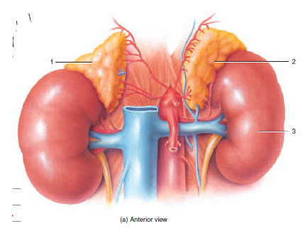
| back 27 1. right adrenal gland
2. left adrenal gland
3. left kidney |
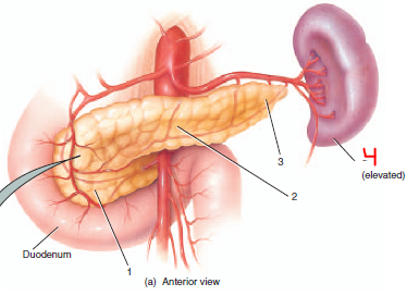
| back 28 1. head of pancreas
2. body of pancreas
3. tail of pancreas |
front 29 name the structure and label its parts | back 29 4. exocrine or acinar cells
5. pancreatic islet |
front 30 label the white blood cells | back 30 a. neutrophils
b. eosinophils
c. basophils
d. lymphocytes
e. monocytes |
front 31 trace a drop of blood through the heart
and lung by listing in order all vessels, heart chambers, and valves through which the blood passes, starting and ending with the right atrium: | back 31 1. right atrium
2. tricuspid valve
3. right ventricle
4. pulmonary (semilunar) valve
5. pulmonary trunk
6. pulmonary arteries
7. pulmonary capillaries
8. pulmonary veins
9. left atrium
10. bicuspid (mitral) valve
11. left ventricle
12. aortic (semilunar) valve
13. aorta
14. systemic arteries
15. systemic capillaries
16. systemic veins
17. venae cavae and coronary sinus
18. right atrium |
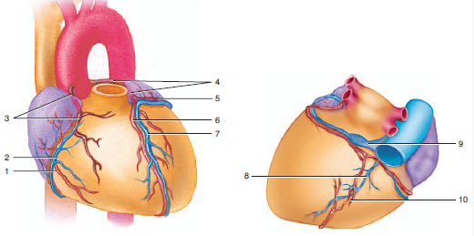
front 32 label the elements of coronary circulaion | back 32 1. marginal branch
2. small cardiac vein
3. right coronary artery
4. left coronary artery
5. circumflex branch
6. anterior interventricular branch (LAD)
7. great cardiac vein
8. middle cardiac vein
9. coronary sinus
10. posterior interventricular branch |
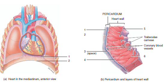
front 33 label the layers of the pericardium and heart wall | back 33 1. fibrous pericardium
2. diaphragm
3. fibrous pericardium
4. parietal layer of serous pericardium
5. pericardial cavity
6. visceral layer of serous pericardium (epicardium)
7. endocardium
8. myocardium |
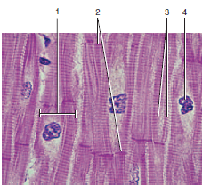
front 34 name the tissue and label the components | back 34 cardiac muscle fibers
1. cardiac muscle fiber
2. intercalated discs
3. branching fiber
4. nucleus |
front 35 label the components of the cardiac conduction system | back 35 1. Purkinje (conduction) fibers in right ventricle
2. right bundle branch
3. AV bundle (of His)
4. Atrioventricular (AV) node
5. Sinoatrial (SA) node
6. Left bundle branch
7. Purkinje (conduction) fibers in left ventricle |
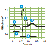
front 36 name and describe each part of the wave | back 36 1. P wave- atrial depolarization
2. P-Q Interval: time it takes for the electrical conduction (excitation) to travel through the atria and AV node to the purkinje fibers
3. QRS Complex: ventricular depolarization that spreads from the AV node, AV Bundle, right and left bundle branches, and to the purkinje (conduction) fibers just before the ventricles contract. Atrial repolarization
4. S-T Segment: time the ventricular fibers are fully depolarized
5. T wave: ventricular repolarization (just before ventricles relax)
6. Q-T interval:beginning of ventricular depolarization until the end of ventricular repolarization |
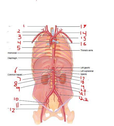
front 37 label the arterial branches and label them as ascending or descenting | back 37 1. right common carotid
2. right subclavian
3. brachiocephalic trunk
4. ascending aorta
5. right coronary
6. celiac trunk
7. right suprarenal
8. right renal
9. right gonadal
10. internal iliac
11. external iliac
12. femoral
13. left common carotid
14. left subclavian
15. aortic arch
16. left coronary
17. superior mesenteric
18. left renal
19. left gonadal
20. abdominal aorta
21. inferior mesenteric
22. left common iliac |
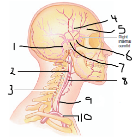
front 38 label the major arteries of the head and neck from the lateral view | back 38 1. basilar
2. internal carotid
3. vertebral
4. middle cerebral
5. anterior cerebral
6. posterior communicating
7. posterior cerebral
8. external carotid
9. common carotid
10. brachiocephalic trunk |
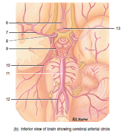
front 39 label the major arteries of the brain from the inferior view | back 39 6. anterior cerebral
7. middle cerebral
8. internal carotid
9. posterior communicating
10. posterior cerebral
11. basilar
12. vertebral
13. anterior communicating |
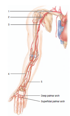
front 40 Label the arteries of the upper extremity | back 40 1. subclavian
2. axillary
3. brachial
4. radial
5. ulnar |
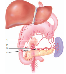
| back 41 1. left gastric
2. celiac trunk
3. common hepatic
4. splenic |
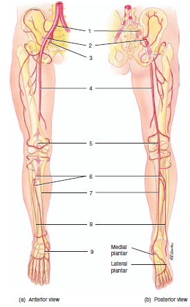
front 42 label the arteries of the lower extremity | back 42 1. common iliac
2. external iliac
3. internal iliac
4. femoral
5. popliteal
6. anterior tibial
7. posterior tibial
8. fibular
9. dorsal artery of the foot |
| back 43 1. superior vena cava
2. coronary sinus
3. inferior vena cava |
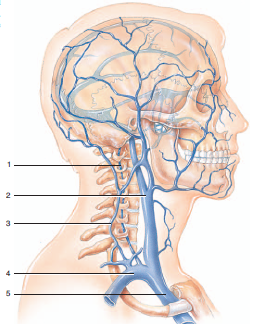
front 44 label the veins of the head and neck | back 44 1. vertebral
2. internal jugular
3. external jugular
4. subclavian
5. brachiocephalic |
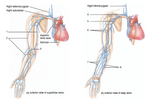
front 45 label the veins of the upper extremity | back 45 1. basilic
2. cephalic
3. median cubital
4. subclavian
5. axillary
6. brachial veins
7. radial
8. ulnar veins |
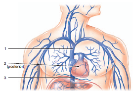
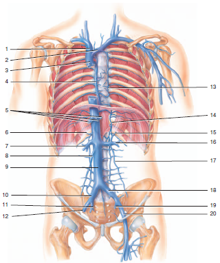
front 46 label the major veins of the abdomen, thorax, and pelvis | back 46 1. right brachiocephalic
2. left brachiocephalic
3. superior vena cava
4. azygos
5. hepatic veins
6. right suprarenal
7. right renal
8. right gonadal
9. inferior vena cava
10. right common iliac
11. right internal iliac
12. right external iliac
13. accessory hemiazygos
14. hemiazygos
15. left suprarenal
16. left renal
17. left gonadal
18. left common iliac
19. left internal iliac
20. left external iliac |
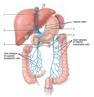
| back 47 1. hepatic
2. hepatic portal
3. superior mesenteric
4. splenic
5. inferior mesenteric |
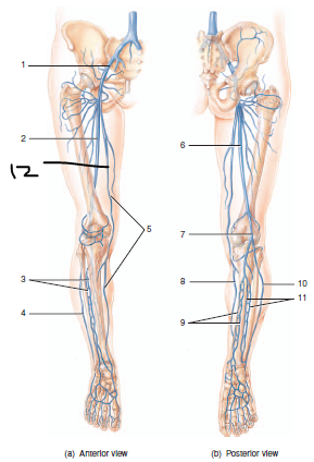
front 48 label the veins of the lower limb | back 48 1. external iliac
2. femoral
3. anterior tibial
4. small saphenous
5. great saphenous
6. femoral
7. popliteal
8. great saphenous
9. posterior tibial
10. small saphenous
11. fibular
12. great saphenous |
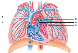
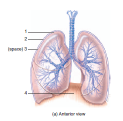
front 49 label the veins of pulmonary circulation | back 49 1. right pulmonary artery
2. right pulmonary vein
3. left pulmonary vein
4. pulmonary trunk
5. left pulmonary vein |
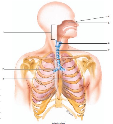
front 50 label the organs of the respiratory system | back 50 1. pharynx
2. right pulmonary bronchus
3. lungs
4. nose
5. nasal cavity
6. larynx
7. trachea |
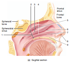
front 51 label the structures of the nasal cavity | back 51 1. internal naris
2. superior nasal concha
3. middle nasal concha
4. inferior nasal concha
5. superior nasal meatus
6. middle nasal meatus
7. inferior nasal meatus
8. external naris
9. hard palate |
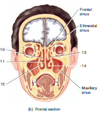
front 52 label the structures of the nasal cavities | back 52 10. middle meatus
11. nasal septum
12. inferior meatus
13. middle concha
14. inferior concha |
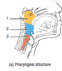
front 53 label the divisions of the pharynx | back 53 1. nasopharynx
2. oropharynx
3. laryngopharynx |
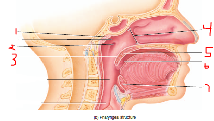
front 54 label the pharyngeal structures | back 54 1. pharyngeal tonsil
2. orifice of auditory tube
3. uvula
4. internal naris
5. soft palate
6. palatine tonsils
7. lingual tonsils |
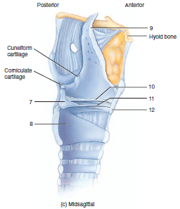
front 55 label the larynx and structures | back 55 1. epiglottis
2. thyroid cartilage
3. cricoid cartilage
4. epiglottis
5. thyroid cartilage
6. cricoid cartilage |
front 56 label the larynx structures | back 56 7. arytenoid cartilage
8. cricoid cartilage
9. epiglottis
10. ventricular fold
11. vocal fold
12. thyroid cartilage |
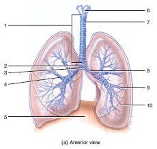
| back 57 1. trachea
2. right primary bronchus
3. carina
4. right secondary bronchus
5. diaphragm
6. larynx
7. tracheal cartilage
8. left primary bronchus
9. left tertiary bronchus
10. bronchiole |
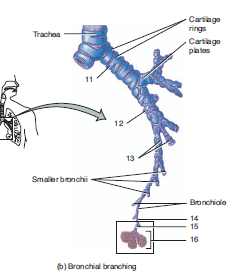
front 58 label the bronchial branching | back 58 11. primary bronchus
12. secondary bronchus
13. tertiary bronchus
14. terminal bronchus
15. respiratory bronchiole
16. alveolar sac |
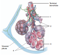
| back 59 1. respiratory bronchiole
2. alveolar ducts
3. alveolar sac
4. alveoli |
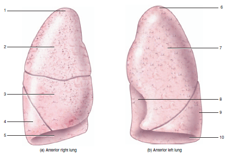
front 60 label the anterior side of the lungs | back 60 1. apex of right lung
2. superior lobe of right lung
3. middle lobe of right lung
4. inferior lobe of right lung
5. base of right lung
6. apex of left lung
7. superior lobe of left lung
8. cardiac notch of left lung
9. inferior lobe of left lung
10. base of left lung |
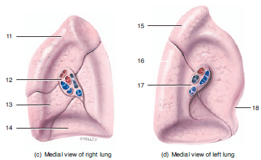
front 61 label the medial view of the lungs | back 61 11. superior lobe of right lung
12. hilus lobe of right lung
13. middle lobe lobe of right lung
14. inferior lobe lobe of right lung
15. superior lobe of left lung
16. inferior lobe of left lung
17. hilus of left lung
18. cardiac notch of left lung |
| back 62 1. visceral pleura
2. parietal pleura
3. pleural cavity
14. diaphragm |
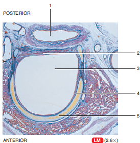
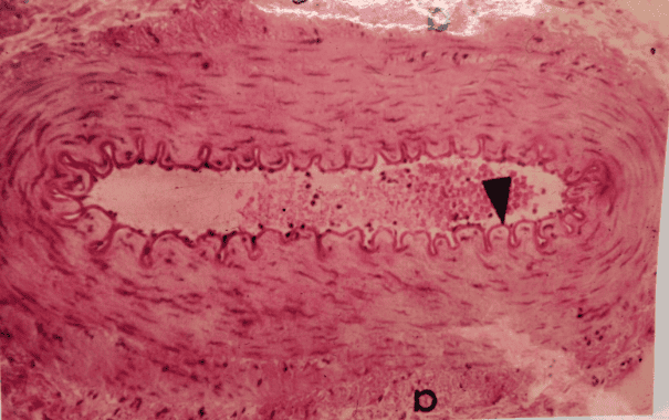
front 63 label the parts of the structures | back 63 1 is esophagus, 3 is trachea
1. lumen of the esophagus
2. trachealis muscle
3. lumen of trachea
4. epithelial lining of trachea
5. tracheal cartilage |
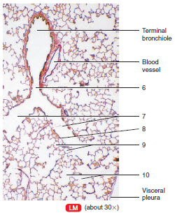
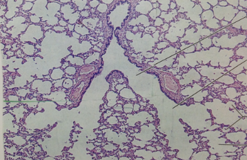
front 64 label the structure and its parts | back 64 lung
6. respiratory bronchiole
7. alveolar ducts
8. simple squamous epithelium
9. alveoli
10. alveolar sac |
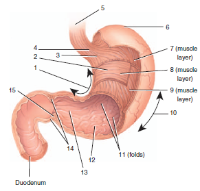
front 65 name the organs and label its parts | back 65 1. lesser curvature
2. body
3. cardia
4. lower esophageal sphincter
5. esophagus
6. fundus
7. longitudinal muscle layer
8. circular muscle layer
9. oblique muscle layer
10. greater curvature
11. rugae
12. pyloric antrum
13. pyloric canal
14. pyloric sphincter
15. pylorus |
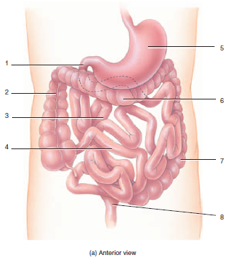
front 66 label the parts of the organs | back 66 1. duodenum
2. ascending colon
3. jejunum
4. ileum
5. stomach
6. transverse colon
7. descending colon
8. rectum |
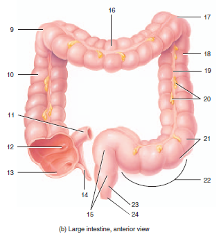
| back 67 9. right colic flexure
10. ascending colon
11. ileum
12. ileocecal sphincter
13. cecum
14. appendix
15. rectum
16. transverse colon
17. left colic flexure
18. descending colon
19. teniae coli
20. epiploic appendages
21. haustra
22. sigmoid colon
23. anal canal
24. anus |
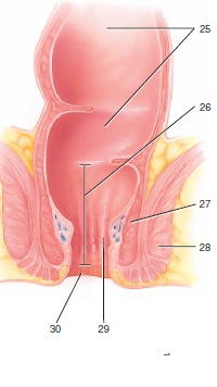
front 68 label the structure and its parts | back 68 25. rectum
26. anal column
27. internal anal sphincter
28. external anal sphincter
29. anal canal
30. anus |
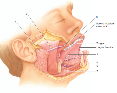
| back 69 1. parotid gland
2. parotid duct
3. opening of parotid duct
4. opening of sublingual duct
5. sublingual gland
6. submandibular duct
7. submandibular gland |
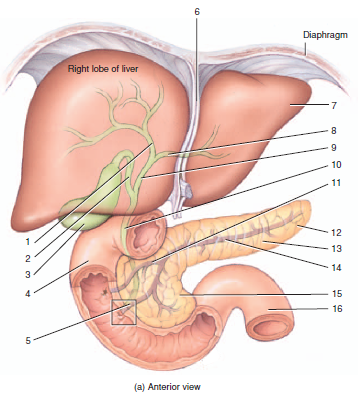
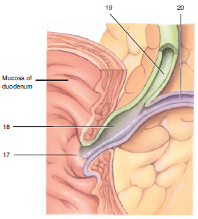
front 70 label the anterior view of the structures | back 70 1. right hepatic duct
2. cystic duct
3. gallbladder
4. duodenum
5. hepatopancreatic ampulla
6. faliciform ligament
7. left lobe of liver
8. left hepatic duct
9. common hepatic duct
10. common bile duct
11. accessory pancreatic duct
12. tail of pancreas
13. body of pancreas
14. pancreatic duct
15. head of pancreas
16. jejunum |
| back 71 17. duodenal papilla
18. hepatopancreatic ampulla
19. common bile duct
20. pancreatic duct |
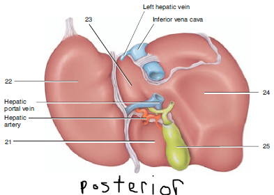
front 72 label the posterior view of the structures | back 72 21. quadrate lobe
22. left lobe
23. caudate lobe
24. right lobe
25. gall bladder |
front 73 name the organ and label its parts | back 73 esophagus
1. stratified squamous epithelium
2. lamina propria
3. circular layer of smooth muscle fibers
4. longitudinal layer of smooth muscle fibers
5. mucosa
6. submucosa
7. muscularis
8. serosa |
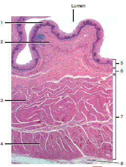
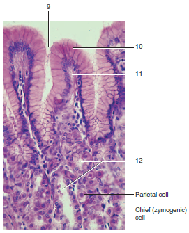
front 74 name the organ and label its parts | back 74 stomach
9. gastric pit
10. simple columnar epithelium
11. lamina propria
12. gastric glands |
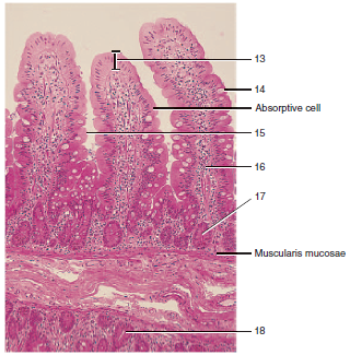
front 75 name the organ and label its parts | back 75 duodenum of small intestine
13. simple columnar epithelium
14. goblet cell
15. villus
16. lamina propria
17. intestinal gland
18. duodenal gland in submucosa |
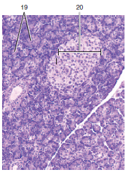
front 76 name the organ and label its parts | back 76 pancreas
19. acini
20. pancreatic islet |
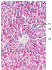
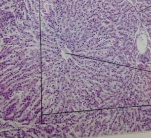
front 77 name the organ and label its parts | back 77 liver lobule
21. hepatocytes
22. sinusoids
23. central vein |
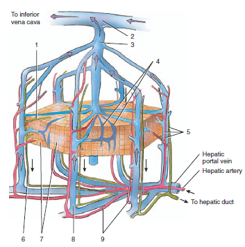
| back 78 1. bile canaliculus
2. hepatic vein
3. central vein
4. sinusoids
5. portal triad
6. bile ducts
7. hepatocytes
8. branch of hepatic portal vein
9. branch of hepatic artery |
| back 79 1. cremaster muscle
2. dartos muscle
3. scrotum
4. tunica albuginea |
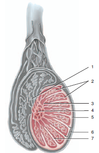
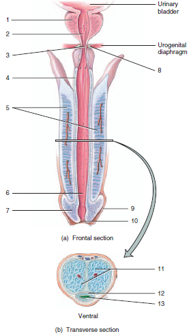
| back 80 1. efferent duct
2. seminiferous tubules
3. straight tubules
4. rete testis
5. tunica albuginea
6. lobule
7. septum |
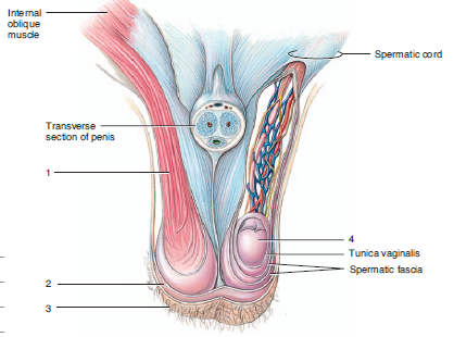
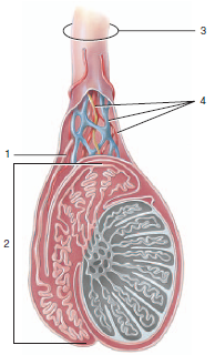
| back 81 1. ductus deferens
2. epididymis
3. spermatic cord
4. blood vessels and nerves |
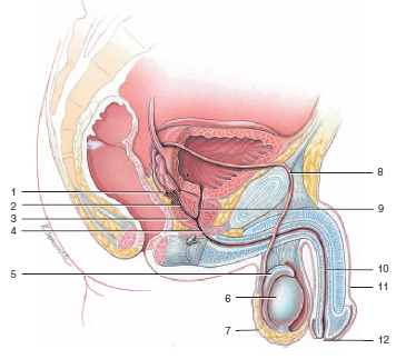
front 82 label the male reproductive organs | back 82 1. ampulla of ductus deferens
2. ejaculatory duct
3. prostate gland
4. membranous urethra
5. epididymis
6. testis
7. scrotum
8. ductus deferens
9. prostatic urethra
10. spongy urethra
11. penis
12. external urethral orifice |
front 83 label the structures (posterior view) | back 83 1. seminal vesicle
2. prostate gland
3. prostatic urethra
4. membranous urethra
5. ductus deferens
6. ampulla of ductus deferens
7. ejaculatory duct
8. bulbourethral gland
9. spongy urethra |
front 84 label the sagittal section of the male accessory sex glands | back 84 1. seminal vesicle
2. prostate gland
3. bulbourethral gland |
| back 85 Penis
1. prostate gland
2. prostate urethra
3. membranous urethra
4. corpus spongiosum penis
5. corpus cavernosa penis
6. spongy urethra
7. glans penis
8. bulbourethral gland
9. prepuce
10. external urethral orifice
11. corpora cavernosa
12. corpora spongiosum
13. spongy urethra |
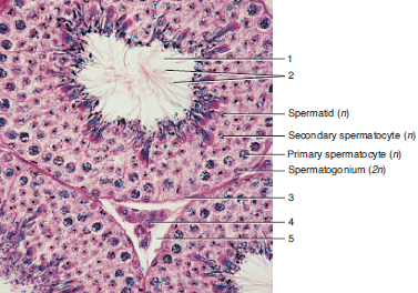
front 86 name and label the structures | back 86 seminiferous tubules
1. lumen
2. tails of sperm
3. basement membrane
4. leydig cell
5. interstitial space |
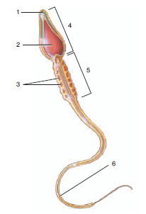
front 87 name the structure and label the parts | back 87 sperm
1. acrosome
2. nucleus
3. mitochondria
4. head
5. midpiece
6. tail |
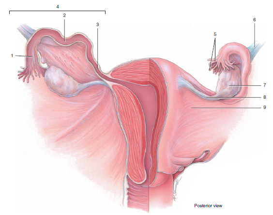
| back 88 1. uterine tube
2. infundibulum
3. ampulla
4. isthmus
5. fimbriae
6. suspensory ligament
7. ovary
8. ovarian ligament
9. broad ligament |
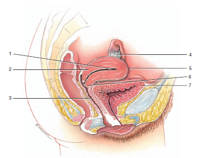
| back 89 1. uterine cavity
2. myometrium of uterus
3. vaginal canal
4. ovary
5. round ligament
6. cervix
7. urinary bladder |
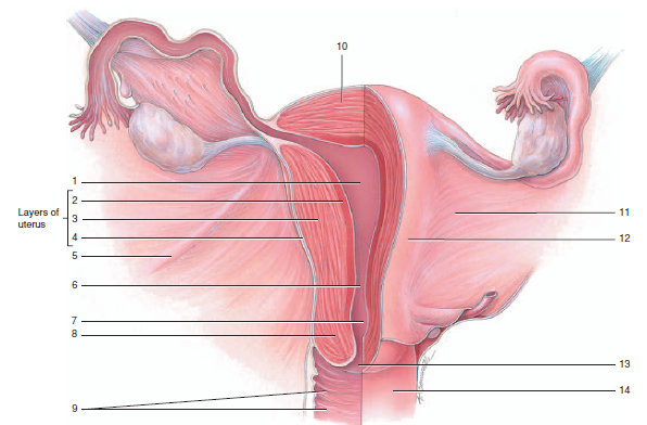
front 90 label the posterior structures | back 90 1. uterine cavity
2. endometrium
3. myometrium
4. perimetrium
5. round ligament
6. internal os
7. cervical canal
8. cervix
9. rugae of vagina
10. fundus of uterus
11. broad ligament
12. body of uterus
13. external os
14. vagina |
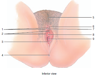
| back 91 1. anus
2. vaginal orifice
3. pubic symphysis
4. mons pubis
5. clitoris
6. external urethral orifice
7. labium minus
8. labium majus |
| back 92 1. labia majora
2. labia minora
3. hymen
4. anus
5. mons pubis
6. prepuce
7. clitoris
8. external urethral orifice
9. vaginal orifice |
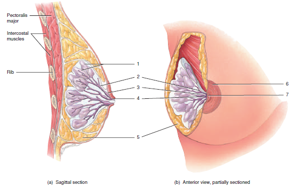
| back 93 1. lobule with alveoli
2. mammary duct
3. lactiferous sinus
4. lactiferous duct
5. adipose tissue
6. areola
7. nipple |
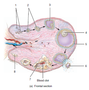
front 94 name the structure and label its parts | back 94 ovary
1. follicles
2. graafian (mature) follicle
3. ovulated oocyte
4. corpus luteum
5. corpus albicans |
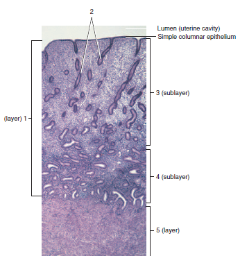
front 95 label the organ and its parts | back 95 uterus
1. endometrium
2. endometrial glands
3. stratum functionalis
4. stratum basalis
5. myometrium |
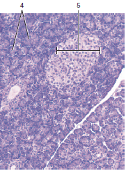
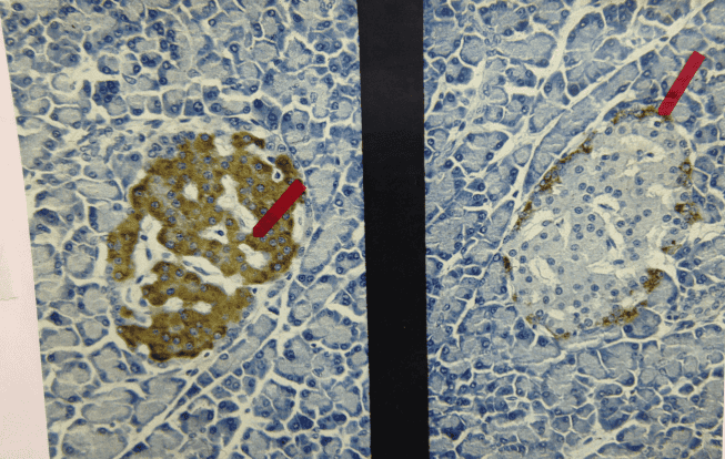
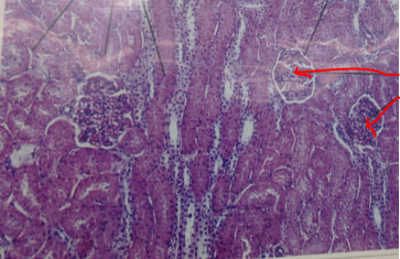
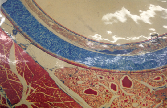
front 96 name the organ and Identify the structure the red arrow is pointing towards | back 96 organ: endocrine pancreas
arrow pointed towards: islet stained with different stains |
| |
| |
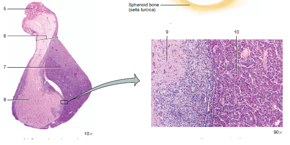
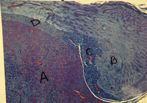
front 99 name and label the structures | back 99 pituitary gland
A= pars distalis
B= pars nervosa
C= pars tuberalis
C= pars intermedia |
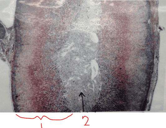
front 100 name and label the structures | back 100 adrenal glad
1- cortex
2- medulla |
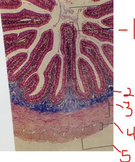
front 101 name and label the structures | back 101 small intestine
1. Villus in mucosa
2. submucosa
3. muscularis externa: inner circular layer
4. muscularis externa: outer longitudinal layer
5. serosa |
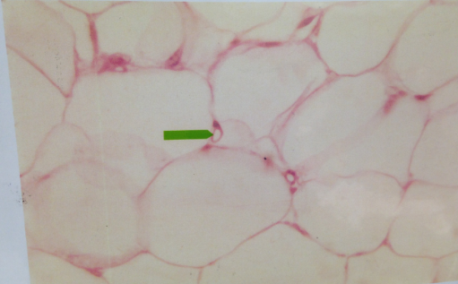
front 102 name the tissue and the structure that the green arrow is pointing towards | back 102 tissue: adipose
green arrow: capillary |
| |
front 104 name the tissue and what the arrow is pointing to | back 104 tissue: kidney (cortex)
arrow: renal corpuscles |
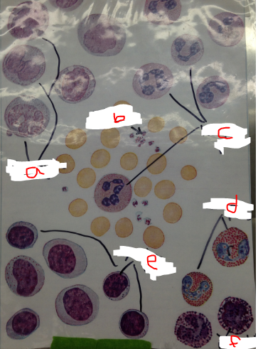
front 109 name the structures (category) and label each type | back 109 white blood cells
a. monocytes
b. platelets
c. neutrophils
d. eosinophils
e. lymphocytes
f. basophils |
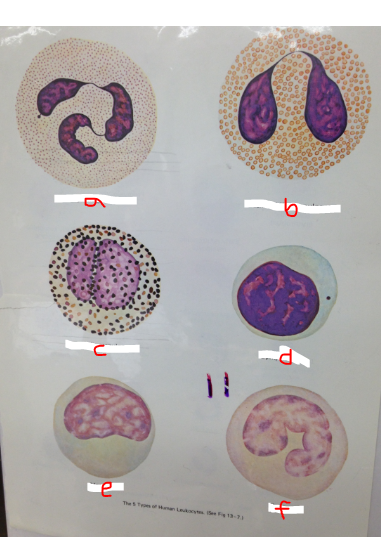
front 110 name the structures (category) and label each type | back 110 white blood cells
a. neutrophilic granulocyte
b. eosinophilic granulocyte
c. basophilic granulocytes
d. lymphocyte
e. monocyte
f. monocyte |