| back 1 o Cardiac
Striated muscle (regular array of actin and myosin in the sarcomeres)
Each muscle has a single, centered nucleus
Cells are connected by intercalated discs
o Skeletal
Hundreds of randomly located nuclei per cell (cells fuse together in development)
Striated
o Smooth
Single nuclei
Not striated |
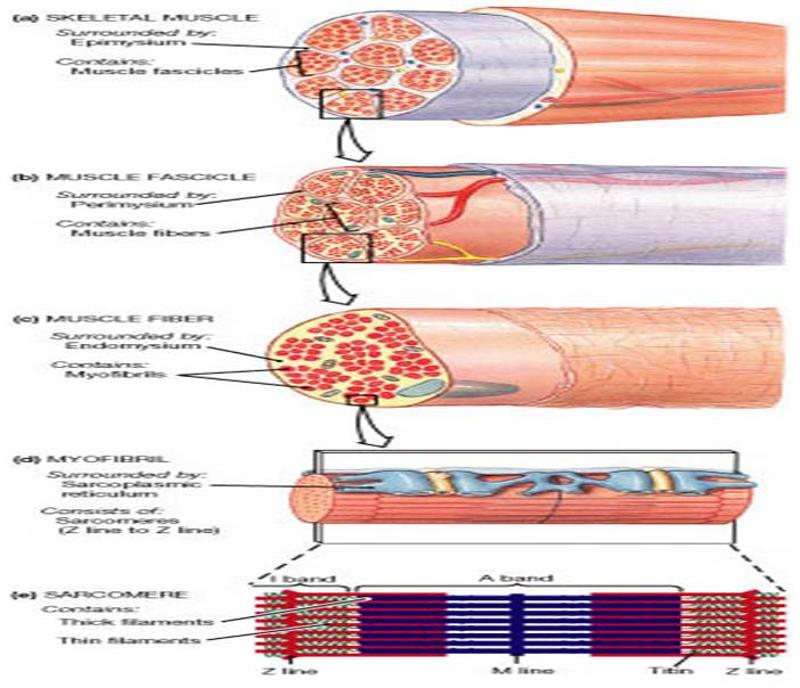
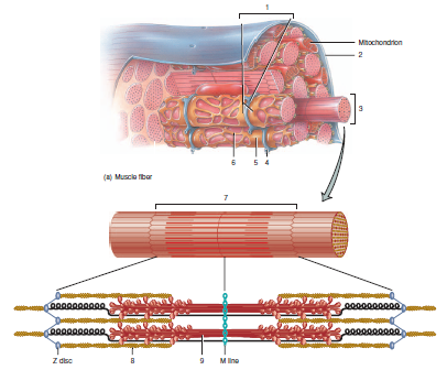
front 2 describe the muscle levels of organization and components (largest to smallest) | back 2 muscle covered with epimysium
made up of fascicles (bundle of cells) covered in perimysium
made up of muscle fibers (cells) covered in endomysium
myofibrils (made of molecular motors) make up each muscle fiber and sarcomeres are contained in each myofibril
blood vessels line in between the connective tissue coverings in between the fascicles
sarcolemma is the cell membrane of muscles with invaginations (t tubules) that go into the cells
sarcoplasmic reticulum stores calcium |
front 3 Muscle Connective tissue coverings | back 3 epimysium- covers whole muscle (allows muscles to contract with respect to each other without friction between them)
perimysium- covers the fascicles
endomysium- covers each muscle fiber
tendons- formed by the fusion of these 3 connective tissues |
| back 4 too much calcium is released and not stored in the sarcoplasmic reticulum so muscle keeps contracting and generating heat |
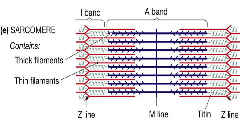
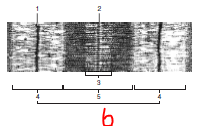
| back 5 • Actin filaments (thin filaments) are held at the Z line
• Myosin (molecular motor) makes up the thick filament and is located on the A band
o I band only has thin filaments
o A band- mostly thick filaments with some areas where thick and thin filaments overlap
o Z line are thick vertical lines
o H zone/band- only thick filaments
o M line- holds thick filaments together |
front 6 What changes in a sarcomere when a muscle contracts? | back 6 o The entire sarcomere decreases in width
o A band is the same width
o Z lines are closer together
o I band decreases in width
o Whole sarcomere decreases in width |
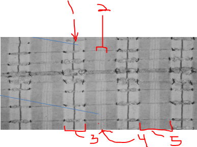
front 7 name the tissue and label the numbers | back 7 skeletal muscle
1- Z line
2- H zone
3- I band
4- M line
5- A band |
front 8 name the tissue and label the numbers | back 8 skeletal muscle
1. Z line
2. M line
3. H zone
4. I bands
5. A band
6. Sarcomere |
front 9 Uncovering the myosin binding site for muscle contraction | back 9 Actin (thin) filaments are made of G proteins with binding sites for myosin that are covered up with tropomyosin when the muscle is relaxed
sarcolemma receives an electrical impulse that releases acetylcholine which causes a change in membrane potential that travels to the t tubules that connects directly to the sarcoplasmic reticulum
sarcoplasmic reticulum then releases calcium
calcium causes a conformation change which causes tropomyosin to be released and the binding site to be uncovered |
front 10 muscle contraction -> relaxation
(starting after the myosin binding site is uncovered) | back 10 myosin binding sites are uncovered
ATP oxidation (ADP + Pi -> ATP)causes myosin to flip up in the activated position so actin can bind and begin the power stroke (pulls thin filaments in towards the center)
ATP hydrolysis (ATP-> ADP + Pi) causes ACh to be removed
sarcoplasmic reticulum recaptures calcium
myosin binding site on the actin filament are covered by tropomyosin
contraction ends |
| back 11 no more ATP is left 8-12 hours after death to replace the ADP on the myosin head, so there is no relaxation in muscle tissues |
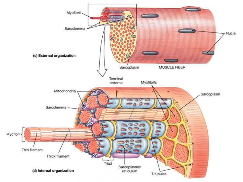
| back 12 1. triad (2 terminal cisternae and a t tubule in between)
2. sarcolemma (plasma membrane)
3. myofibril (bundled thick and thin filaments, contains a chain of sarcomeres)
4. t tubule (transmit the action potential deep inside the fiber to the terminal cisternae)
5. terminal cisternae (widened sarcoplasmic reticulum that receive t tubule signals)
6. sarcoplasmic reticulum (endoplasmic reticulum that stores calcium)
7. sarcomere (smallest structural and contractile unit of a muscle myofibril)
8. thin filament (mostly actin)
9. Thick filament (mostly myosin) |
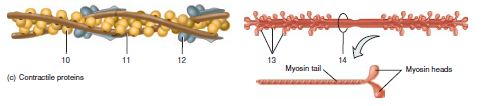
| back 13 10. actin (thin proteins)
11. tropomyosin (covers the myosin binding site until calcium releases it)
12. troponin
13. myosin crossbridges (heads project towards actin, myosin cross bridges attach to actin and pull the thin filaments past the thick filaments)
14. myosin tails |
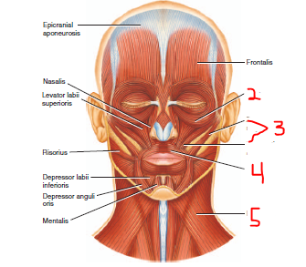
front 14 label and name the nerve that innervates them | back 14 muscles of facial expression
facial nerve
2. orbicularis oculi
3. zygomaticus
4. orbicularis oris
5. platysma |
front 15 name and the muscles of facial expression and describe how to test their nerve | back 15 facial nerve
to test, have the patient:
orbicularis oculi- shut their eyes tightly
zygomaticus- smile
orbicularis oris- purse their lips
buccinators- whistle (buccinator presses cheek inward)
platysma- depresses mandible (pouting) |
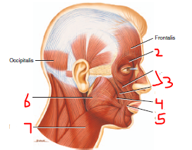
front 16 label each muscle, its nerve, and which muscle group it belongs to | back 16 2. Orbicularis Oculi (facial expression, facial nerve)
3. Zygomaticus (facial expression, facial nerve)
4. Buccinator (facial expression, facial nerve)
5. Orbicularis oris (facial expression, facial nerve)
6. Masseter (mastication, trigeminal nerve (mandibular))
7. Platysma (facial expression, facial nerve) |
| back 17 muscles of facial expression
infrahyoid (strap) muscles (raise and lower hyoid)
abdominal muscles
splenius and semispinalis capitis (deep neck muscles that hold your head up)
erector spinae (longitudinal deep back muscles responsible for posture) |
| back 18 pectoral girdle- holds the upper extremity to the trunk
upper extremity- mostly in charge of controlling the hand
pelvic girdle- holds the lower extremity to the trunk
lower extremity |
front 19 flexors and extensor organization for the upper and lower extremities | back 19 upper extremity
flexors are anterior
extensors are posterior
lower extremity
extensors are anterior
flexors are posterior |
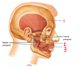
front 20 label each muscle, its nerve, and which muscle group it belongs to | back 20 1. Temporalis (trigeminal nerve, muscles of mastication)
2. Buccinator (facial nerve, facial expression)
3. Orbicularis oris (facial nerve, facial expression) |
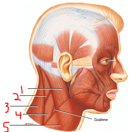
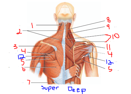
front 21 label each muscle and which muscle group it belongs to | back 21 1. Sternocleidomastoid (Neck muscles)
2. Splenius Capitis (Neck Muscles)
3. Trapezius (Scapula movers and stabilizers)
4. Levator Scapulae (scapula movers and stabilizers)
5. Platysma (facial expression) |
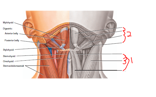
front 22 label each muscle and which muscle group it belongs to | back 22 1. Infrahyoid muscles (neck muscles)
2. Suprahyoid Muscles (neck muscles)
3. sternocleidomastoid (neck muscles) |
front 23 list the muscles of mastication, what they do, and their nerve | back 23 trigeminal nerve (mandibular)
Temporalis- elevates and retracts mandible
Masseter- elevates and retracts mandible |
front 24 list the neck muscles, what they do, and their nerve | back 24 innervated by the cervical plexus
sternocleidomastoid (flexes head (looking down), rotates "no")
infrahyoids (depresses the hyoid bone during speaking and swallowing)
suprahyoids (elevates the hyoid bone during speaking and swallowing)
splenius capitis (holds your head up, helps to turn your neck) |
front 25 label, name the muscle group | back 25 1. Platysma (facial expression)
2. Pectoralis Major (movers of the shoulder joint)
3. Latissimus Dorsi (movers of the shoulder joint)
4. Serratus Anterior (Scapula movers and stabilizers)
5. External oblique (abdominal wall)
6. sternocleidomastoid (neck muscles)
7. Trapezius (scapula movers and stabilizers)
8. Internal intercostals (respiratory muscles)
9. External intercostals (respiratory muscles)
10. Internal Oblique (abdominal wall)
11. Transverse abdominus (abdominal wall)
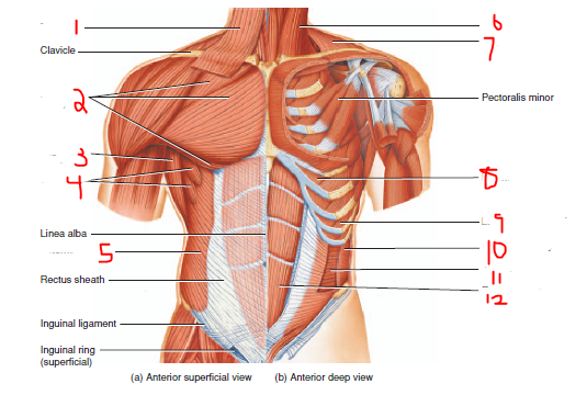
12. Rectus Abdominis (abdominal wall) |
front 26 describe the abdominal wall muscles and name their nerve | back 26 innervated by the lumbar plexus
• External oblique- most superficial side, runs down your sides (down and medially)
• Internal oblique- run up and medially under the external oblique and deeper
• Transverse abdominus-comes across (run transversely)
• rectus (straight) abdominalis- 6 pack muscles |
front 27 describe the scapula mover and stabilizer muscles and name their nerve | back 27 anterior: innervated by the brachial plexus
serratus anterior- abducts scapula and rotates it upward (throwing a punch), holds the scapula to the trunk, wings scapula back if it is injured
posterior
trapezius- shoulder shrugging, test by having a patient shrug their shoulders against resistance (accessory nerve)
levator scapulae- elevates scapula and rotates it downward (dorsal rami)
rhomboids- elevates and adducts scapula and rotates it downward, stabilizes scapula (dorsal rami) |
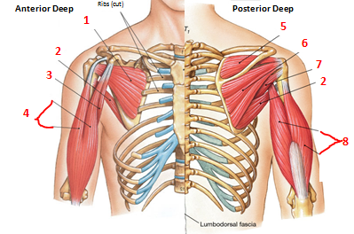
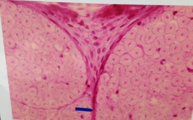
front 28 describe the movers of the shoulder joint (not including the rotator cuff) and name their nerve | back 28 all are innervated by the brachial plexus
Posterior and anterior
deltoid- powerful abductor (raises shoulder and arm away from the body, anterior medically rotates arm, posterior laterally rotated the arm)
Anterior
Pectoralis Major- flexor (moves the humerus towards the trunk), stretchs from the chest to the humerus
posterior
Teres Major- extends, adducts, and medially rotates the arm
latissimus dorsi- pulling the arm down if it is above your head (swimming)
lateral
coracobrachialis- flex and medially rotate the arm |
front 29 label each muscle and its muscle group | back 29 1. sternocleidomastoid (neck muscles)
2. Trapezius (scapula movers and stabilizers)
3. Deltoid (movers of the shoulder joint)
4. Infraspinatus (movers of the shoulder joint, rotator cuff)
5. Teres Major (movers of the shoulder joint)
6. Latissimus Dorsi (movers of the shoulder joint)
7. External Oblique (abdominal wall)
8. Splenius Capitis (neck muscles)
9. Levator Scapulae (scapula movers and stabilizers)
10. Rhomboids (scapula movers and stabilizers)
11. Supraspinatus (movers of the shoulder joint, rotator cuff)
12. Teres Minor (movers of the shoulder joint, rotator cuff) |
front 30 describe the vertebral column muscles and their nerve | back 30 erector spinae- group of longitudinal deep back muscles, responsible for posture, helps to turn the neck
innervated by the dorsal rami |
front 31 label each muscle and its muscle group | back 31 1. Subscapularis (movers of the shoulder joint, rotator cuff)
2. Teres Major (movers of the shoulder joint)
3. Coracobrachialis (movers of the shoulder joint)
4. Biceps Brachii (movers of the elbow joint)
5. Supraspinatus (movers of the shoulder joint, rotator cuff)
6. Infraspinatus (movers of the shoulder joint, rotator cuff)
7. Teres Minor (movers of the shoulder joint, rotator cuff)
8. Triceps Brachii (movers of the elbow joint) |
front 32 describe the rotator cuff muscles | back 32 SITS
innervated by brachial plexus and branches
supraspinatus- posterior, assists the deltoid in abducting the arm
infraspinatus- posterior, laterally rotates and adducts the arm
teres minor- posterior, laterally extends, adducts, and rotates the arm
subscapularis- anterior, medially rotates the arm |
| back 33 little branches that have motor and sensory rami (mixed spinal nerves)
only go to the deep back muscles and skin on the back
innervates the erector spinae and splenius capitis |
| back 34 when people break their humerus they can damage thier radial nerve (part of the brachial plexus)
cannot extend their arms/wrist, everything is flexed |
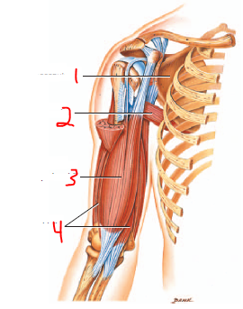
front 35 describe the movers of the elbow joint | back 35 triceps brachii- major extensor of the upper extremity
biceps brachii- flexes the arm
brachialis- most powerful flexor of the forearm
brachioradialis- weak flexor of the elbow |
front 36 label the muscles and name their muscle group | back 36 1- subscapularis (movers of the shoulder joint, rotator cuff)
2- teres major (movers of the shoulder joint)
3- biceps brachii (movers of the elbow)
4- brachialis (movers of the elbow) |
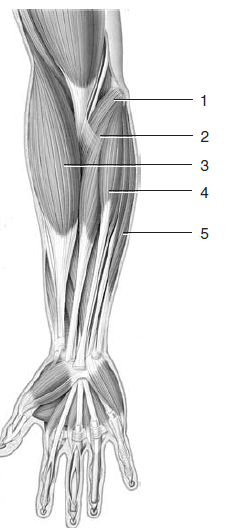
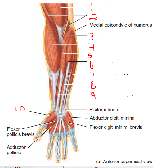
front 37 label the muscles and the muscle group | back 37 1- pronator teres (movers of the wrist and fingers)
2- flexor carpi radialis (movers of the wrist and fingers)
3- brachioradialis (movers of the elbow)
4- palmaris longus (movers of the wrist and fingers)
5- flexor carpi ulnaris (movers of the wrist and fingers) |
front 38 label and describe the muscles | back 38 All flexors or abductors
1. biceps brachii (flexes arm)
2. brachialis (flexes forearm)
3. pronator teres (pronates and weakly flexes forearm)
4. brachioradialis (flexes forearm)
5. flexor carpi radialis (flexes wrist)
6. palmaris longus (weakly flexes wrist)
7. flexor carpi ulnaris (flexes and adducts hand)
8. flexor digitorum (flexes the digits-IMPORTANT)
9. flexor pollicis longus (flexes distal phalanx of thumb)
10. Abductor Pollicis brevis (abducts the thumb) |
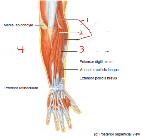
front 39 label and describe the muscles | back 39 1- extensor carpi radialis (extend and adduct hand)
2- extensor carpi ulnaris (extends and adducts hand)
3- extensor digitorum (extends hand and phalanges)
4- flexor carpi ulnaris (flexes and adducts hand) |
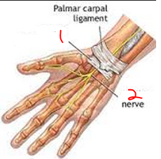
| back 40 space in the wrist containing flexor digitorum tendons and median nerve
carpal tunnel syndrome- overuse of the hand causes the tendons to become inflamed which compresses the median nerve |
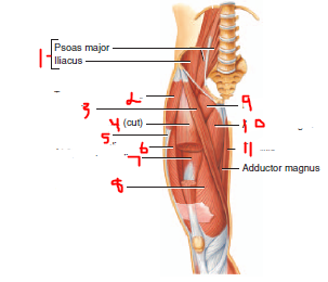
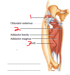
front 41 describe the movers of the hip joint | back 41 Adductor Longus
pectineus
iliopsoas- major hip flexor
tensor fascia lata- flexes and abduct the thigh
gluteus maximus
gluteus medius
piriformis
sartorius- flexes leg and flexes, abducts and laterally rotates the thigh (allows us to cross our legs)
gracilis |
| back 42 changes in the hand depending on where the brachial plexus was injured
claw hand
Crutches push on the brachial plexus and can cause numbness in the hands when the brachial plexus is compressed (affects all nerves of the upper extremity) |
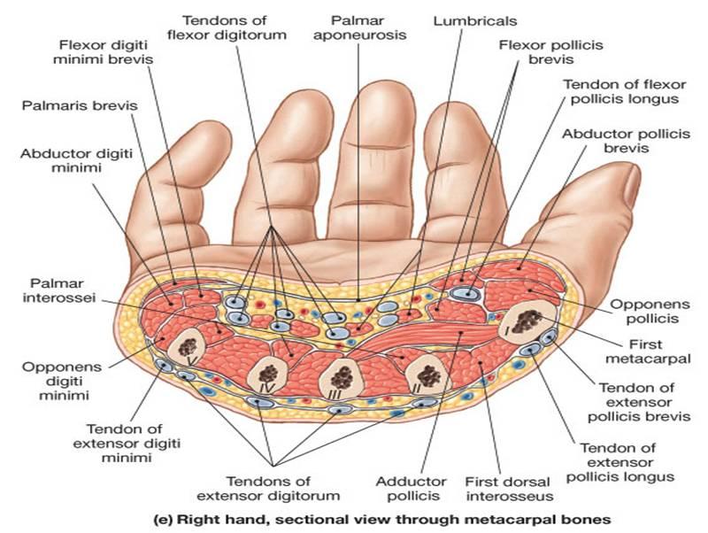
| back 43 innervated by the brachial plexus
lumbricals- little worms, flex the metacarpal joint
abductor pollicis- abducts the thumb
flexor retinaculum- carpal tunnel is underneath
interossei- muscle group in hand |
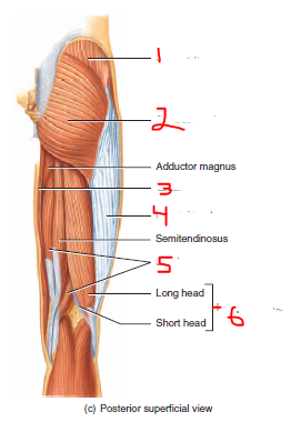
| back 44 gluteus maximus- powerful extensor of the hip joint
gluteus medius- abducts and medially rotates the thigh
test gluteal region strength by having someone sit in a chair and and stand up without using their hands or by walking up stairs |
front 45 label the anterior superficial thigh muscles | back 45 1- iliopsoas
2- tensor fasciae latae
3- sartorius
4- rectus femoris (Quadriceps)
5- iliotibial tract (tendon, abducts the thigh)
6- vastus lateralis (Quadriceps)
7- vastus intermedius (Quadriceps)
8- vastus medialis (Quadriceps)
9- pectineus
10- adductor longus
11- gracilis |
front 46 describe the movers of the hip joint | back 46 medial adductors in the groin region are innervated by the obturator nerve
adductor longus (anterior, medial adductor)
pectineus (anterior, adducts and flexes thigh)
iliopsoas (anterior, major hip flexor)
tensor fascia lata (lateral, adductor of the thigh)
gluteus maximus (posterior, powerful extensor of the hip, laterally rotates the thigh)
gluteus medius (posterior, adducts and medially rotates the thigh)
piriformis (deep posterior, laterally rotates and extends the thigh, sciatic nerve is underneath it)
sartorius (anterior, allows us to cross our legs, innervated by the femoral nerve)
gracilis (anterior, adducts and medially rotates thigh, flexes leg)
hip adductors get injured in a groin pull |
| back 47 largest nerve in the body
branches into the tibial nerve (posterior) and the fibular nerve (anterior)
sciatic nerve and its branches innervates mostly the whole lower extremity (except parts of the thigh)
piriformis syndrome- an overactive piriformis (sciatic nerve sits underneath the piriformis)caused a compressed sciatic nerve which causes major lower extremity problems |
front 48 describe the movers of the knee joint | back 48 Quadriceps (rectus femoris, vastus lateralis, vastus medialis, vastus intermedius)- knee extensor, innervated by the fibular nerve
hamstrings (bicep femoris, semimembranosus)- flexes knee (more important) and extends the hip |
front 49 label the anterior deep thigh muscles or ligaments | back 49 1- inguinal ligament
2- gracilis
3- adductor longus |
| back 50 1- gluteus medius
2- gluteus maximus
3- gracilis
4- iliotibial tract (tendon, abducts the thigh)
5- semimembranosus
6- biceps femoris (hamstring) |
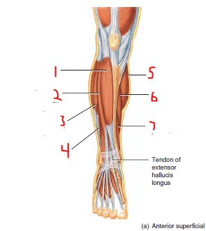
front 51 describe the movers of the ankle and toes | back 51 gastrocnemius- posterior, plantar flexes the foot and flexes the leg
soleus- posterior, plantar flexes the foot
fibularis (peroneus) longus- lateral anterior, plantar flexes and everts the foot
fibularis brevis
tibialis anterior- lateral anterior, dorsiflexes and inverts the foot (innervated by the fibular nerve)
tibialis posterior- inverts the ankle
flexor digitorum longus- posterior, flexes the toes
flexor hallucis longus- posterior, flexes the big toe
extensor digitorum- lateral anterior, dorsiflexes the foot and extends the toes (innervated by the fibular nerve)
extensor hallucis longus
anterior leg muscles dorsiflex (extend) the ankle
tibialis anterior and posterior (together) invert the ankle
deeper posterior leg muscles cause plantar flexion of toes
lateral leg muscles are everters |
front 52 label the muscles of the anterior superficial leg | back 52 1= tibialis anterior
2= extensor digitorum
3- fibularis (peroneus) longus
4= fibularis (peroneus) brevis
5= gastrocnemius
6= soleus
7= flexor digitorum longus |
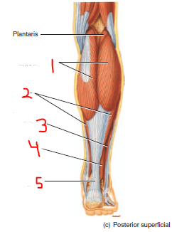
front 53 label the muscles or tendons of the posterior superficial leg | back 53 1- gastrocnemius
2- soleus
3- fibularis (peroneus) longus
4- flexor hallucis longus
5- calcaneal (Achilles) tendon |
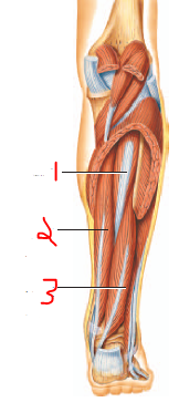
front 54 label the muscles of the posterior deep leg | back 54 1- tibialis posterior
2- flexor digitorum longus
3- flexor hallucis longus |
| back 55 injury to the fibular nerve causes the foot to stay plantarflexed (lack of dorsiflexion, toes point down)
fibular nerve is the most commonly injured nerve in the body |
front 56 Central nervous system (CNS) vs peripheral nervous system (PNS) | back 56 CNS- brain and spinal cord, develops from neural tube
integrates sensory (afferent) and motor (efferent) information
PNS- formed from neural crest cells that migrated from the neural tube
Somatic (innervated skeletal muscle), autonomic (innervates smooth muscle, cardiac muscle, and glands), and sensory ganglia brings information into the nervous system
connected to receptors and effectors |
| back 57 PNS
develop from neural crests
myelinate peripheral axons (form protective covering for all axons)
disease that affect peripheral myelin usually go away |
| back 58 cells that myelinate axons of the CNS
diseases of the CNS myelin are usually contiguous (ex. multiple sclerosis goes on and off but is usually progressive) |
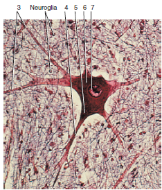
| back 59 motor neuron
3- dendrites
4- axon
5- axon hillock
6- cell body
7- nucleus |
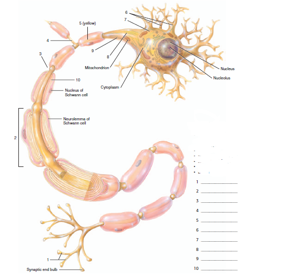
| back 60 1. axon terminal
2. schwann cell
3. node of ranvier
4. axon collateral
5. axon
6. dendrites
7. cell body
8. axon hillock
9. trigger zone
10. myelin sheath of schwann cell |
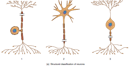
front 61 functional classifications of neurons | back 61 sensory neurons (afferent)- conduct signals from receptor to CNS when environmental changes are detected, unipolar neuron
interneuron (association)- confined to CNS, multipolar neurons
motor neuron (efferent)- conduct signals from the CNS (spinal cord) to effectors such as muscles and glands, multipolar neurons |
front 62 classify the neuron structure | back 62 1- unipolar (sensory (afferent) neuron)
2- multipolar (interneuron)
3- bipolar (motor (efferent) neuron) |
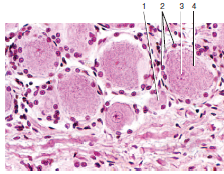
front 63 label the cells and components | back 63 dorsal root ganglia, unipolar cells
1= process
2= satellite cells
3= nucleus
4= neuron cell body |
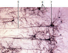
front 64 label the cells and components | back 64 cerebral cortex, multipolar neurons
5- neuron cell body
6- nucleus
7- process |
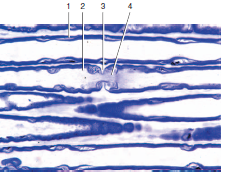
front 65 name the structure and label | back 65 1- neurolemma
2- myelin sheath
3- node of ranvier
4- axon |
front 66 Peripheral Nervous system (PNS) | back 66 Spinal nerves (below the neck)- sensory and motor
Autonomic Nervous System (ANS, innervates things that are automatic, spinal or cranial nerves, ex. salivation)- sympathetic and parasympathetic
Cranial nerves (generally innervate the things in the head, ex. salivation)- come off the brain but innervate structures outside of the CNS |
| back 67 (“horse’s tail”)= nerve roots at the lower part of the spinal cord (lumbar and sacrum)
At birth the spinal cod has equal length as the vertebra (spinal cord and spinal nerve labels match their places in the vertebral column)
As an adult the spinal cord ends at the L1/L2 vertebrae since the spinal cord grows much slower then the vertebral column. Vertebrae pulls nerve roots down with it during development so the nerve roots still exit at their appropriate vertebrae
best place for a Lumbar puncture/spinal tap |
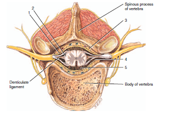
front 68 lumbar puncture/spinal tap | back 68 Best place to get cerebral spinal fluid (CSF) is between L4 or L5 in the cauda equina because you can get CSF without puncturing the spinal cord since there is lots of CSF and only small nerve roots (like sticking a fork into cooking spaghetti) |
front 69 name the structure and label the parts | back 69 1. posterior root
2. lateral white column
3. central canal
4. posterior median sulcus
5. posterior white column
6. posterior gray horn
7. posterior root ganglion
8. anterior root
9. anterior gray horn
10. anterior white column
11. Gray commissure
12. Anterior median fissure |
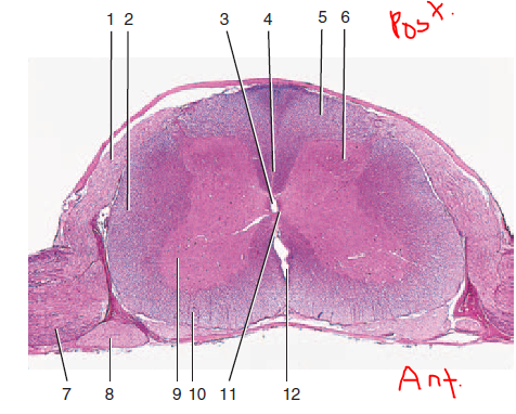
front 70 name the structure and label and describe each number | back 70 cross section of spinal cord
1- pia mater- delicate covering where small blood vessels are located
2- dura mater- tough, plastic like outer covering
3- epidural space- space in between the dura mater and vertebral bone where epidural anesthesia is injected for pelvis and lower extremity surgeries
4- subarachnoid space- where cerebral spinal fluid is located
5- arachnoid matter- spider web-like covering |
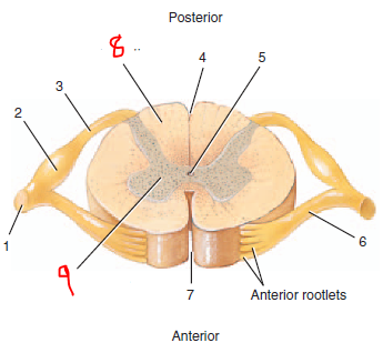
front 71 name the structure and label each number | back 71 cross section of spinal cord
1- spinal nerve
2- posterior root ganglion
3- posterior root
4- posterior median sulcus
5- central canal
6- anterior root
7- anterior median fissure
8- white matter
9- gray matter |
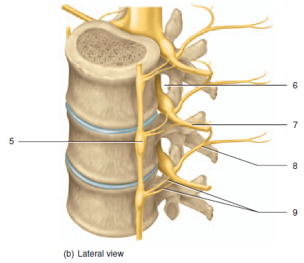
| back 72 5- sympathetic ganglion
6- spinal nerve
7- intervertebral foramen
8- anterior ramus
9- posterior ramus
10- rami communicantes |
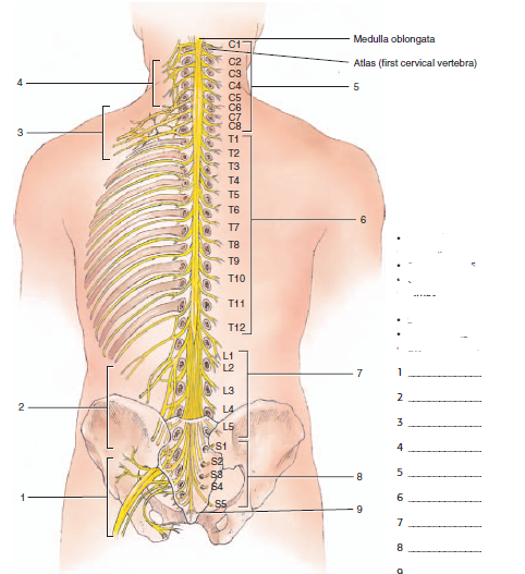
| back 73 1- sacral plexus
2- lumbar plexus
3- brachial plexus
4- cervical plexus
5- cervical nerve
6- thoracic nerve
7- lumbar nerve
8- sacral nerve
9- coccygeal nerve |
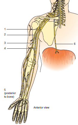
front 74 label the nerves that come from the cranial and brachial plexuses | back 74 1- axillary nerve
2- musculocutaneous nerve
3- median nerve
4- ulnar nerve
5- radial nerve
6- phrenic nerve |
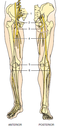
front 75 label the nerves that cam from the lumbar and sacral plexuses | back 75 1- obturator nerve
2- pudendal
3- femoral nerve
4- sciatic
5- tibial
6- common fibular |
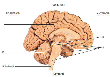
front 76 label the 4 major brain regions | back 76 1- cerebrum
2- cerebellum
3- diencephalon
4- brain stem |
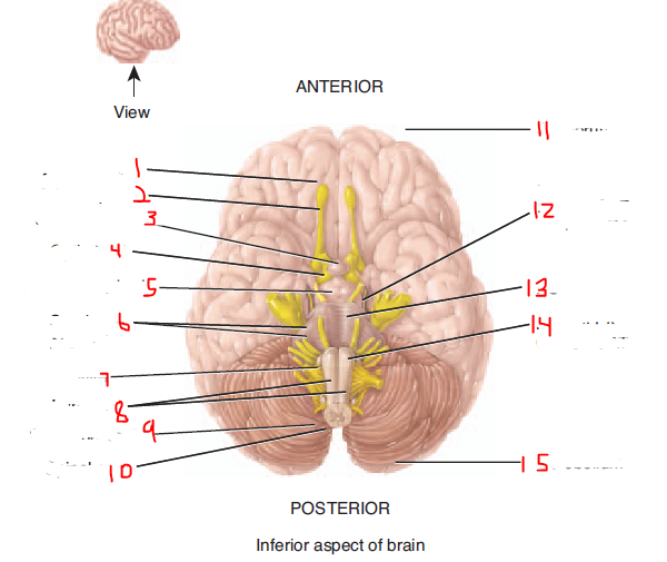
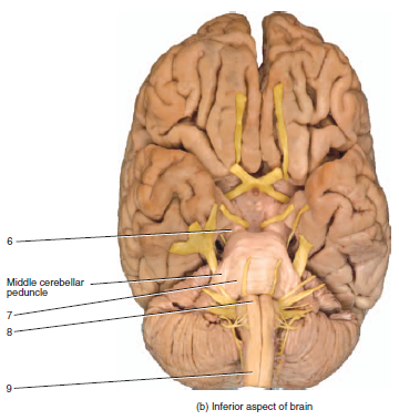
front 77 label the inferior side of the brain | back 77 1- olfactory bulb
2- olfactory tract
3- pituitary gland
4- optic tract
5- mammillary body
6- cerebellar peduncles
7- olive
8- pyramids
9- spinal nerve C1
10- spinal cord
11- cerebrum
12- cerebral peduncle of midbrain
13- pons
14- medulla oblongata
15- cerebellum |
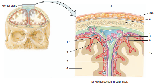
| back 78 blood vessels that run between the dura mater and the skull
if one breaks they bleed between the dura and the skull- epidural hematoma (creating a space that shouldn't be there, arterial bleed so it compresses the brain pretty rapidly)
Middle meningeal artery- between the dura and the temporal bone
• Artery that breaks if someone gets hit in the temporal part of the head |
| back 79 normally occurring and blood drains from bridging veins from the brain and the subarachnoid space into these sinuses that eventually form the jugular vein
If a brain shrinks (can happen with aging and dementia) then the bridging veins stretch and if someone hits their head, then the veins will rupture and cause a subdural hematoma
• Subdural hematoma- slow bleed, person becomes confused, major problems later, serious but difficult to diagnose
• Bleeds into the subdural space |
| back 80 o Arteiries in brain also travel through the subarachnoid space
Artery break- subarachnoid hematoma
• Weakness in walls of arteries of high blood pressure |
| back 81 consists of the midbrain (processes visual and auditory data), pons (relays sensory information to cerebellum and thalamus) and medulla (relays sensory information to thalamus)
origin of all cranial nerves (except I and II)
many nervous pathways travel through the brainstem |
front 82 levels of the spinal cord | back 82 31 levels of the spinal cord (each level gives off mixed spinal nerves (motor and sensory))
8 cervical spinal cord levels (7 cervical vertebrae)
12 thoracic spinal cord levels (1 for each rib)
5 lumbar levels
5 sacral levels
1-2 coccygeal levels (usually considered to have 1) |
front 83 white vs gray matter in the spinal cord | back 83 White matter (axons) are on the outside of the spinal cord carrying sensory info up to the brains and motor info down to the limbs
Gray matter-cell bodies (sensory info enters at the dorsal root via the dorsal horn (dorsal root ganglia are sensory cell bodies), ventral part of gray matter contains motor neuron cell bodies that exit the spinal cord at the ventral root) |
| back 84 Dorsal root ganglia- sensory cell bodies
Dorsal root- bringing info into the spinal cord (sensory neurons, unipolar)
Ventral root- axons of motor neurons, goes directly to muscles
Mixed spinal nerves- signals to and from the goal location (everywhere neck down), travel in the same nerve in opposite directions like a highway, made by dorsal and ventral roots joining, split into dorsal and ventral rami
Dorsal rami-only innervates deep back muscles, erector spinae muscles, skin of midline of the back
Ventral rami- much larger than dorsal, innervates everything else that isn’t covered by dorsal rami (neck and below), can go off on their own or form plexi (how upper and lower extremities are innervated) |
| back 85 all from the brachial plexus
musculocutaneous- flexors of arm (biceps and brachialis)
median- most flexors of the wrist, lateral phalanges, most thumb muscles. passes through the carpal tunnel
ulnar- some flexor of the wrist, all flexors of medial phalanges (funny bone, medial side of humerus)
radial- all extensors of the upper extremity
axillary- deltoid and part of the rotator cuff |
| back 86 o dermatome- sensory information from a single dorsal root
If you injure a dorsal root you get a dermatome pattern of sensory losses
shingles-the virus lives in a dorsal root and becomes activated and comes out of a dorsal root to cause painful symptoms along a dermatome (single stripe pattern on skin) |
front 87 major nerve plexi and what they innervate | back 87 all are made from ventral rami
cervical plexus: C1-C5, innervates strap muscles
brachial plexus: C5-T1, innervates the entire upper extremity, has 5 branch nerves
T2-T12 we don't form plexi, just intercostal nerves
Lumbar plexus: L1-L5, innervates abdominal muscles
Sacral Plexus: S1-S4
Lumbosacral plexus- gives rise to sciatic nerve which innervates the entire lower extremity |
| back 88 five rise to the following nerves
femoral nerve- innervates quadriceps
obturator nerve- innervates adductors
sciatic nerve- L4 to S3 (largest nerve in the body)
innervates hamstrings
gives rise to: tibial nerve (innervates all flexors), and fibular nerve (innervates all extensors)
sacral nerve damage- bladder and bowel problems due to parasympathetic innervations |
front 89 autonomic nervous system (ANS) | back 89 all glands and smooth muscle innervations and cardiac muscle
parasympathetic and sympathetic nervous systems
both parasymp and symp divisions have 2 motor neurons between the spinal cord and the effectors (the first motor neuron (preganglionic) synapses with the second motor neuron (postganglionic) in the ANS ganglia |
front 90 parasympathetic vs sympathetic nervous systems | back 90 Sympathetic- go everywhere (innervated sweat glands in skin that are located everywhere), preganglionic sympathetics located in 14 thoracolumbar (T1-L2 in CNS), in chain that extends the entire length of the trunk, Every spinal nerve from C1 to coccygeal must have sympathetic innervations
Parasympathetic- in head in places that drip [pupil of eye, lacrimal gland (tears), salivary gland], vagus nerve (heart, lungs), sacral nerve (bowel and bladder)
go to oculomotor nerve (constrict pupils) and facial nerve (glands( |
front 91 3 things every spinal nerve has | back 91 motor fibers for muscle
postganglionic sympathetic fibers to innervate sweat glands in skin
sensory fibers for sensation |
front 92 label the inferior portions of the brain and name the part of the brain formed by 6, 7, and 8 | back 92 brainstem (formed by 6,7,8)
6- midbrain
7- pons
8- medulla oblongata
9-spinal cord |
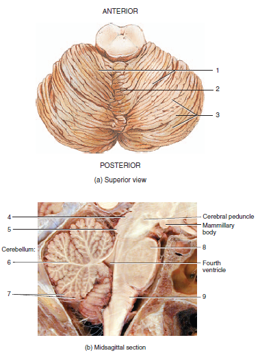
front 93 name the part of the brain and label | back 93 cerebellum
1- cerebellar hemispheres
2- vermis
3- folia
4- superior colliculus
5- inferior colliculus
6- arbor vitae (white matter)
7- cerebellar cortex (gray matter)
8- pons
9- medulla oblongata |
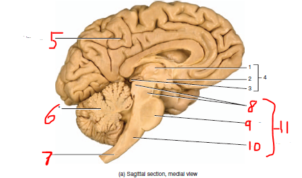
front 94 label the parts of the sagittal brain | back 94 1- pineal gland
2- thalamus
3- hypothalamus
4- diencephalon
5- cerebrum
6- cerebellum
7- spinal cord
8- midbrain
9- pons
10-medulla oblongata
11- brainstem |
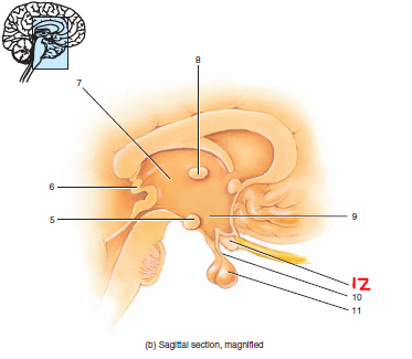
front 95 label that parts and name the structure | back 95 diencephalon
5- mammillary body
6- pineal gland
7- thalamus
8- intermediate mass of thalamus
9- hypothalamus
10- infundibulum
11- pituitary gland
12- optic chiasm |
front 96 name the part of the brain and label | back 96 cerebrum
1- internal capsule
2- cerebral cortex
3- white matter
4- corpus callosum
5- fornix
6- basal nuclei |
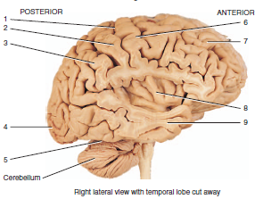
| back 97 1- central sulcus (shallow groove separating the frontal lobe and parietal lobe)
2- postcentral gyrus (elevation located just posterior to the central sulcus)
3- parietal lobe
4- occipital lobe
5- transverse fissure (deep groove separating the cerebrum from the cerebellum in the posterior/ inferior part of the brain)
6- precentral gyrus (elevation located just anterior to the central sulcus)
7- frontal lobe
8- insula (inner lobe deep to the lateral cerebral fissure)
9- temporal lobe (cut) |
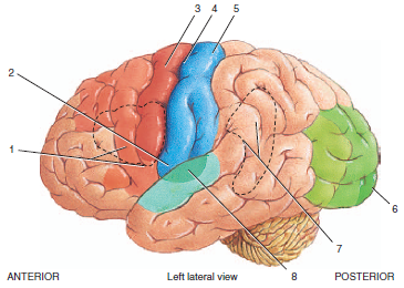
front 98 label the functional areas of the cerebral cortex | back 98 1- Broca's speech area
2- primary gustatory area
3- primary motor area
4- central sulcus
5- primary somatosensory area
6- primary visual area
7. Wernicke's area
8. primary auditory area |
front 99 label the cranial meninges | back 99 1- subarachnoid space
2- arachnoid villus
3- falx cerebri
4- white matter
5- superior sagittal sinus
6- parietal bone
7- dura mater
8- arachnoid mater
9- pia mater
10- cerebral cortex |
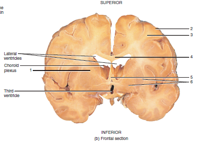
front 100 label the ventricles of the brain | back 100 8- lateral ventricles
9- interventricular foramen
10- 3rd ventricle
11- cerebral aqueduct
12- 4th ventricle
13- central canal of the spinal cord |
| back 101 14-superior sagittal sinus
15- arachnoid villus
16- subarachnoid space
17- lateral ventricle
18- choroid plexus
19- 3rd ventricle
20- cerebral aqueduct
21- 4th ventricle
22- central canal |
| back 102 groove that separates the frontal and parietal lobes |
| back 103 part of the motor areas to the cerebral cortex
located in the precentral gyrus of each frontal lobe
controls impulses to muscles
• A lot of cortex controls the face, fingers
• Medially controls toes and lower extremities
• Lateral cerebral cortex stroke- effects of facial expression, speech (left side), movements of hands and fingers
o Problems with motor control on the opposite side
Premotor (or motor association) cortex- programs of motor movement are made |
| back 104 part of the motor areas to the cerebral cortex
located anterior to the primary motor cortex
initiates impulses that result in speech
• Lateral cerebral cortex stroke- effects of facial expression, speech (left side), movements of hands and fingers |
front 105 primary somatosensory cortex | back 105 part of the sensory areas to the cerebral cortex
determines where on the body the sensory stimulation occurred
mostly devoted to hands and face |
| back 106 part of the sensory areas to the cerebral cortex
in the temporal lobe
interprets auditory stimuli from auditory receptors |
| back 107 part of the sensory areas to the cerebral cortex
receives impulses from taste receptors |
| back 108 judgement and decision making |
| back 109 in occipital lobe
part of the sensory areas to the cerebral cortex
receive information from the retina and interprets the visual stimulu |
| back 110 located on in the temporal lobe
part of the sensory areas to the cerebral cortex
receives impulses from olfactory receptors |
| back 111 part of the association areas to the cerebral cortex
recognize spoken words, translates words into thoughts, and possibly helps us sound out strange or new words |
front 112 somatosensory, visual, and auditory association areas | back 112 part of the association areas to the cerebral cortex
adjacent to their corresponding sensory cortex
integrate sensory information from the sensory cortex with past experiences |
| back 113 crossing pathways from one side of the brain to the other
o Corpus callosum (connects 2 cerebral hemispheres)- major commissure
Almost everything in the right cortex contributes info to the left cortex via crossing fibers
o Optic chiasm is another example |
| back 114 o Projects from higher to lower
o Like for your cerebral cortex (motor cortex) to tell your muscles what to do you need fibers that go from the cortex to the spinal cord (motor neurons for muscles are in the spinal cord, for these neurons to fire they need a signal from the cortex)
o Projection pathways- north to south or south to north |
| back 115 connect parts of our brain on the same side
ex. seeing and interpreting what your hand is writing |
| back 116 huntington's disease
the patient cannot suppress unwanted movements
• Basal ganglia (deeper nuclei, gray matter)- suppresses unwanted movement |
| back 117 2 hemispheres with the vermis connecting them
regulates posture and balance, smooths and coordinates skilled skeletal muscle movements |
| back 118 2 main regions: thalamus, hypothalamus, and epithalamus
thalamus- gets all sensory infor except olfaction (olfaction needs to be faster for survival)
suppresses unwanted sensory infor- allows for focus, selects what sensory info is important and sends it to the cerebral cortex
hypothalamus- control many bodily functions and homeostasis
contains the optic chiasm- where the optic nerve crosses
epithalamus- contains some glands |
| back 119 head trauma (brain swelling, broken artery) causes the brain to squish to the only space available which compresses the oculomotor nerve
uncus is a piece of the temporal lobe that herniates if the brain swells which pushes on the oculomotor nerve
o Pupils cannot compress and stay dilated and are not responsive to light |
| back 120 tumor of Schwann cells (peripheral nervous system cells), compresses nerve VIII (vestibulocochlear and facial VII), and can affect the cerebellopontine angle (angle between the cerebellum (important for coordination) and the pons)
• Nerve damage causes uncoordinated gate, droopy face, hearing loss, ringing in the ears can only all happen together at the cerebellopontine angle |
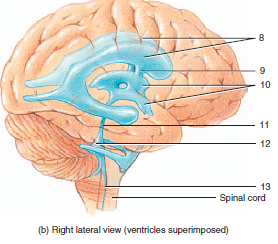
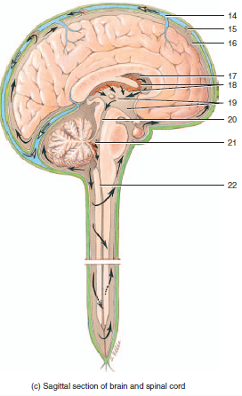
front 121 path of cerebral spinal fluid (CSF) | back 121 CSF leaks out of the choroid plexus blood vessels
ependymal cells with cilia push the CSF in one direction into the lateral ventricle
the CSF then moves into the 3rd ventricle then into the 4th ventricle via cerebral aqueducts
the CSF then goes into the subarachnoid space surrounding the brain and spinal cord (CSF follows the central canal of the spinal cord then goes back into the subarachnoid space)
CSF it then returned to the blood through arachnoid villi located at the superior sagittal sinus |
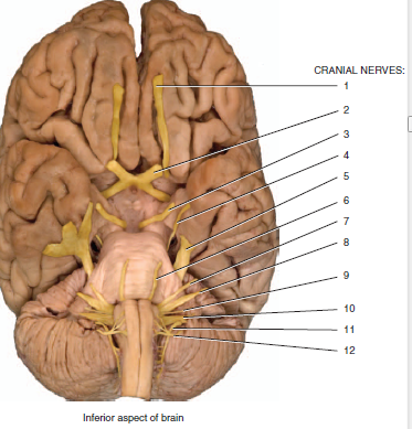
front 122 list all the cranial nerves in order | back 122 I Olfactory
II Optic
III Oculomotor
IV Trochlear
V Trigeminal (Ophthalmic, Maxillary, Mandibular)
VI Abducens
VII Facial
VIII Vestibulocochlear
IX Glossopharyngeal
X Vagus
XI Accessory
XII Hypoglossal
Oh, Oh, Oh To Touch And Feel Very Good Velvet, AH |
| back 123 1. olfactory
2. optic
3. Oculomotor
4. Trochlear
5. Trigeminal (Ophthalmic, Maxillary, Mandibular)
6. Abducens
7. Facial
8. Vestibulocochlear
9. Glossopharyngeal
10. Vagus
11. Accessory
12. Hypoglossal |
| back 124 • Usually if you have a problem with one olfactory nerve then the other one will take over
You may regenerate olfactory neurons (but not any others)
Olfactory nerves are tiny branches that are attached to receptors, axons go through the cribriform plate then form into the olfactory nerve
What we often call the olfactory nerve is actually the olfactory tract (CNS)
Test by having a patient smell something through one nostril at a time
purely sensory
goes straight to the cerebrum for processing (only sensory nerve that doesn't go through the thalamus) |
| back 125 Optic nerve starts in the retina ((retinal ganglion cells give rise to the optic nerve [myeleinated by an oligodendrocyte which speeds up conduction in the CNS]). Optic nerve is very fast conducting nerve
Actually a CNS tract
Right visual field process on the left side of the brain, visa versa
Tested using visual fields, doctors move fingers from lateral to medial parts of visual fields |
| back 126 CNS disease affecting myelination and oligodendrocytes
deteriorates CNS myelin and is first noticed in the optic nerve since conduction is usually so fast |
| back 127 when the optic nerve crossed over in the brain above the pituitary gland (commissure). Optic nerve comes from the lateral part of your visual field.
Pituitary gland tumors causes “tunnel vision” which limits your vision into the center of your visual field due to damage to the optic chiasm, damages the crossing fibers |
front 128 Oculomotor, Trochlear, and Abducens (III, IV, and VI) cranial nerves | back 128 Always tested together, more extra-ocular muscles
Oculomotor- innervates the levator palpebrae (oculomotor lesion would cause trouble elevating the eyelid) and 4 extra ocular muscles and pupil (parasympathetic, constrict)
Oculomotor Damage- causes droopy eyelid (ptosis) and a dilated pupil
Trochlear lesion- Can’t look down and in (trouble reading)
Abducens- abducts the eye, cannot abduct both eyes are the same time (one abducts and the other stays neutral)
test these by observing eyelids (ptosis is abnormal), moving eye in different directions, and making sure pupil constricts |
| back 129 main sensory nerve to the head, motor to muscles of mastication (gives you sensation on your face)
• Ophthalmic nerve (afferent, corneal blink reflex with facial nerve)- innervates forehead and cornea
o Test afferent by putting a whisp of cotton on their eye to get them to blink or tap forhead
o Afferent=ophthalmic, efferent=facial -> V-VII reflex
• Maxillary nerve- innervates the upper teeth, cheek, around the nose (purely sensory)
• Mandibular division- lower teeth, chin, motor component
o Trigeminal motor component- muscles of mastication (chewing, temporalis, masseter)
test by tapping chin |
| back 130 motor to muscles of facial expression, taste, anterior 2/3 of tongue, ANS to lacrimal submandibular and (sublingual)salivary glands
Innervates muscles of facial expression- test it by having the patient make faces or by testing the corneal blink reflex
Carries taste, autonomic to the lacrimal gland and 2 of the salivary glands
corneal blink refllex (V-VII)
shutting the eye is a muscle (nerve VII) for facial expression (open with III-oculomotor), puff of air causes patient to blink which tests facial nerve |
front 131 vestibulocochlear (VIII) nerve | back 131 purely sensory
Tumor on VII cranial nerve- dizziness, ringing in ears, hearing loss
Acoustic Schwannomas |
| back 132 sensory to pharynx (back or throat, or motor to 1 small pharynx muscle)
taste, posterior 1/3 of tongue
visceral sensory
Test using the gag reflex (IX-X reflex)- sticks a tongue depressor on the tongue to push it down and see the back of the throat and make you gag
Afferant- IX (vagus), constricts pharynx
Efferent- causes gag (X) |
| back 133 Motor:larynx and pharynx
ANS: visceral organs
Sensory:visceral organs
Taste
Vagabond: wanders out of the head to innervate the pharynx and larynx (swallowing and speaking)
Parasympathetic to all the organs down to the the small intestine
Tested using the gag reflex (IX-X reflex) |
front 134 Accessory and hypoglossal nerves (XI and XII) | back 134 Accessory (XI)- innervates sternocleidomastoid (test by having the look from side to side) and trapezius (check by putting hands on pts shoulder and having them shrug against resistance)
Hypoglossal (XII)- innervates the muscles on the tongue, test by sticking the tongue out and having the patient move it from side to side, damage on one side has the tongue pointed towards the side of the lesion |
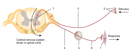
| back 135 1. interneuron
2. sensory neuron (afferent)
3. sensory receptor
4. preganglionic motor neuron
5. autonomic ganglion
6. postganglionic motor neuron
7. visceral effector |
| back 136 1. superior oblique
2. superior rectus
3. lateral rectus
4. medial rectus
5. inferior oblique
6. inferior rectus |
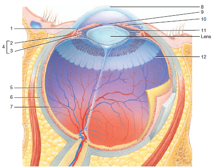
front 137 label the parts of the eye | back 137 1. scleral venous sinus
2. ciliary muscle
3. ciliary process
4. ciliary body
5. choroid
6. sclera
7. retina
8. cornea
9. pupil
10. iris
11. suspensory ligaments
12. ora serrata |
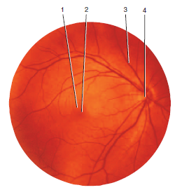
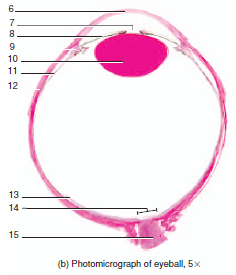
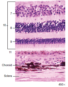
front 138 label the tissue and the numbers | back 138 retina
1. macula lutea
2. central fovea
3. blood vessel
4. optic disc |
| back 139 6. cornea
7. pupil
8. iris
9. ciliary body
10. lens
11. choroid
12. sclera
13. retina
14. optic disc
15. optic nerve |
| |
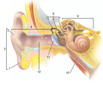
| back 141 1. lobule
2. auricle
3. helix
4. external ear
5. malleus
6. incus
7. stapes attached to oval window
8. middle ear
9. internal ear
10. auditory tube
11. tympanic membrane
12. external auditory canal |
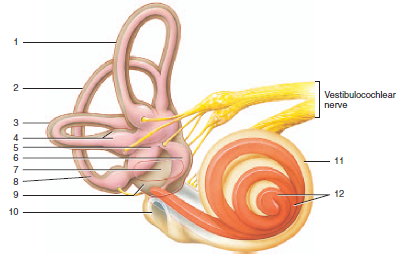
| back 142 1. anterior semicircular canal
2. posterior semicircular canal
3. lateral semicircular canal
4. ampulla of semicircular canal and duct
5. utricle
6. saccule
8. membranous semicircular duct
9. vestibule
10. round window
11. cochlea
12. cochlear duct |
| back 143 fibrous (cornea=clear layer, sclera= opaque layer)
choroid (highly vascular, gives rise to iris and ciliary body)
retinal layer (inner, sensory (photo) receptors) |
| back 144 connective tissue layer that lines the eyelid, makes a bend and reflects over the sclera, does not go over the cornea (or you wouldn’t be able to see)
Contact lenses can get stuck behind the ledge |
front 145 path of light through the eye | back 145 o Light goes through the cornea, through the anterior chamber of the eye through fluid, through the pupil, through the lens where its focused, through the vitreous (eye jelly) then to the retina |
| back 146 o Aqueous humor- circulating fluid, produced by cells in Ciliary body, circulated then is reabsorbed in the canal of schlemm
If the canal of schlemm is blocked then pressure builds in the eye and causes glaucoma |
| back 147 o Middle ear has ossicles (tiny bones) that vibrate with sound waves and that vibration is transduced to pressure waves which is then transduced to an electrical signal in the cochlea
o Hair cells run against the tectorial membrane to transmit sound
o 2 tiny muscles (tensor tipany, )that are in the ear that contract with very large sounds to dampen the sound and damaging the cochlea |
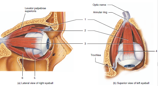
| back 148 • innervated by the cranial nerves III (oculomotor), IV (trochlear), VI (abducens)
• ocular motor nerve- droopy eye lid, opens the eye (lavatory palpebrae)
• lateral rectus- abducens nerve, that abducts, lets the eye move laterally |
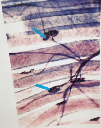
front 149 name the structure and what the arrows are pointing to | back 149 axon terminals on a neuromuscular junction
arrows point to motor end plates |
| |
| |
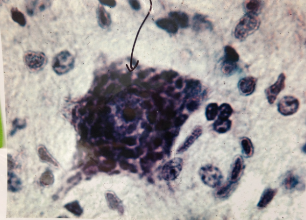
front 152 name the structure and what the arrow is pointing to | back 152 neuron
arrow pointing to nissl substance |
front 153 name the structure and what the arrow is pointing to | back 153 peripheral nerve in cross section
arrow is pointing to perneurium |
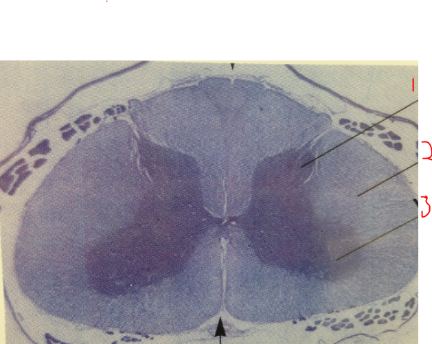
front 154 name the structure and label | back 154 spinal cord
1. dorsal horn (gray matter)
2. white matter
3. Ventral horn (gray matter) |
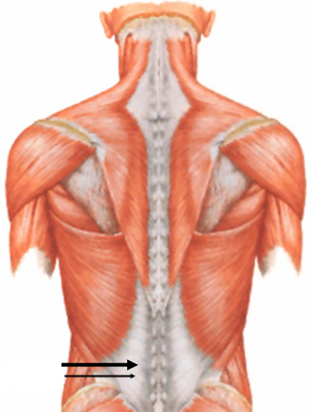
front 155 name the tendon indicated by the arrow | back 155 lumbodorsal (thoracolumbar) fascia (tendon) |
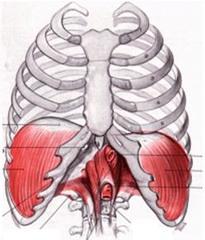
front 156 name the white part of the muscle | back 156 central tendon of the diaphragm |
front 157 name the tendon and nerve | back 157 1. flexor retinaculum of the wrist (tendon)
2. median (nerve) |