| back 1 Regulate ECF volume & blood pressure
Regulate osmolarity
Maintain ion balance
Regulate pH
Excrete wastes
Regulate hormone production
filter, reabsorb, secrete |
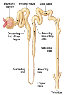
front 2 nephron anatomy (picture) | back 2 Normally highly twisted
Blood flows out of the nephrons into the efferent arterioles |
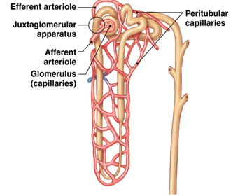
front 3 Kidney portal system (picture) | back 3 blood flows from the renal artery then into the cortex, then from the afferent arteriole into the glomerulus (ball-like network of capillaries). Then blood flows into the efferent arteriole then into the peritubular capillaries, then into the renal vein and out of the kidneys
In juxtamedullary nephrons, long peritubular capillaries that dip into the medulla are vasa recta.
Function: filter fluid out of the blood and into the lumen of the nephron at the glomerular capillaries, then to reabsorb fluid from the tubule back into the blood at the peritubular capillaries |
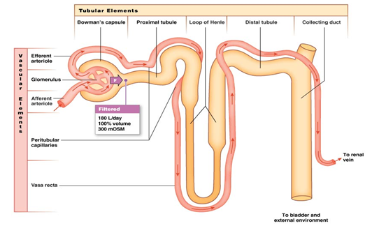
front 4 kidney filtration (picture) | back 4 o Happens in glomerulus
o 300 mOsmol
o Filter fluid from plasma
o Leave behind plasma proteins & RBCs
o only 20% of the plasma that passes through the glomerulus is filtered and less than 1% of the filtered fluid is excreted (the other 99% return to systemic circulation
o takes place in the renal corpsule
net driving pressure causes filtration |
front 5 renal corpuscle filtration | back 5 o first barrier is the capillary endothelium which contains glomerular fenestrated capillaries (large pores- RBC and most proteins still cannot pass)
o second filtration barrier is the basal lamina (Extensive extracellular matrix to exclude plasma proteins from the fluid)
o third filtration barrier is the epithelium of the bowmans capsule (podocyte foot processes leave narrow slits through which filtration takes place |
front 6 3 pressure components acting within the renal corpuscle | back 6 Blood pressure – blood moving into glomerulus (provides hydrostatic pressure) forces fluid through leaky fenestrated capillaries
Osmotic pressure – Fluid moving back due to protein concentration differences
Capsule fluid pressure – hydrostatic fluid pressure of the enclosed space opposes fluid movement in and is in competition with blood and osmotic pressure |
front 7 Glomerular filtration rate (GFR) | back 7 volume of liquid that filters into bowman's capsule that depends on 2 things: filtration pressure and the filtration coefficient (which depends on surface area of capillaries, permeability of membranes)
stays relatively constant even across ranges in blood pressure
regulated by blood flow in the renal arterioles or by autonomic regulation (sympathetic control changes the resistance in arterioles bu using NE to vasoconstrict as necessary) |
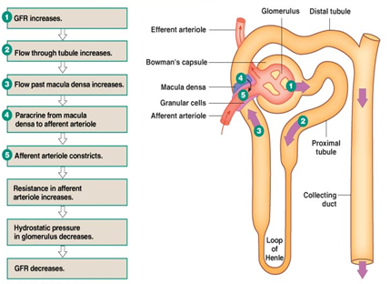
front 8 regulation of the GFR by blood flow in the renal arterioles (picture) | back 8 Increase in blood pressure= vasoconstriction to keep GFR constant
• Myogenic response: Vascular smooth muscle responds to pressure changes (Increase in blood pressure causes vasoconstriction, decreases causes vasodilation)
• Tubuloglomerular feedback: Paracrine signaling to change fluid flow through the loop of Henle
Passes by afferent and efferent arterioles which get direct feedback and twisting causes ascending limb to pass between afferent & efferent arterioles |
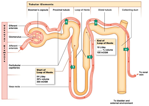
front 9 nephron absorption (picture) | back 9 70% gets reabsorbed almost immediately
Loop of Henle is where solutes change concentrations
• About 100mOsmol at the end of the loop of Henle
• Must remove stuff without removing water
• Only 18L/day reaches the end of the loop
• Excrete about 1.5 L/day at the end of the tube
pressure gradient favors reabsorption |
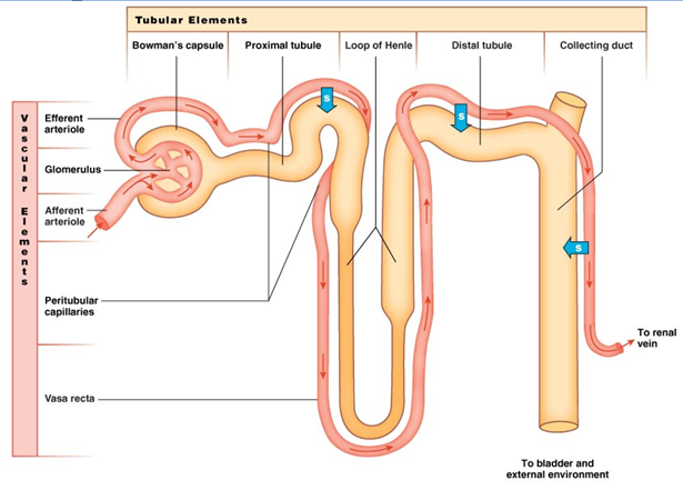
front 10 Nephron secretion (picture) | back 10 o After filtration, to adjust concentrations
o Happens via aided transport
o transfers solutes from the ECF to the lumen
o Na+ is the driving molecule with the sodium potassium pump
o uses transport molecules (active transport) and concentration gradients to fine scale adjust the concentrations in the lumen |
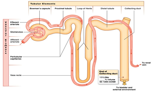
front 11 nephron excretion (picture) | back 11 o about 1.5 L/day
o Amount Filtered- Amount reabsorbed+ amount secreted= amount of solute excreted
o most useful metabolites have been removed at this point
o ion and water concentration is highly variable depending on needs |
front 12 Sodium (Na+) in kidney reabsorption | back 12 o Na+ is the main player because it sets up the electrical gradient (active transport)
o water equalizes concentrations by moving to the nephron from the lumen
o ECF has higher Na+ concentration than the ICF so Na+ follows its concentration gradient into cells and is actively pumped out of cells via the sodium-potassium pump |
front 13 urea reabsorption in the kidneys (picture) | back 13 transports proteins into the epithelium and breaks them down |
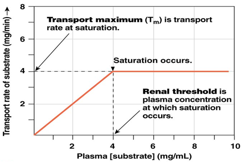
front 14 solute transport in the kidneys (picture) | back 14 Saturation – Maximum rate of transport due to maximum carrier use (receptors and channels)
Filtration has a linear increase in the amount of glucose in the plasma and the glucose filtration rate
you can reabsorb up to 300 mg/mL and everything else gets excreted |
| back 15 easiest method to determine the Rate at which a solute disappears from the body due to excretion or metabolism
Provides a method for estimating GFR as well
Determines how well a nephron handles a filtered solute |
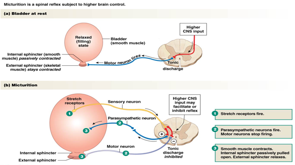
| back 16 urination
what leaves the collecting duct must be excreted and the concentrations cannot be adjusted any further
Normal conditions: Motor neuron is leading to tonic contraction of the external sphincter
When you have to urinate
• Spinal reflex tells stretch receptors to fire-As you gain control of your bladder, more stretch receptors are present so that you can notice how full your bladder is instead of just whether you have to urinate or not |
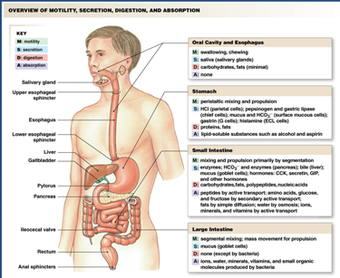
front 17 primary goal of the digestive system | back 17 move nutrients, water, and solutes from the external environment to the internal evironment
4 Basic Processes: Digestion- breaking down of food particles, Absorption, Motility, Secretion |
front 18 gastrointestinal tract and secretion | back 18 hollow tube (lumen) passing through the body
the primary way pathogens can get into the body
• Secretions come from enzymes
• Substantial amount of fluid from secretions enter the digestive system
• 7.5/9 L of liquid is reabsorbed, the rest is excreted |
front 19 Gastrointestinal tract anatomy | back 19 mouth/ salivary glands- chewing and the secretion of saliva
stomach- mixes food with acid and enzymes to create a soupy mixture called chyme
small intestine- Ducts pour in enzymes from the liver to help break down and absorb the fluid and particles and nearly all the digested nutrients and secreted fluids are absorbed here |
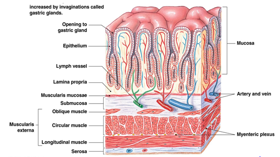
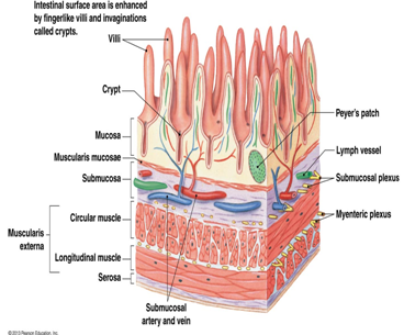
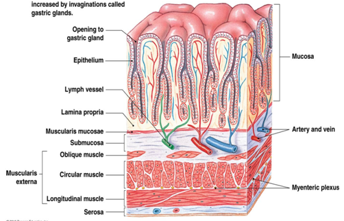
front 20 GI tract wall structure (picture) | back 20 The stomach and wall function very similarly (only a few small differences)
Basic structure is similar in stomach and small intestine
• Mucosa- interfacing with the food and lumen
• Submucosa
• Muscularis externa
• Serosa |
| back 21 • layer of the GI Tract wall
o Gastric glands- Deep folds in mucosa
o Single epithelial tissue layer (enterocytes)
o Lots of goblet calls and other paracrine cells than excrete
o Makes up part of the intestinal space
o First line of defense by having many immune cells on the inside of the interstitial space
o villi- play a big role in absorption and mobility
o epithelial cell layer- function depends on their location in the tract (Transporting – enterocytes in small intestine, Secreting – endocrine & exocrine, Stem cells) |
| back 22 • layer of the GI Tract wall
- muscle layer
• Muscle rings- change the diameter of the lumen
• Longitudinal muscle- changes the length of the tube
o Longitudinal and muscle rings do not always agree
• Innervation for smooth muscle tissue is directly next to the layer |
| back 23 • layer of the GI Tract wall
- convergence of smaller vessels into larger muscles
•Basically the same for stomach and small intestine |
| back 24 • layer of the GI Tract wall
- outermost layer
• Anchors the intestine and other organs within the abdominal cavity
o Must be anchored because the 2 muscle layers (muscle rings and longitudinal) must be working together so if they can move, then the muscles would just get tied into a knot |
front 25 purpose of motility in the GI tract | back 25 o Two main purposes:
Move food through the GI tract
Mix food and break down into small pieces
• Food moves around a lot to absorb as many enzymes as possible to get broken down |
front 26 GI smooth muscle contraction- migrating motor complex | back 26 • Normal pattern of contraction between meals
• Contraction begins in stomach and ends in large intestine
o Keeps things moving all in one direction
o Important in preventing infection
• Sweeps remnants to large intestine |
front 27 GI smooth muscle contraction- peristalsis | back 27 • smooth muscle in the bottom 1/3 of the esophagus (the top 2/3 is skeletal muscle)
• peristaltic contraction pushed the food (bolus) into the receiving segment which pushes it forward more down the esophagus and into the stomach
• segmental contractions are responsible for mixing by alternating contracting segments so that there is little or no net forward movement and the bolus is exposed to as much intestinal wall (where enzymes are located) as possible |
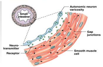
front 28 single unit smooth muscle in the GI tract (picture) | back 28 o Groups of cells connected by gap junctions
o Signal passes quickly through gap junctions
o Tonic or phasic contractions |
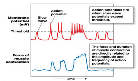
front 29 GI tract contraction- slow wave potential | back 29 • Dependent on Ca2+ signaling
• Graded contractions
• More Ca2+ àstronger and/or longer contraction |
front 30 Things the digestive system secretes | back 30 Ions & Water, Digestive enzymes, Mucus, Saliva, Bile
• Wherever we move ions, water wants to follow (osmotic equilibrium) |
front 31 acid secretion in digestion | back 31 Parietal cells secrete acid
1-3 L of acid per day
Lumen of stomach can reach pH of 1(very acidic)
Parietal cell cytoplasm has pH=7.2 (pretty normal)
• Pumping out H+ extremely against its concentration gradient to transition from a pH of 1 to 7.2
• Very active process (uses ATP)
o Lumen net gain is a lot of chloride
o Bicarbonate goes into the interstitial fluid and eventually into the blood
o pancreas secretes bicarbonate to neutralize the acid that gets pumped into the lumen and enters the small intestine because we don't want a very acid fluid going into other tissues |
front 32 bicarbonate secretion in digestion | back 32 Bicarbonate, water, Na+ & Cl- move into the lumen
H+ moves into ECF à blood
Sets up electrical gradient due to pumping of negative ions into the lumen
• Causes Na+ and water to pump through too |
front 33 NaCl secretion in digestion | back 33 • Cryp cells from the small intestine and colon secrete salt solution
• Creates gradient for water to follow which creates an isotonic salt solution |
front 34 enzyme secretion in digestion | back 34 o secreted by exocrine glands (Salivary glands and pancreas) or by epithelial cells in the mucosa of the stomach and small intestine
o Stored as proenzyemes |
front 35 mucus secretion for digestion | back 35 o Made up of glycoproteins
o Forms protective coating over the GI mucosa and lubricates the contents of the gut
o Secreted by mucous cells (stomach), serous cells (salivary glands), and goblet cells (intestine)
o Also secretes salt solution |
front 36 bile secretion for digestion | back 36 o Secreted by the liver (hepatocytes)
o Made up of:
o Bile salts – facilitate fat digestion (main goal)
o Bile pigments like bilirubin
o Cholesterol
o Drugs & other waste products |
front 37 locations of digestive secretions | back 37 • Ions (acid, bicarbonate, salt)
• Digestive enyzmes (salivary glands & pancreas)
• Mucus (stomach, salivary glands, & intestine)
• Saliva (salivary glands)
• Bile (liver) |
| back 38 • Villi increase surface area for absorption
• Brush Border creates a border for protection
Not directly regulated
• Regulated by motility, feelings of hunger, etc.
3 major nutrients: carbohydrates, proteins, fats |
| back 39 • Starch, sucrose, glycogen, cellulose, lactose, maltose, glucose, fructose
o Must break giant polymers into smaller pieces (monosaccharides) before you can move it across a cellular border
o Amylase- breaks polymers into simpler molecules
Found in the saliva and gets secreted by the pancreas
o Enterocytes don’t use glucose (they use glucogen) so they don’t mess up the gradient |
| back 40 • Most ingested protein are large molecules
• Not all proteins are processed equally (80% of protein is absorbed from eggs, only 30-40% absorbed from beans
• 30-60% of protein in lumen comes from dead cells & secretion
• endopeptidases and exopeptidases |
front 41 endopeptidases vs exopeptidases | back 41 • Endopeptidases- breaks up initial peptide bonds
o Proteases are a more common name
o Proenzymes
o Ex: pepsin, trypsin
• Exopeptidases- break off amino acids from the amino or carboxyl ends of smaller proteins that have been broken up
o Carboxypeptidases are most important |
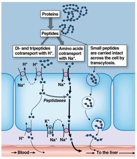
front 42 protein transport in digestion | back 42 • Many different transport systems for the different amino acids
• Large proteins can be brought in via transcytosis
o Stimulates immune response and allergy responses
o Larger peptides are that are transported through the cell are more likely to cause an immune response so if you wait until a baby has more mature villi then they are less likely to transport those larger molecules and cause an allergic response |
| back 43 • Triglycerides
• Cholesterol
• Phospholipids
• Long-chain fatty acids
• Fat-soluble vitamins
• Not water soluble
o Fat groups into large droplets
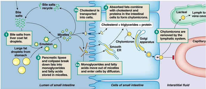
o Need to break up the droplets into smaller ones via Bile salts |
| back 44 break up fats into smaller pieces
Bile salts interact with water
Fat droplets alone are relatively unstable (break into smaller ones)
• Bile salt ringed fat droplets are relatively stable
• Broken into smaller and smaller pieces
• Secreted into the lumen |
| back 45 combination of all the things that have been broken up into the lumen
• Small, disk-shaped droplets
• Diglycerides & monoglycerides, Phospholipids, Fatty acids, Cholesterol
• Migrate towards the microvilli and brush border for secretion
• Water soluble
o Does not need transport protein and will go through the membrane without a problem
• Once they pieces get through the membrane into the cytoplasm they reform into larger molecules that get secreted by the golgi apparatus
• Too big to go into the capillaries or normally secrete (A few fatty acids can pass to the capillaries but it is fairly infrequent) |
front 46 fat digestion and absorption (picture) | |
| back 47 • DNA & RNA polymers are small part of diet
• Broken down into nitrogenous bases & monosaccharides
• Nitrogenous bases require active transport for absorption |
| back 48 • Fat-soluble vs. water-soluble
• Fat-soluble vitamins are packaged into micelles and absorbed (vitamins A, D, and K)
• Water-soluble are absorbed by mediated transport (all B and C vitamins)
• Olestra- fat that cannot be absorbed
o Was added to chips but caused negative side effects when too much was ingested |
| back 49 • Generally occurs via active transport
• Iron & calcium are actively regulated |
| back 50 • Most absorption occurs in small intestine
• Na+ & Cl- absorbed by multiple pathways
• K+ & water absorbed via paracellular transport |
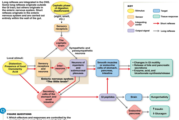
front 51 digestive system regulation signaling | back 51 o Neural, endocrine & local signals
Long reflexes (integrated in CNS)
Short reflexes (integrated within GI tract)
Peptide hormones & reflexes
• Paracrine system (stomach) |
front 52 integration of digestive reflexes | back 52 Everything is happening within GI tract\same responses we would normally see just input from the CNS is not required |
front 53 Function of components of the GI tract in digestive signaling | back 53 Muscularis are in charge of secretion and regulates motility and control
o Peptides are a major class of signaling molecule
o Act as hormones or paracrine signals
Could affect other hormones as well
o Inhibit or excite secretion & motility
o Can work on either side of epithelial layer |
| back 54 Acts as an independent integrating center like the brain or spinal cord
Does not have a specific bundle or ganglia of nerve cells like the CNS for processing specific signals
Nerve network responds to sensory stimuli and initiates reflex
• Everything is reflex responses since there is no integrating center |
front 55 shared characteristics between the enteric nervous system and the CNS | back 55 • Intrinsic neurons à interneurons
• Neurocrine molecules
• Glial support cells
• Tight capillaries around ganglia à blood-brain barrier
• Acts as an integrating center |
front 56 digestive system summary (picture) | |
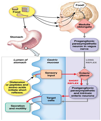
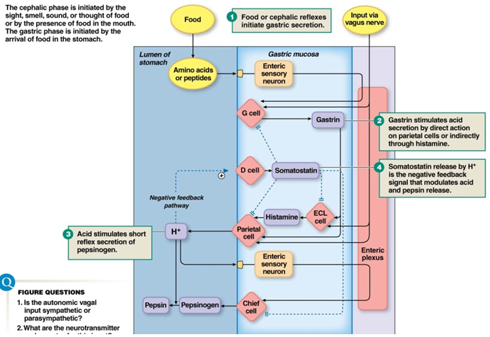
front 57 cephalic phase of digestion | back 57 Feedforward response
• Smelling, seeing, or thinking about food triggers digestion
Anticipation or stimulus of food in mouth causes stimulation via medulla
Food in mouth causes: Salivary response (Lubricates, Digests - salivary amylase (starts breaking down carbohydrates), Protects – Lysozyme), Mastication = chewing, Deglutition= swallowing |
| back 58 tongue pushes bolus against soft palate
breathing is inhibited as the bolus passes the closed airway (epiglottis folds down to help keep swallowed material out of the airways and upper esophagael sphincter relaxes)
food moves downward into the esophagus propelled by peristaltic waves and |
front 59 gastric phase reflex (picture) | |
front 60 Stomach function- storage | back 60 o Bring in 3.5L of food, liquid, & saliva per day
Relaxed stomach holds 45mL of liquid, after a normal meal it expands to 1.5L, after a large meal it can stretch to 2.5-3L
o Regulates amount of food that goes into small intestine for digestion
o Upper stomach (fundus) stores food |
front 61 stomach function- digestion | back 61 o Lower stomach (antrum) continues digestion
o Peristalsis mixes food with enzymes & acid to break down food
Only 10% of digestion happens in the stomach, the rest happens in bile, etc. |
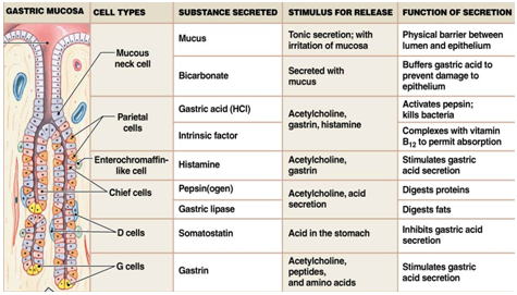
front 62 secretory cells of the gastric mucosa | |
front 63 integration of cephalic and gastric phase secretion | |
front 64 stomach function- protection | back 64 o Mucous neck cells secrete mucuous- bicarbonate barrier
o Gastric juice pH = 2
o Enterocyte cell surface pH = 7 |
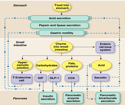
| back 65 Chyme enters small intestine
Started to break down proteins by pepsin in stomach
Rate of entry of chyme determines intestinal response (secretion, motility, nutrient use) |
front 66 intestinal phase secretion | back 66 • Bicarbonate (pancreas)- Neutralizes stomach acid
• Mucus (goblet cells)- Lubricates & protects epithelium
• Bile (gallbladder)- Fat digestion
• Digestive enzymes (enterocytes & pancreas)- Break down food particles
• 5.5L of total stuff in the lumen after secretion |
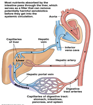
front 67 hepatic portal system (picture) | back 67 • after the transport of organic nutrients and ions through the duodenum and jejunum most absorbed nutrients move into the capillaries in the villi and then into the hepatic portal system.
• specialized region of the circulation that has 2 sets of capillary beds: one that picks up absorbed nutrients in the intestine and another that delivers the nutrients to the liver |
front 68 absorption in the small intestine | back 68 7.5L of 9L reabsorbed
Most happens in duodenum & jejunum
Move into capillaries & then the hepatic portal system
As you move solutes water will follow |
front 69 digestion in the intestines | back 69 • most digestion occurs in the small intestine
• Protein -Broken down by many peptidases & proteases à small peptides & amino acids
• Carbs- Digestible polysaccharides broken down into monosaccharides & absorbed
• Fats- Bile salts enter at the duodenum to allow digestion & absorption
• By large intestine down to 1.5 L of stuff |
front 70 differences between large intestine and small intestine walls | back 70 Large intestine has:
Colonocytes
Mucosa (Smooth, no villi)
Muscularis (Discontinuous longitudinal layer) |
front 71 large intestine digestion and absorption | back 71 o Traditionally thought that little to no digestion/absorption occurs here
o Recent research increasingly points to importance of gut bacteria for digestion |
| back 72 o Around 500 species of bacteria in your gut
o Key for breaking down nutrients
o Three main categories: Prevotella (Carbs & simple sugars), Bacteroides (Proteins),Ruminococcus
Absorption of nutrients is dramatically affected if these bacteria are missing
o Byproduct: gas production
o babies develop a gut microbiome which varies depending on the way the birth was given, how much the baby is interacted with (exposure) and whether the baby drank breast milk ir formula
o stool transplant |
| back 73 o Chyme enters colon & mixes via segmental contractions
o Forward movement is minimal
o Occurs via mass movement
Decreases diameter of colon
Pushes bolus forward
Gastrolic reflex
o Pushes bolus into rectum |
| back 74 • Distension of rectal wall initiates
• Internal anal sphincter relaxes
• Peristalsis pushes material forward
• External anal sphincter relaxes & feces move out of anus
• Emotions and the limbic system can affect defecation |
| back 75 • Abnormal water absorption causes watery feces
o Osmotic-Unabsorbed solutes pull water into feces (Ex: Lactose, sorbitol, Olestra, polyethylene glycol)
o Secretory-Bacterial toxins enhance Cl- secretion, Increased fluid secretion causes increased motility |
| back 76 • Specialized epithelial cells
• Sample gut content
• Transport content to interstitial fluid
• If bad stuff present, launch immune response
o Cytokines are released and trigger an immune response
• Irritable bowel syndrome- M cells can misrecognize normal foods and cause an immune response to normal food |
| back 77 Protective reflex
Force contents out of stomach and duodenum
Regulated by medulla
Caused by: Cytokines, drugs, pain, disturbed equilibrium
Reverse peristalsis- smooth muscle wave moves from small intestine, through the stomach, up the esophagus, and out the mouth |
| back 78 Regulated by two hypothalamic centers
• Feeding center
o Tonically active
o Removal results in stopping eating
• Satiety center
o Inhibits feeding center
o Removal results in overeating which causes obesity |
| back 79 o a theory about how food uptake is regulated
o Glucose metabolism regulates food intake
o Low blood glucose levels causes a suppression of satiety center which causes hunger |
| back 80 o Lipid level regulates food intake
o Body seeks to maintain an “ideal” weight
o Low lipid level à hunger
o Some research support for this theory
Leptin – protein hormone made in adipocytes that indirectly regulates food intake
Neuropeptide Y directly regulates food intake |
| back 81 • Transport work- Moving molecules between compartments, moving materials into/out of body, Includes gradients for solutes
• Mechanical work, Muscle contraction, movement of vesicles down filaments
• Chemical work, Growth, maintenance, & storage |
| back 82 o Oxygen consumption (indirect calorimetry)
Easiest way to measure
Measure O2 used and convert that into glucose using the glucose pathway
o Estimate of glucose metabolism indirectly
o Glucose metabolism is most efficient using aerobic pathways
• Metabolic rate (kcal/day)= L O2 consumed/day * kcal/L O2 |
| back 83 • Get energy from proteins, carbohydrates & fats
• Store energy for immediate use primarily as glycogen (Glycogen is more compact for storage than glucose)
• Long term storage as fat |
| back 84 o Sum of all chemical reactions in the body
Extract energy from nutrients
Use energy for work
Store energy for later work
anabolism (small to larger molecules) vs. catabolism (larger to smaller molecules)
fed state vs. fasted state |
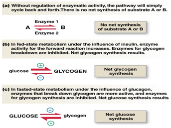
front 85 Fed State vs Fasted State | back 85 o Fed state
AKA absorptive state
Following a meal
Anabolic pathways (smaller to larger molecules) active
most glucose is stored as glycogen in fats
o Fasted state
AKA postabsorptive state
Used up nutrients from food already
Catabolic pathways active (larger to smaller molecules)
most glucose is used right away (in the form of glycogen)
o Body turns incoming proteins into amino acids that can be used where they are needed
If there is too much protein the proteins can be turned into glucose and used for energy |
front 86 hormone regulation of metabolism | back 86 insulin drives the production of glycogen from glucose |
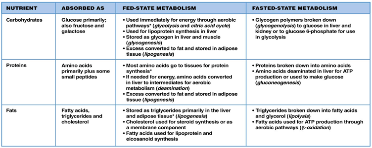
front 87 fed-state metabolism- carbohydrates | back 87 • Absorbed by intestine as monosaccarides (glucose mostly)
o About 30% of the glucose that goes through the liver is immediately metabolized
o The remaining 70% is sent out to the blood and circulated to the areas where it is needed
o If it is not needed it is stored as glycogen |
front 88 fed-state metabolism- protein | back 88 • Absorbed by intestine as amino acids
• Diffuse into blood via hepatic portal vein
• Sent to the liver
• Excess amino acids get stored as fat |
front 89 fed-state metabolism- fats | back 89 o Triglycerides + phospholipids + cholesterol + lipid-binding proteins
o More protein = heavier
o Range from VLDL (very low density lipoproteins) to HDL (high density lipoproteins)
o Protein bound with lipids makes them more soluble to cell membranes
o cholesterol |
| back 90 o Range from VLDL (very low density lipoproteins) to HDL (high density lipoproteins)
Cholesterol levels are determined by the number of lipoproteins in the lipoprotein-cholesterol complex
o Most found as low-density lipoprotein cholesterol (LDL-C)
“Lethal cholesterol”
Uses apoprotein B to diffuse through membranes
o Second most common is high-density lipoprotein cholesterol (HDL-C)
“Healthy cholesterol”
Uses apoprotein A to diffuse through membranes
o Elevated LDL-C level is largest risk factor associated with heart disease
Can manage through diet, exercise, drugs |
| back 91 • Diabetes mellitus – abnormal glucose metabolism
• Kidney & liver disorders – abnormal plasma protein levels
• Heart disease – abnormal cholesterol levels
o No studies that show causation between high dietary cholesterol and blood cholesterol levels
o Is a correlation between high blood cholesterol and heart disease |
front 92 fasted vs fed state metabolism nutrients (picture) | |
| back 93 After all nutrients from a meal have been cleared from digestive tract & plasma glucose concentrations start to fall
• After a meal blood glucose rises and fasted state begins when the glucose begins to fall
Plasma glucose level a key regulator
Catabolism hallmark of postabsorptive state |
front 94 glycogenolysis (carbohydrate fasted state metabolism) | back 94 glycogen can be converted directly to glucose 6-phosphate by the addition of a phosphate
glycogen that is broken down first to glucose then phosphorylated "costs" the cell an extra ATP
most glycogen is found in the liver |
front 95 amino acid catabolism (protein fasted state metabolism) | back 95 deamination- removal of the amino group from an amino acid creates ammonia and an organic acid
body does not like ammonia (toxic) so it gets secreted
free amino acid pool used for energy production
creates pyruvate, acetyl CoA, etc. that can be used in glycolysis and the citric acid cycle |
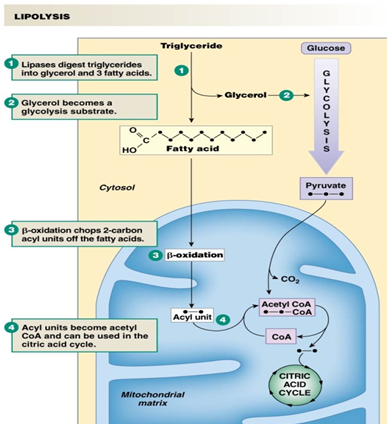
front 96 lipolysis (fat fasted state metabolism) | back 96 • Primary fuel-storage molecule
• Excess acetyl CoA production à ketones
o Useful in the citric acid cycle
• Basis for fad diets (Atkins, South Beach) |
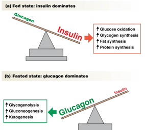
front 97 metabolism regulation (picture) | back 97 o Primarily regulated by endocrine cells
o Pancreas secretes insulin & glucagon
o Ratio of two hormones is what drives metabolism
o Insulin and glucagon are both always present so the ratios of them are what matter
o Short half lives, so they need to be replaced regularly |
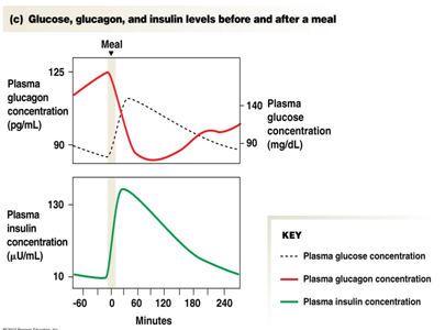
front 98 GLucose, glucagon, and insulin levels before and after a meal (picture) | |
| back 99 Peptide hormone
Stimuli for insulin secretion:
• Increased plasma glucose
• Increased plasma amino acids
• Feedforward effects of other hormones involved in digestion
• Autonomic division activity
o “rest and digest” |
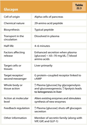
| back 100 o Secreted by alpha cells in pancreas
o Whatever insulin does, glucagon does the opposite
o Low plasma glucose causes glucagon release
o Triggers catabolism
o Glycogenolysis & gluconeogenesis to generate new glucose
o Liver is primary target |
front 101 metabolism regulation dysfunction | back 101 o Diabetes mellitus- Abnormally elevated plasma glucose levels (AKA hyperglycemia)
o Type 1 – insulin deficiency due to beta cell destruction
Fasted state: in the absence of insulin, there are no GLUT4 transporters in the membrane, Fed state: insulin signals the cell to insert GLUT 4 transporters into the membrane, allowing glucose to enter the cell
Damaging to blood vessels, eyes, kidneys & nervous system
8.3% of the U.S. population
o Type 2 – insulin resistant
Variety of problems: Atherosclerosis, Neurological changes, Renal failure, Blindness |
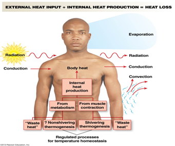
| back 102 conduction, radiation, convection, evaporation |
| back 103 Thermoneutral zone (82-86°F)
Normal metabolism generates enough heat to maintain body temperature
above that range causes gain of heat and the need to cool down, likewise for below that range
sweating |
| back 104 • Evaporative cooling
• Use 2-3 million sweat glands in skin
• Sympathetic control
• Secretion of hypotonic solution
• Dependent on humidity |
| back 105 • Use convection by passing blood close to surface
• Sympathetic control
• Heat conservation- Close to zero blood flow through cutaneous layer
• Heat loss- 1/3 of cardiac output to cutaneous layer |
| back 106 • Shivering
o Skeletal muscle contraction generates heat
• Non-shivering
o Brown fat- fat found in babies between the shoulder blades which is just now being discovered in adults
o More pronounced in babies and non-humans |
front 107 variation in temperature regulation | back 107 • Circadian patterns: Daily, menstrual cycle
• Postmenopausal hot flashes
• Fever-Pyrogens
Cytokines produced by immunocytes
Increase body temperature
Enhances activity of WBCs |
| back 108 Recognize and remove foreign invaders
Recognize and remove abnormal body cells
Remove dead or damaged cells |
front 109 immune system dysfunction | back 109 • Incorrect response- Fail to recognize self from non-self (Ex: Autoimmune diseases)
• Overactive response- Respond at a greater level than threat posed (Ex: Allergies)
• Lack of response- Failure of immune system to respond (Ex: Acquired immunodeficiency syndrome (AIDS)) |
| back 110 • Divides by self replicating
• Circular DNA, Unicellular, Small, Cell wall
• Prokayoric (no nucleus)
• No membrane bound organelles
• Antibiotics work well to kill bacteria |
| back 111 • Needs a host to reproduce
• DNA or RNA
• Use host cell machinery
• Small genomes
• Integration of DNA |
| back 112 • Found throughout body
• Thymus gland
• Bone marrow
• Lymph nodes
• Diffuse tissues |
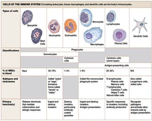
front 113 Cells of the immune system | |
| back 114 • Phagocytes
• Most abundant WBC- 50-70% of white blood cells
• Release pyrogens
o Fever response |
| back 115 • Important for inflammatory & allergic responses
• Granulocytes
o Histamine, Heparin |
| back 116 • Rare
• Allergic & parasitic response
• Granulocytes
o Cytotoxins |
front 117 Monocytes and Macrophages | back 117 • Monocytes à macrophages
• Trash cans of immune system
o Pick up and take out mostly everything
o Anything abnormal, aging
• Antigen-presenting cells |
| back 118 • Key part of acquired immunity
• B lymphocytes
• T lymphocytes and Natural killer (NK) cells- play important roles in defense against intracellular pathogens, such as viruses |
| back 119 • Non-circulating
• Antigen-presenting cells
• Found in skin |
front 120 immunogen, antigen, antibody | back 120 • Immunogen- Anything that triggers the body’s immune response
• Antigen- Immunogen that reacts with immune response
• Antibody- Proteins secreted by immune cells that bind to antigens to make them more visible to immune response |
front 121 goals of an immune response | back 121 • Detection and identification of foreign substances
• Communication to other immune cells
• Coordination of response
• Suppression of target |
| back 122 • Innate Immunity- Present from birth, Nonspecific, immediate response
• Acquired Immunity- Specific response, Slower response |
| back 123 Found throughout body
Thymus gland
Bone marrow
Lymph nodes
Diffuse tissues |
| back 124 Epithelium- the protective barrier of skin and mucous membrane is the body's first line of defense
Secretions- salivary glands and the glands in airways secrete mucus and immunoglobulins to trap and disable inhaled or ingested pathogens
Stomach acid- the low pH of the stomach helps destroy swallowed pathogens |
| back 125 • Innate immunity
• Attracted by chemotaxins
• Secrete own chemotaxins (cytokines) to attract more phagocytes
• Chemotaxins bind to PRRs
• Secrete inflammatory cytokines
• Ingest pathogen |
| back 126 • Innate immunity
• Key for viral infections and cancer cells
• Rapid response
• Trigger apoptosis in infected cells |
front 127 inflammatory response for innate immunity | back 127 • inflammatory response is key for innate immunity
• Attracts immune cells
• Produces physical barrier to prevent spread of infection
• Promotes tissue repair |
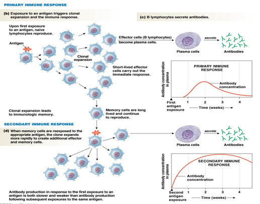
| back 128 • antibody production and antigen presentation, become plasma cells
• memory cells: coordinate immune response much more quickly and strongly when exposed to the same antigen a second time
• effector cells: short lived plasma cells that carry out the immediate immune response and secrete antibodies |
| back 129 o Cytotoxic – Attack & destroy
o Helper – Coordinate immune response |
front 130 major histocompatibility complex (MHC) | back 130 • tissue rejection- want similar HLA antigens for tissue transplant (blood groups) |
front 131 Active and Passive immunity | back 131 Active immunity
• Body responds to pathogen & creates antibodies
• Ex: chicken pox, vaccines
Passive immunity
• Receive antibodies made from another organism
• Ex: Motheràfetus, breast milk |
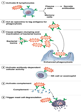
front 132 Antibodies (AKA immunoglobulins) (picture) | back 132 o Activate B lymphocytes
o Act as opsonins
o Enhance phagocytosis
o Trigger cytotoxic responses
o Activate complement proteins
o Activate mast cells |
front 133 Bacteria Immune Response (picture) | |
front 134 virus immune response (picture) | |
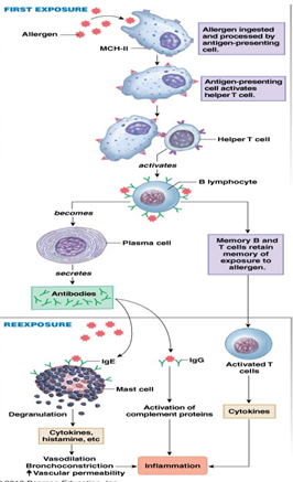
front 135 allergen immune response (picture) | back 135 Inflammatory immune response to a non-pathogenic antigen
Wide range of triggers and severity
Key player: Histamine |
front 136 vaccination immune response (picture) | |