Instructions for Side by Side Printing
- Print the notecards
- Fold each page in half along the solid vertical line
- Cut out the notecards by cutting along each horizontal dotted line
- Optional: Glue, tape or staple the ends of each notecard together
Tissue Identification
front 1  Name this tissue. | back 1 ciliated pseudostratified epithelium |
front 2  Name this tissue. | back 2 pseudostratified columnar epithelium |
front 3  Name this tissue
| back 3 areolar connective tissue
|
front 4  Name this tissue. | back 4 Simple squamous epithelium |
front 5  Name this tissue. | back 5 stratified squamous epithelium |
front 6  Name this tissue. | back 6 Stratified columnar epithelium |
front 7  Name this tissue. | back 7 simple cuboidal epithelium |
front 8 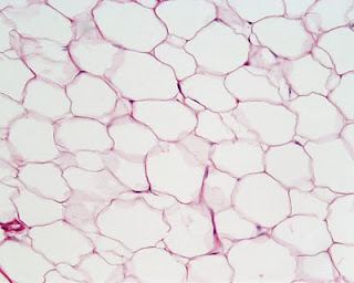 Name this tissue. | back 8 adipose connective tissue |
front 9  Name this tissue. | back 9 Stratified cuboidal epithelium |
front 10  Name this tissue. | back 10 keratinized stratified squamous epithelium |
front 11 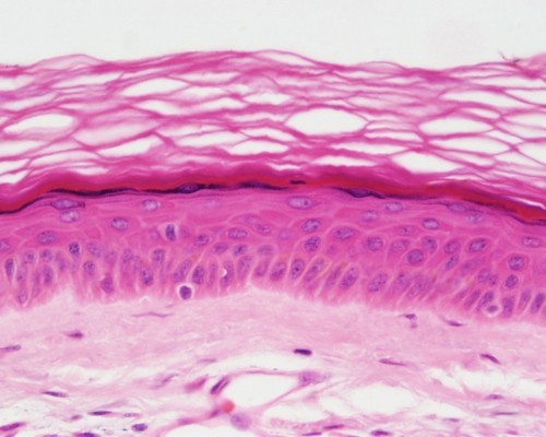 Name this tissue. | back 11 keratinized stratified squamous epithelium |
front 12  Name this tissue.
| back 12 dense irregular connective tissue
|
front 13  Name this tissue.
| back 13 reticular connective tissue
|
front 14  Identify the indicated structures. | back 14 Intercalated disks |
front 15  Name this tissue.
| back 15 dense regular connective tissue
|
front 16 Name this tissue. | back 16 transitional epithelium |
front 17  Name this tissue.
| back 17 Connective Tissue - blood
|
front 18  Identify:
| back 18 a. Volkmann's canal
|
front 19 Name this tissue.
| back 19  Connective Tissue - bone
|
front 20  Name this tissue. | back 20 Muscle Tissue - cardiac muslce |
front 21  Name this tissue.
| back 21 Muscle Tissue - skeletal muscle
|
front 22  Name this tissue.
| back 22 Muscle Tissue - smooth muscle
|
front 23  Name this tissue. | back 23 Connective Tissue - hyaline cartilage |
front 24  Identify:
| back 24 1. chondrocytes
|