Instructions for Side by Side Printing
- Print the notecards
- Fold each page in half along the solid vertical line
- Cut out the notecards by cutting along each horizontal dotted line
- Optional: Glue, tape or staple the ends of each notecard together
IGCSE Biology 3
front 1 List the components of blood as and functions | back 1
|
front 2 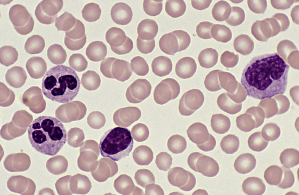 Identify red and white blood cells and lymphocytes and phagocytes | back 2  Identifying Red Blood Cells:
Identifying White Blood Cells:
Identifying Lymphocytes and Phagocytes:
|
front 3 Describe the process of clotting | back 3 1. Clotting Factors and Thrombin:When a blood vessel is damaged, clotting factors are released, which ultimately leads to the activation of the enzyme thrombin. 2. Fibrinogen to Fibrin:Thrombin acts as a catalyst, converting the soluble plasma protein fibrinogen into insoluble fibrin. 3. Mesh Formation:Fibrin molecules then polymerize, forming long, insoluble threads that create a mesh-like network. 4. Clot Formation:This fibrin mesh traps platelets and blood cells, including red blood cells, forming a clot that seals the wound. 5. Scab Formation:Over time, the clot dries and hardens, forming a scab that protects the wound while new skin cells grow underneath. |
front 4 Define a pathogen | back 4 A disease-causing organism. |
front 5 Define a transmissible disease | back 5 A disease in which the pathogen can be passed from one host to another. |
front 6 State how a pathogen is transmitted | back 6 (a) by direct contact, including through blood and other body fluids (b) indirectly, including from contaminated surfaces, food, animals and air |
front 7 Describe the body defences | back 7
|
front 8 Define active immunity | back 8 Defence against a pathogen by antibody production in the body. |
front 9 Define antibodies | back 9 Proteins that bind to antigens leading to direct destruction of pathogens or marking of pathogens for destruction by phagocytes. |
front 10 Outline the process of vaccination | back 10 (a) weakened pathogens or their antigens are put into the body (b) the antigens stimulate an immune response by lymphocytes which produce antibodies (c) memory cells are produced that give long-term immunity |
front 11 Define passive immunity | back 11 Passive immunity is a short-term defence against a pathogen by antibodies acquired from another individual, including across the placenta and in breast milk. |
front 12 Describe cholera | back 12 A disease caused by bacteria which is transmitted in contaminated water. The cholera bacterium produces a toxin that causes secretion of chloride ions into the small intestine, causing osmosis of water into the gut, causing diarrhoea, dehydration and loss of ions from the blood. |
front 13 Describe the features of gas exchange surfaces in humans | back 13 limited to: large surface area, thin surface, good blood supply and good ventilation with air |
front 14 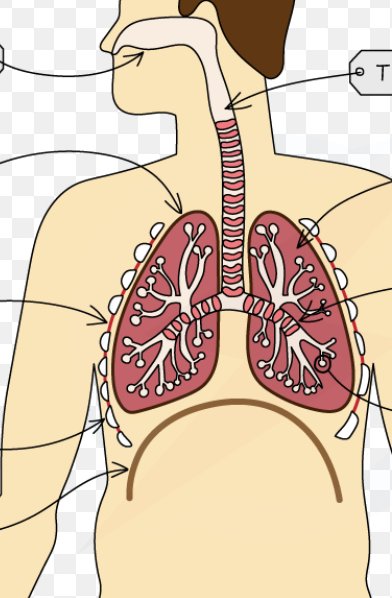 Identify in diagrams and images the following parts of the breathing system: | back 14 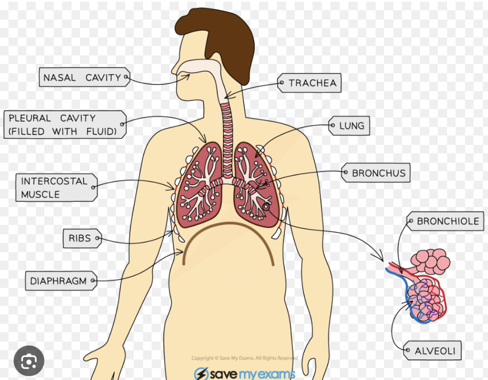 Identify in diagrams and images the following parts of the breathing system: lungs, diaphragm, ribs, intercostal muscles, larynx, trachea, bronchi, bronchioles, alveoli and associated capillaries |
front 15 Explain the role of the ribs, the internal and external intercostal muscles and the diaphragm in producing volume and pressure changes in the thorax leading to the ventilation of the lungs. | back 15 Overall process:
|
front 16 Explain the differences in composition between inspired and expired air | back 16 Inspired air has more oxygen (about 21%) and less carbon dioxide (about 0.04%). Expired air has less oxygen (around 16%) and more carbon dioxide (around 4%), along with more water vapor and a higher temperature. |
front 17 Explain the link between physical activity and the rate and depth of breathing | back 17 An increased carbon dioxide concentration due to muscles working harder and more as a waste product in the blood, which is detected by the brain, leading to an increased rate and greater depth of breathing. |
front 18 Explain the role in protecting the breathing system from pathogens and particles | back 18 Goblet cells produce sticky mucus that traps pathogens and particles, while ciliated cells have hair-like cilia that beat to sweep this mucus, and its trapped contents, up the airways towards the throat to be swallowed and destroyed. |
front 19 State the uses of energy in living organisms, including | back 19
|
front 20 Describe aerobic respiration and equation | back 20 The chemical reactions in cells that use oxygen to break down nutrient molecules to release energy. glucose + oxygen → carbon dioxide + water C6H12O6 + 6O2 → 6CO2 + 6H2O |
front 21 Describe anaerobic respiration and 2 equations | back 21 The chemical reactions in cells that break down nutrient molecules to release energy without using oxygen. *Anaerobic respiration releases much less energy per glucose molecule than aerobic respiration. State the word equation for anaerobic respiration in yeast as: glucose → alcohol + carbon dioxide (C6H12O6 → 2C2H5OH + 2CO2) State the word equation for anaerobic respiration in muscles during vigorous exercise as: glucose → lactic acid *lactic acid builds up in muscles and blood during vigorous exercise causing an oxygen debt To help oxygen debt: (a) continuation of fast heart rate to transport lactic acid in the blood from the muscles to the liver (b) continuation of deeper and faster breathing to supply oxygen for aerobic respiration of lactic acid (c) aerobic respiration of lactic acid in the liver |
front 22 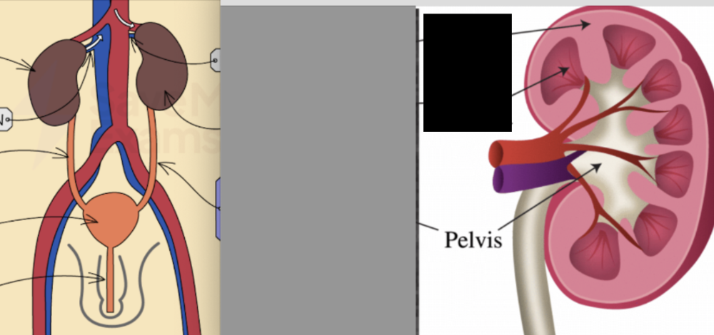 Identify in diagrams and images the structure of the kidney and bladder etc. | back 22 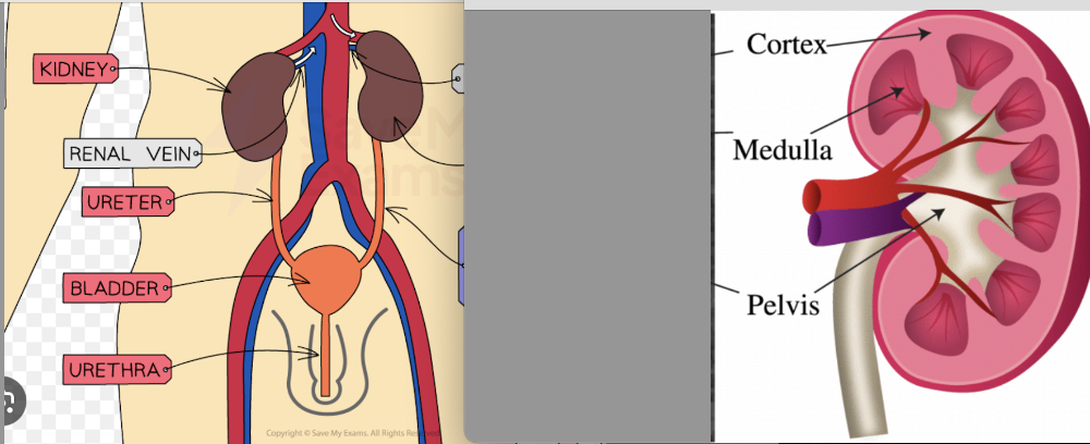 Identify in diagrams and images the kidneys, ureters, bladder and urethra Identify in diagrams and images the structure of the kidney, limited to the cortex and medulla |
front 23 Outline the structure and function of a nephron and its associated blood vessels. | back 23 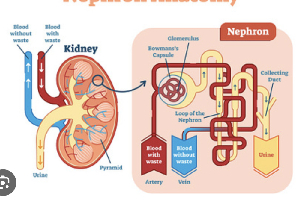 (a) the role of the glomerulus in the filtration from the blood of water, glucose, urea and ions (b) the role of the nephron in the reabsorption of all of the glucose, some of the ions and most of the water back into the blood (c) the formation of urine containing urea, excess water and excess ions |
front 24 Describe role of liver in excretion including definition for deamination. | back 24 Assimilation:
Describe deamination as the removal of the nitrogen-containing part of amino acids to form urea. Excretion of urea is vital because urea's accumulation can poison cells, disrupt metabolic processes, and eventually lead to organ failure. |
front 25 What travels along neurones? | back 25 electrical impulses |
front 26 Describe the mammalian nervous system and its role | back 26 (a) the central nervous system (CNS) consisting of the brain and the spinal cord (b) the peripheral nervous system (PNS) consisting of the nerves outside of the brain and spinal cord *the role of the nervous system as coordination and regulation of body functions |
front 27 Describe the events at a synapse and definition | back 27 A junction between two neurones. (a) an impulse stimulates the release of neurotransmitter molecules from vesicles into the synaptic gap (b) the neurotransmitter molecules diffuse across the gap (c) neurotransmitter molecules bind with receptor proteins on the next neurone (d) an impulse is then stimulated in the next neurone *Synapses ensure that impulses travel in one direction only |
front 28 Define a reflex action | back 28 A means of automatically and rapidly integrating and coordinating stimuli with the responses of effectors (muscles and glands). |
front 29 Describe a simple reflex arc in terms of: receptor, sensory neurone, relay neurone, motor neurone and effector | back 29
|
front 30 Describe the structure of a synapse | back 30 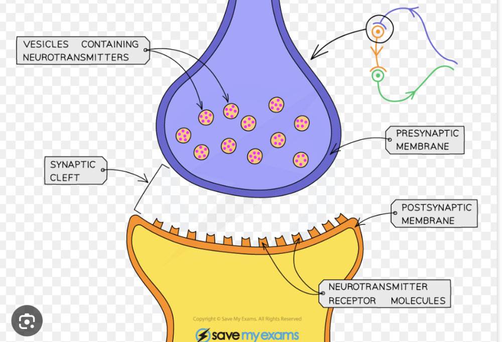 |
front 31 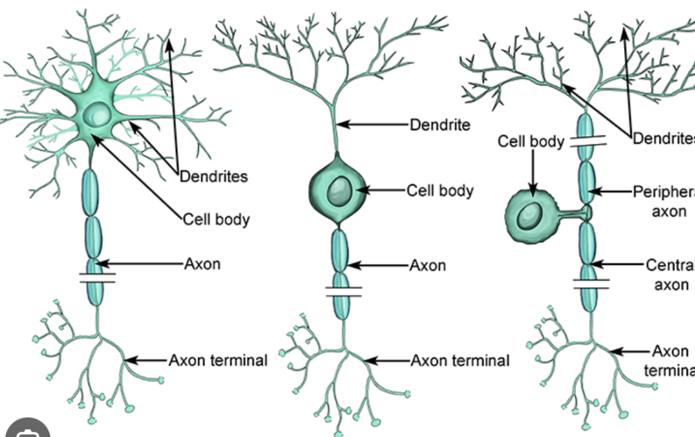 | back 31 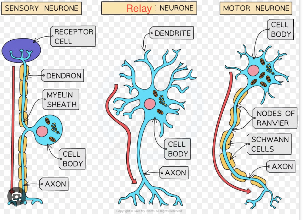 Sensory neurones are long, have a cell body branching from the middle of the axon, and carry impulses from receptors to the CNS; relay neurones are short, found within the CNS, and connect sensory to motor neurones; motor neurones are long, with a cell body at one end and long dendrites, carrying impulses from the CNS to effectors like muscles or glands. |
front 32 Define sense organs | back 32 Groups of receptor cells responding to specific stimuli: light, sound, touch, temperature and chemicals. |
front 33 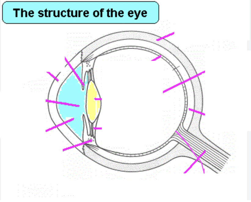 Identify in diagrams and images the structures of the eye | back 33 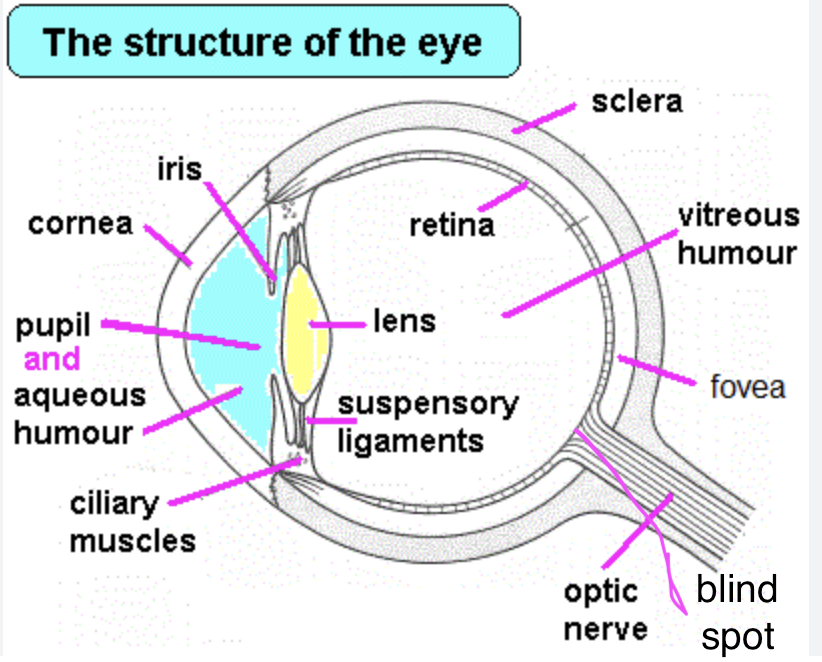 cornea, iris, pupil, lens, retina, optic nerve and blind spot |
front 34 Describe the function of each part of the eye, limited to: (a) cornea (b) iris (c) lens (d) retina (e) optic nerve | back 34 (a) cornea – refracts light (b) iris – controls how much light enters the pupil (c) lens – focuses light on to the retina (d) retina – contains light receptors, some sensitive to light of different colours (e) optic nerve – carries impulses to the brain |
front 35 Explain the pupil reflex | back 35 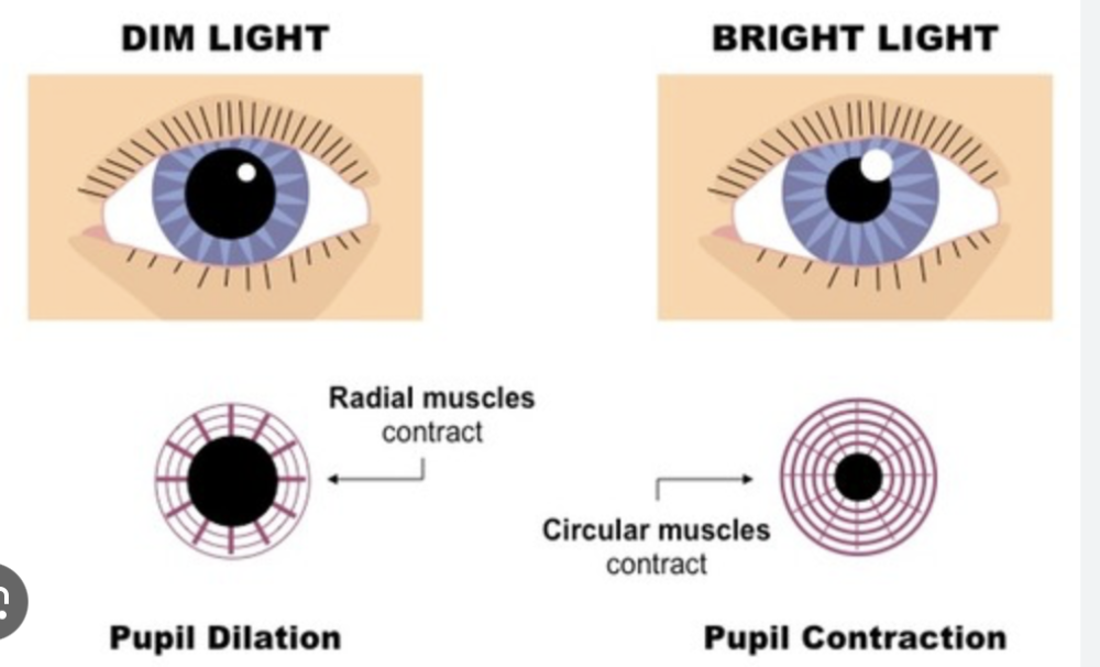 Antagonistic muscles: are pairs of muscles that work in opposite ways; when one contracts, the other relaxes. In Bright Light: The circular muscles contract, while the radial muscles relax which shrinks the pupil allowing less light to enter the eye, protecting the retina. In Dim Light: The radial muscles contract, while the circular muscles relax causing the pupil to dilate (widen). A larger pupil allows more light to enter the eye, helping the eye to see in low-light conditions. |
front 36 Explain accommodation to view near and distant objects, how eye changes | back 36 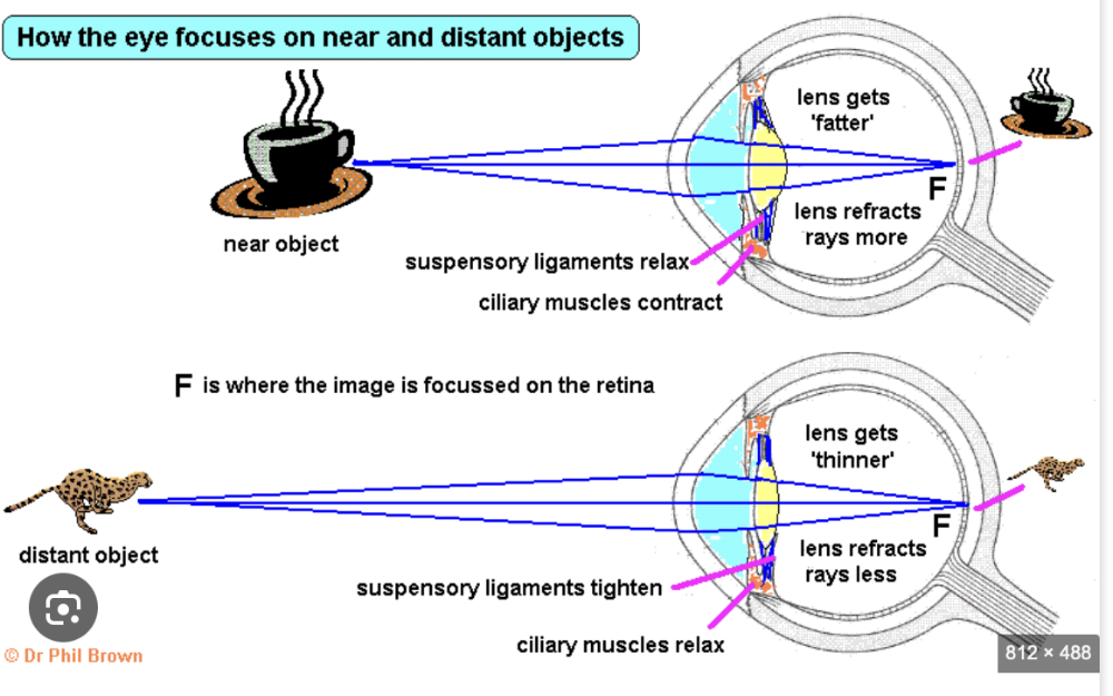 For distant objects, the ciliary muscles relax, tightening the suspensory ligaments and making the lens thinner and flatter, which reduces light refraction. For near objects, the ciliary muscles contract, relaxing the suspensory ligaments and allowing the elastic lens to become thicker and more convex, increasing its refractive power to focus the light. |
front 37 Function and distribution of rods and cones | back 37 Rods
Cones
|
front 38 Define a hormone | back 38 A chemical substance, produced by a gland and carried by the blood, which alters the activity of one or more specific target organs. |
front 39 Identify in diagrams and state the hormones they secrete, limited to: adrenaline, insulin, testosterone, oestrogen and glucagon | back 39 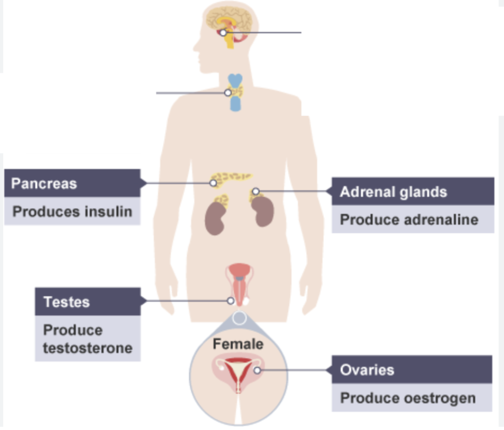 (a) adrenal glands and adrenaline (b) pancreas and insulin (c) testes and testosterone (d) ovaries and oestrogen (e) glucagon is secreted by the pancreas |
front 40 Define adrenaline and its effects, | back 40 the hormone secreted in ‘fight or flight’ situations (a) increased breathing rate (b) increased heart rate (c) increased pupil diameter (d) increasing the blood glucose concentration |
front 41 Compare nervous and hormonal control, limited to speed of action and duration of effect | back 41 Nervous control is characterised by very fast, almost instantaneous responses that are short-lived. Hormonal control is a slower process involving chemical signals (hormones) transported via the bloodstream, resulting in longer-lasting effects on the body's tissues and organs. |
front 42 Define homeostasis and how it works | back 42 The maintenance of a constant internal environment. Homeostatic control uses negative feedback to keep internal body conditions stable around a fixed set point.When a condition, like blood glucose, deviates from its set point, receptors detect the change. This triggers a correction mechanism, such as the release of insulin, that acts in the opposite direction of the initial change to bring the condition back to its set point, after which the mechanism is switched off. |
front 43 what does insulin do and how liver controls blood glucose concentration | back 43 decreases blood glucose concentration The liver controls blood glucose through the action of insulin and glucagon, produced by the pancreas, in a negative feedback loop. When blood glucose is high, insulin stimulates the liver to store excess glucose as glycogen. When blood glucose is low, glucagon stimulates the liver to break down stored glycogen into glucose, which is released into the blood. |
front 44 Outline the treatment of Type 1 diabetes | back 44 Treatment for Type 1 diabetes involves lifelong management to control blood glucose levels, through insulin injections or insulin pump therapy, monitor their blood sugar regularly, follow a balanced diet to manage carbohydrate intake, and engage in regular exercise to help lower blood glucose levels. |
front 45 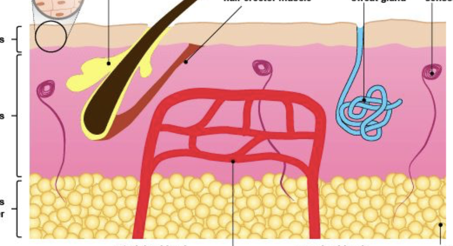 Identify in diagrams and images of the skin: hairs, hair erector muscles, sweat glands, receptors, sensory neurones, blood vessels and fatty tissue | back 45 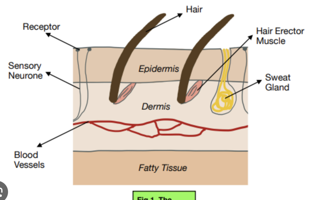 |
front 46 Describe methods for the maintenance of a constant internal body temperature | back 46 Mammals maintain a constant internal temperature (thermoregulation) through a feedback system controlled by the hypothalamus in the brain, using mechanisms like insulation (fur/fat to trap air), sweating (evaporative heat loss) shivering (muscle activity to generate heat) vasodilation (widening of blood vessels for heat loss) : This increases blood flow to the skin, allowing more heat to be radiated, conducted, or convected away from the body. vasoconstriction (narrowing of blood vessels for heat conservation) of skin capillaries : This reduces blood flow to the skin, minimizing heat loss to the environment and conserving the body's core heat. |
front 47 Define gravitropism | back 47 A response in which parts of a plant grow towards or away from gravity. |
front 48 Define phototropism | back 48 A response in which parts of a plant grow towards or away from the direction of the light source. |
front 49 Explain phototropism and gravitropism | back 49 Phototropism
Gravitropism
It is a chemical control of plant growth |
front 50 Describe auxin | back 50 (a) auxin is made in the shoot tip (b) auxin diffuses through the plant from the shoot tip (c) auxin is unequally distributed in response to light and gravity (d) auxin stimulates cell elongation |
front 51 Describe experiment for phototropism and gravitropism | back 51
|
front 52 How temperature effects yeast respiration | back 52 A moderate temperature increase accelerates the rate by increasing enzyme kinetic energy and reaction speed. However, beyond an optimal point (often around 25-35°C, depending on the yeast strain), enzymes denature, and the rate of respiration rapidly slows and eventually stops. |