Instructions for Side by Side Printing
- Print the notecards
- Fold each page in half along the solid vertical line
- Cut out the notecards by cutting along each horizontal dotted line
- Optional: Glue, tape or staple the ends of each notecard together
BMED-4440-CB Final
front 1 The Enterotube II / EnteroPluri-Test is designed to identify ___ bacteria and other select oxidase-______, Gram-_____ bacteria. | back 1 1. Enteric 2. Negative 3. Negative |
front 2 How many chambers are there? Name the mediums present in each one. | back 2 12 chambers
|
front 3 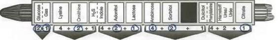 E. coli Positives: Lys, Ind, Adon, Arab, Sorb E. aerogenes Positives: Lys, Orn, Arab, Sorb, VP, Citrate K. pneumoniae Positives: Lys, Arab, Sorb, VP, Urea, Citrate What are the five digit codes? | back 3 E. coli: 04660 E. aerogenes: 06061 K. pneumoniae: 04063 |
front 4 For the Indole Test, what reagent is used? Positive result is what color? | back 4 Kovac's Reagent Red |
front 5 For the VP test, what reagents are used? Positive result is what color? | back 5 KOH Reagent & alpha-Naphthol Reagent Red |
front 6 Steps for performing the Enterotube Test? | back 6
|
front 7 ELISA is an acronym for | back 7 Enzyme Linked Immunosorbent Assay |
front 8 It is used to ______ | back 8 detect antigen or antibody in a sample |
front 9 What are the steps to the ELISA test? | back 9
|
front 10 Why is the secondary antibody needed? | back 10 It will be what gives us a positive or negative result. |
front 11 What is the enzyme used and why is itneeded? What color is a positive result? | back 11 The enzyme is HRP and it will bind secondary antibody to indicate its presence. Blue indicates a positive result. |
front 12 Where does the Kirby-Bauer method get its name? | back 12 Of the names who published it |
front 13 What is the meaning of MIC? | back 13 Minimum inhibitory concentration |
front 14 How is the antibiotic used in the Kirby-Bauer method? | back 14 Antibiotic-impregnated paper disks |
front 15 What is the drug that kills bacteria? | back 15 Bactericidal |
front 16 What is the drug that stops the bacteria from dividing? | back 16 Bacteriostatic |
front 17 What do you read with McFarland turbitidy? | back 17 Cell Density |
front 18 We diluted the cultures with sterile saline until ____ McFarland. | back 18 0.5 |
front 19 If a bacteria is susceptible to an antibiotic, the zone diameter would be ___. What if it was resistant? | back 19 Large Small |
front 20 What bacteria did we use for the Kirby-Bauer lab? | back 20 E. coli and S. aureus |
front 21 What antibiotics did we use for the Kirby-Bauer lab? | back 21 Chloramphenicol, Ciprofloxicin, Trimethoprim, Penicillin |
front 22 What is bacterial transformation? | back 22 The process by which competent bacterial cells pick up DNA from the environment and make use of the genes it carries. |
front 23 What are the structural and functional genetic units of prokaryotes? | back 23 Operons |
front 24 Each operon minimally includes a ______ ______ and two or more ______ _______ coding for enzymes in the same metabolic pathway. | back 24 promoter site structural genes |
front 25 What is used as a vector in the pGLO lab? | back 25 a plasmid |
front 26 Plasmids are _______ | back 26 small, naturally occurring, circular DNA molecules that possess only a few genes. |
front 27 What does the pGLO plasmid contain? | back 27 araC - allows RNA polymerase to bind to promoter Pbad - arabinose promoter GFP - green fluorescent protein, makes it glow bla - antibiotic resistance gene, produces beta-lactamase which hydrolyzes ampicillin ori - replication origin |
front 28 Viruses that attack bacteria are called | back 28 bacteriophages |
front 29 Once assembly is complete, the cell lyses and releases the phages, which then attack other bacterial cells. What is this process called? | back 29 Lytic Cycle |
front 30 Lysis of bacterial cells growing in a lawn on an agar plate produce a clearing, what are these clearings called? | back 30 Plaques |
front 31 Why are plaques important? | back 31 They allow us to calculate the phage concentration in a given sample. |
front 32 What is the formula to calculate phage titer? | back 32 Phage titer = PFU / Original Sample Volume |
front 33 Why is the soft agar, the tube that contains phage-host mix, important? | back 33 The consistency of it is sufficient enough to immobilize the bacteria while allowing the smaller bacteriophages to diffuse short distances and infect surrounding cells. |
front 34 Which parasite causes an infection that involves a significant amount of surface area of the small intestine and can cause chronic diarrhea, dehydration, abdominal pain and other symptoms? | back 34 Giardia lamblia |
front 35 What parasite causes malaria? | back 35 Plasmodium spp. |
front 36 Review bacteria shapes & stains: What does gram positive cocci look like? What does gram positive bacilli look like? What does gram negative spirochetes look like? Be aware of difference between spirochetes, spirilla, and vibrio. | back 36 Purple circles Purple rods / pill shaped Pink coils look up differences on google |
front 37 Steps for Gram Staining | back 37
|
front 38 Steps for a Wet Mount | back 38 1. Place a loopful of water on a clean glass slide. |
front 39 What are the chemicals that develop color as they oxidize? | back 39 Chromogenic reducing agents |
front 40 What kind of culture do you have if you only have a single species? | back 40 pure culture |
front 41 What kind of culture do you have if you have two or more species? | back 41 mixed culture |
front 42 What is an important thing to do with the metal material when transferring microorganisms? | back 42 sterilize using bunsen burner |
front 43 When do you use petroleum jelly? | back 43 Hanging Drop |
front 44 What color should a positive and negative result for a McConkey agar look like? | back 44 Pink for Positive, Yellow for Negative |
front 45 What enzyme transforms hydrogen peroxide into water and gaseous oxygen? | back 45 Catalase |
front 46 In the Oxidase Reaction, what enzyme turns the color? | back 46 cytochrome c oxidase |
front 47 What substance is designed to reduce the number of pathogens in living tissue? | back 47 Antiseptics |