Instructions for Side by Side Printing
- Print the notecards
- Fold each page in half along the solid vertical line
- Cut out the notecards by cutting along each horizontal dotted line
- Optional: Glue, tape or staple the ends of each notecard together
A&P I Lab Final
front 1 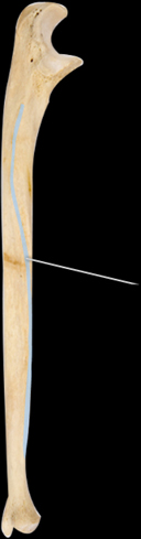 Which structure is highlighted? | back 1 interosseous border |
front 2 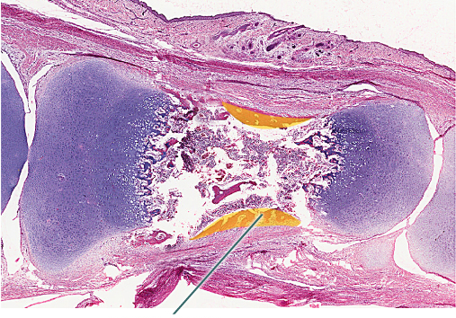 Which structure is highlighted? | back 2 perisoteal bone collar |
front 3 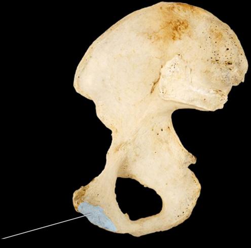 Which structure is highlighted? | back 3 pubic symphyseal fossa |
front 4  Which tissue is highlighted? | back 4 elastic cartilage |
front 5 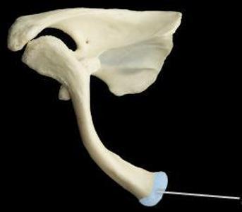 Which structure is highlighted? | back 5 sternal end |
front 6 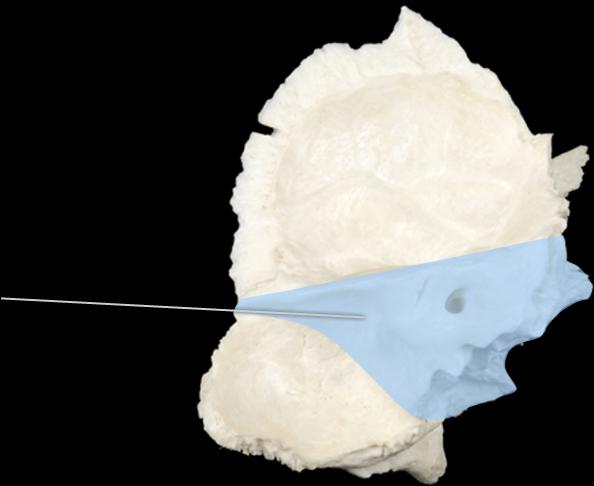 Which region is highlighted? | back 6 petrous |
front 7 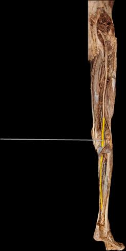 Two major muscles innervated by the highlighted nerve are the ________. | back 7 vastus lateralis and vastus medialis |
front 8 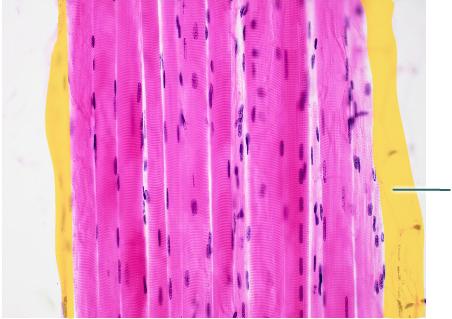 What muscle structure is continuous with the highlighted tissue? | back 8 tendon |
front 9 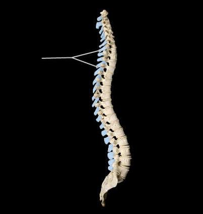 Which structures are highlighted? | back 9 spinous processes |
front 10  The highlighted organ secretes which of the following hormones in response to growth hormone? | back 10 IGF-1 |
front 11 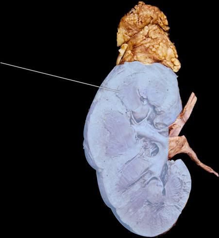 Which hormone does the highlighted organ secrete in response to hypoxia? | back 11 erythropoietin |
front 12 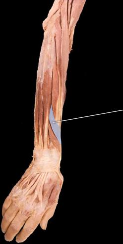 What are the actions of the highlighted muscle? | back 12 abduction and extension of thumb |
front 13 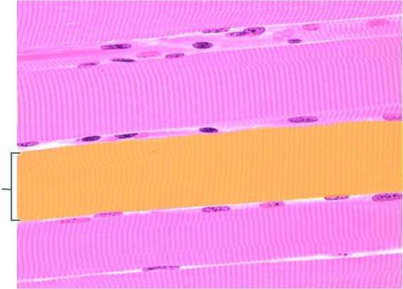 What mechanism allows for a contraction of the highlighted cell? | back 13 sliding filament |
front 14 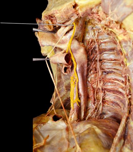 Which structure is highlighted? | back 14 sympathetic trunk |
front 15 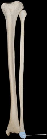 The highlighted structure articulates with which of the following bones? | back 15 talus |
front 16 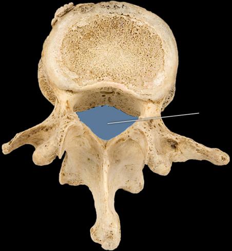 Which structure is highlighted? | back 16 vertebral foramen |
front 17 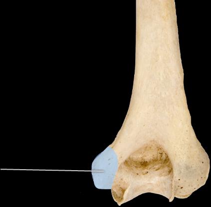 Which structure is highlighted? | back 17 medial epicondyle |
front 18 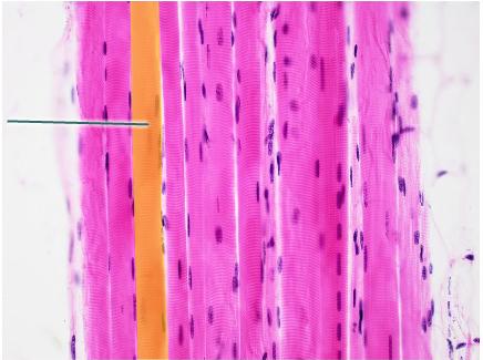 Which structure is highlighted? | back 18 skeletal muscle fiber |
front 19  Which structure is highlighted? | back 19 costal groove |
front 20  The highlighted structure articulates with which structure and bone? | back 20 radial notch of the ulna |
front 21 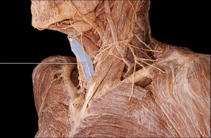 Which muscle is highlighted? | back 21 omohyoid |
front 22 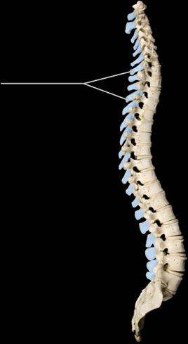 ________ is a condition characterized by the exaggerated lateral bending of the highlighted structure. | back 22 Scoliosis |
front 23 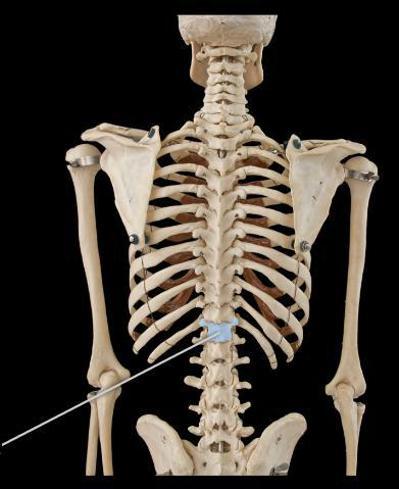 True or false. The highlighted bone articulates with a thoracic and lumbar vertebra. | back 23 True |
front 24 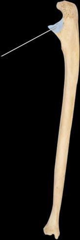 Which structure is highlighted? | back 24 coronoid process |
front 25  Which of the following muscles originates on the highlighted bone? | back 25 deltoid |
front 26 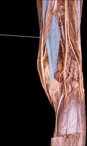 What is an action of the highlighted muscle? | back 26 extends thigh at hip |
front 27 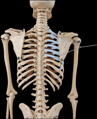 Which structures are highlighted? | back 27 true ribs |
front 28 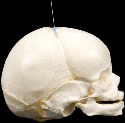 Which structure is highlighted? | back 28 anterior fontanelle |
front 29 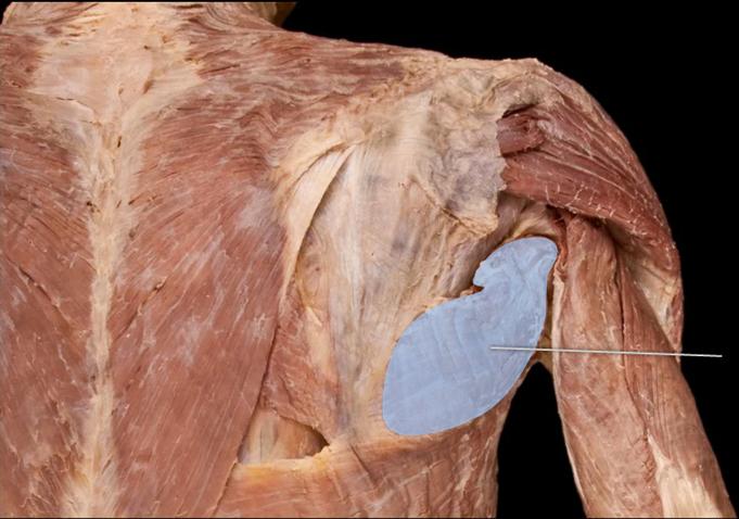 Which muscle is highlighted? | back 29 teres major |
front 30 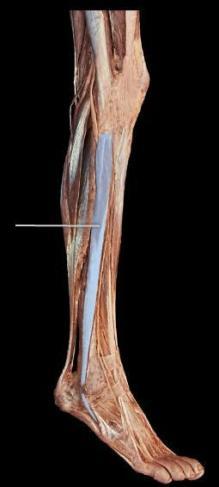 Which muscle is highlighted? | back 30 fibularis longis |
front 31 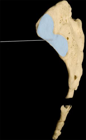 Which structure is highlighted? | back 31 auricular surface |
front 32  Which muscle is highlighted? | back 32 extensor digiti minimi |
front 33 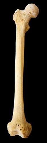 Which bone is in the image? | back 33 femur |
front 34 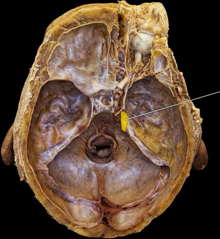 Which cranial nerve is highlighted? | back 34 trigeminal nerve |
front 35 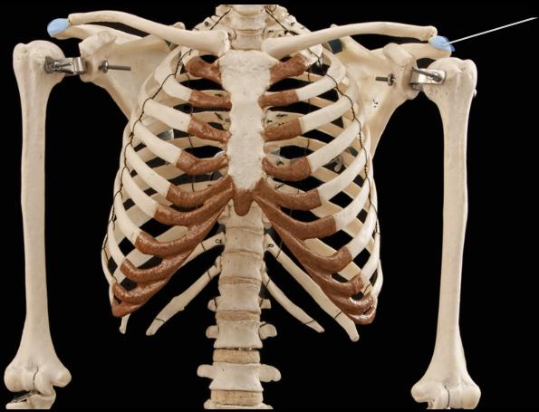 Which structures are highlighted? | back 35 acromia |
front 36 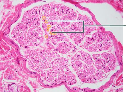 Which tissue is highlighted? | back 36 endoneurium |
front 37 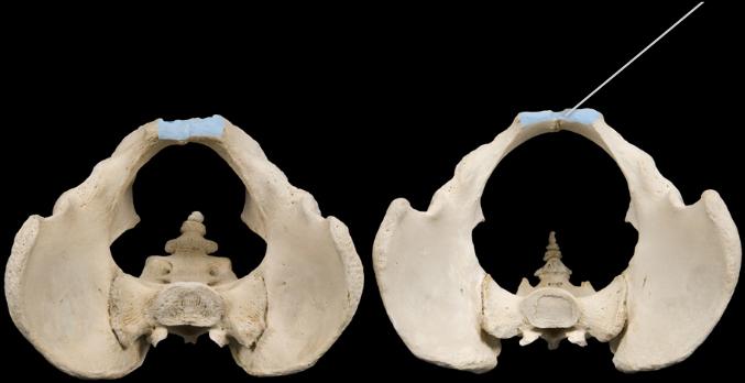 Which of the following muscles originates on the highlighted structure? | back 37 rectus abdominis |
front 38 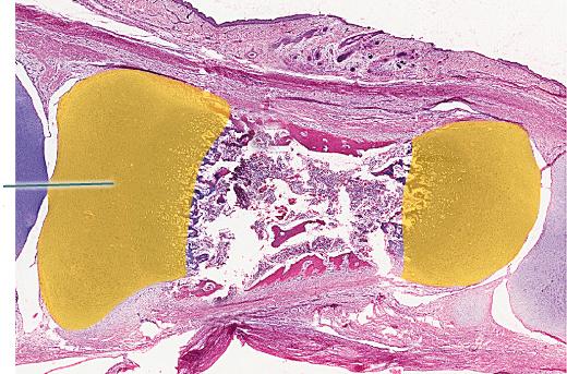 What structure is highlighted? | back 38 cartilaginous epiphysis |
front 39 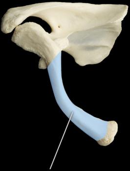 Which of the following is true regarding the highlighted structure and the associated bone? | back 39 develops from the formation of ossification centers within mesenchymal tissue |
front 40 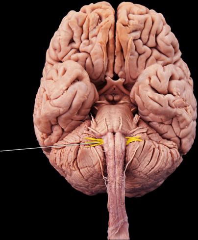 Which two cranial nerves are highlighted? | back 40 glossopharyngeal and vagus |
front 41 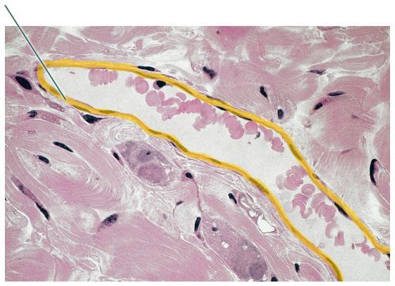 What term is used to refer to the highlighted epithelium in a blood vessel? | back 41 endothelium |
front 42 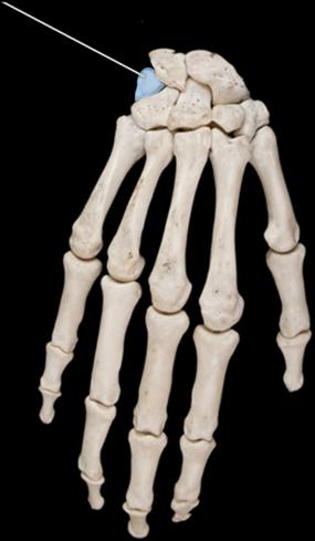 True or false. The highlighted bone articulates with the ulna. | back 42 False |
front 43 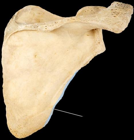 Which structure is highlighted? | back 43 medial border |
front 44 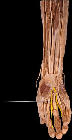 Which structures are highlighted? | back 44 palmar digital branches of the median nerve |
front 45 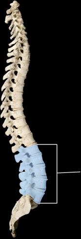 Which structure is highlighted? | back 45 lumbar curvature and vertebra |
front 46 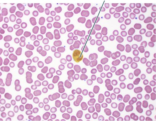 Which of the following is a function of the highlighted cell? | back 46 phagocytosis |
front 47 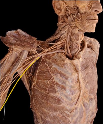 Which nerve is highlighted? | back 47 ulnar |
front 48 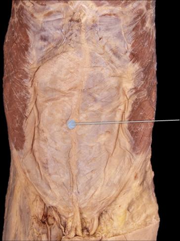 Which structure is highlighted? | back 48 umbilicus |
front 49 Lack of thyroid hormone in a newborn infant leads to what type of disorder? | back 49 cretinism |
front 50 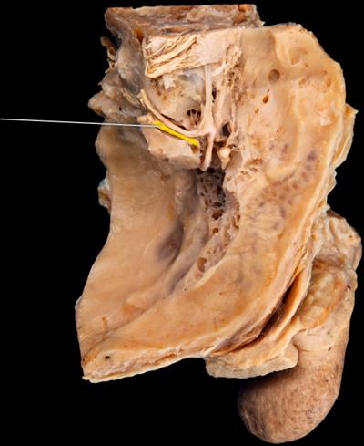 Which nerve is highlighted? | back 50 vestibular |
front 51 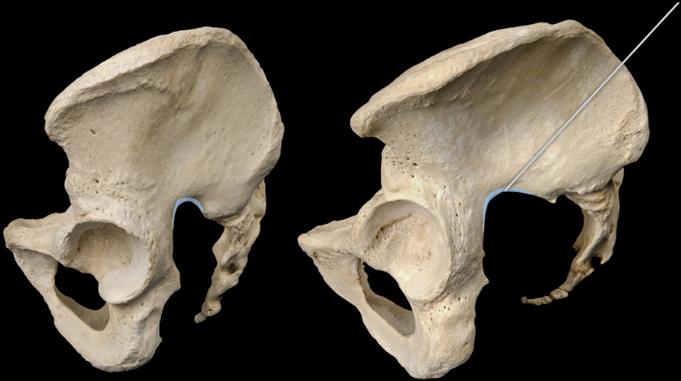 Which structure is highlighted? | back 51 greater sciatic notch |
front 52 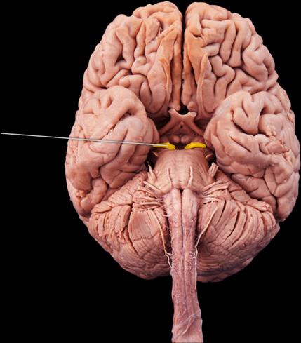 The highlighted nerve plays what role in the parasympathetic nervous system? | back 52 pupil constriction |
front 53 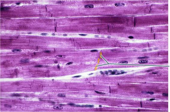 What is a function of the highlighted structures? | back 53 allows action potentials to quickly travel from one cell to another |
front 54 The highlighted bone and the hip bone form what type of synovial joint? | back 54 ball and socket |
front 55 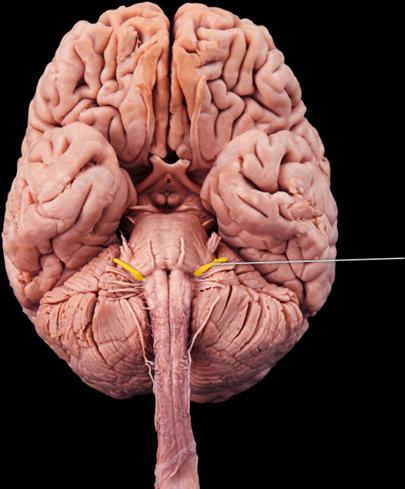 Which cranial nerve is highlighted? | back 55 facial |
front 56 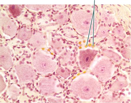 Which cells are highlighted? | back 56 satellite cell |
front 57 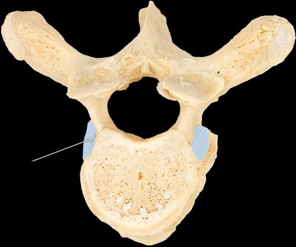 The highlighted structure articulates with which of the following? | back 57 head of the rib |
front 58 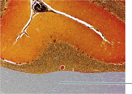 Which tissue is highlighted? | back 58 white matter |
front 59 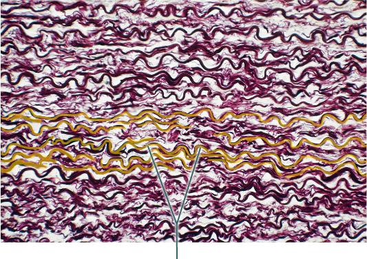 The highlighted fibers are produced by what cell type? | back 59 fibroblast |
front 60 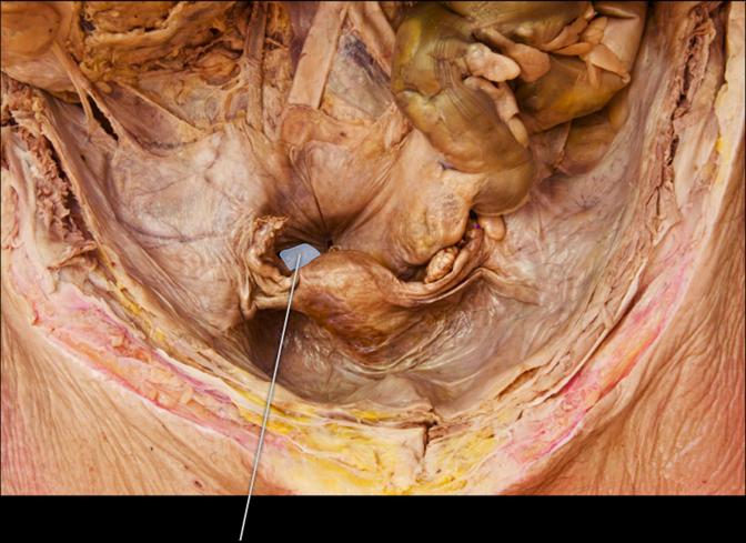 Which structure is highlighted? | back 60 ovary |
front 61 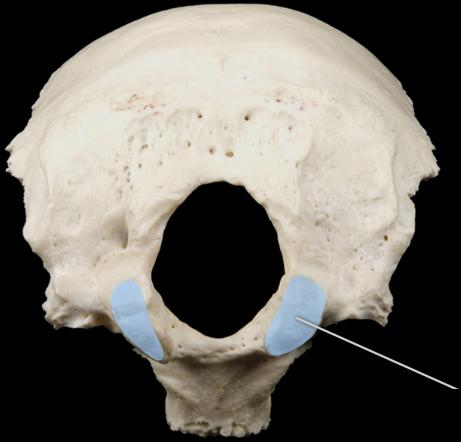 Which structures are highlighted? | back 61 occipital condyles |
front 62  Which cranial nerve is highlighted? | back 62 abducens nerve (VI) |
front 63  Which structures are highlighted? | back 63 nuclei |
front 64 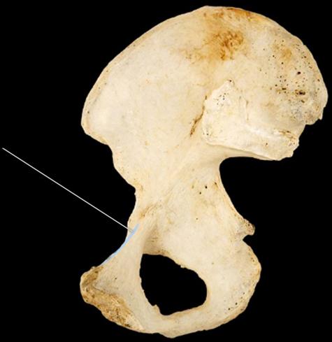 The highlighted structure is continuous with the ________ of the ilium and the ________ of the pubis to form the pelvic brim. | back 64 arcuate line; pubic crest |
front 65  Which structure is highlighted? | back 65 olfactory tract |
front 66 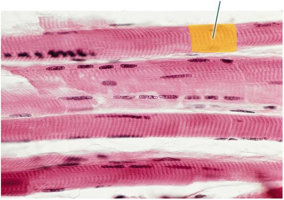 Which structure is highlighted? | back 66 striation |
front 67 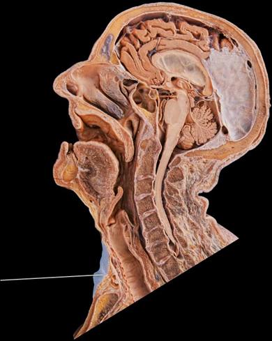 Which gland is highlighted? | back 67 thyroid |
front 68 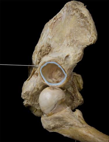 Which structure is highlighted? | back 68 acetabular labrum |
front 69 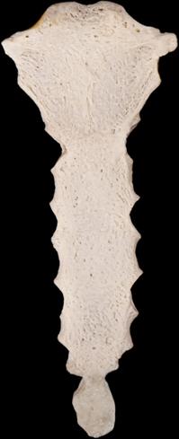 True or false. The bone in this image directly articulates with all 12 ribs. | back 69 False |
front 70 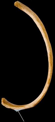 Which surface is highlighted? | back 70 articular tubercle |
front 71 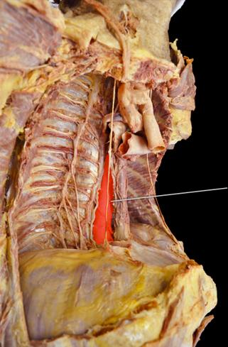 Which vessel is highlighted? | back 71 thoracic aorta |
front 72 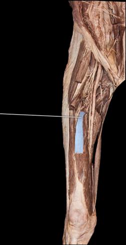 Which muscle is highlighted? | back 72 vastus intermedius |
front 73 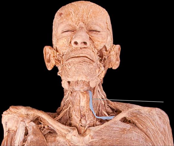 What is an action of the highlighted muscle? | back 73 depresses hyoid |
front 74 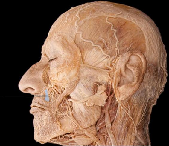 Which muscle is highlighted? | back 74 levator labii superioris |
front 75 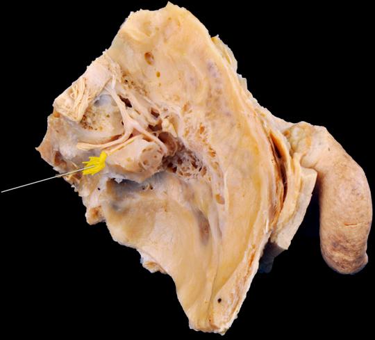 Which nerve is highlighted? | back 75 vestibulocochlear |
front 76 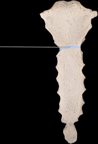 The highlighted structure is an important clinical marker. What is its significance? | back 76 aligns with the superior border of the heart |
front 77 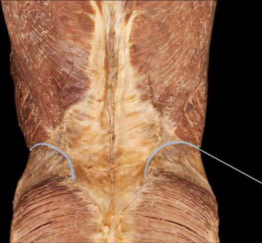 Which structure is highlighted? | back 77 iliac crest |
front 78 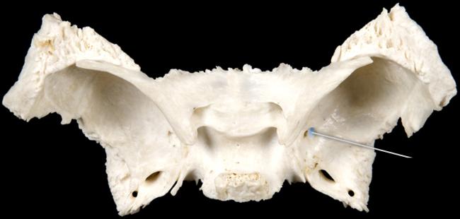 Which structure is highlighted? | back 78 foramen rotundum |
front 79 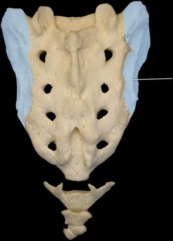 True or false. The highlighted structure directly articulates with the ischium of the coxal bone. | back 79 False |
front 80 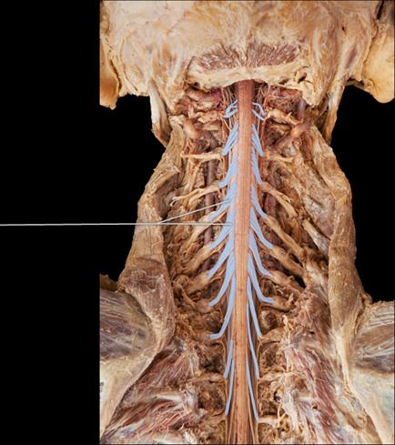 Which structures are highlighted? | back 80 dorsal roots of spinal cord |
front 81 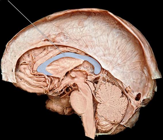 Which structure is highlighted? | back 81 corpus callosum |
front 82 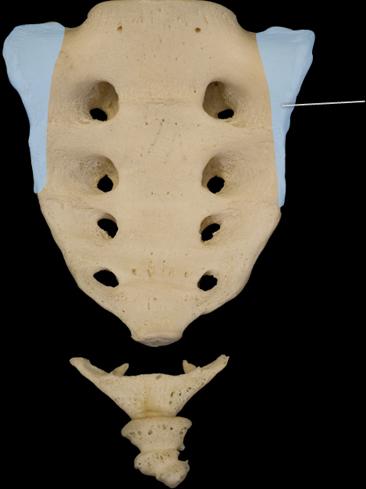 Which structure is highlighted? | back 82 ala |
front 83 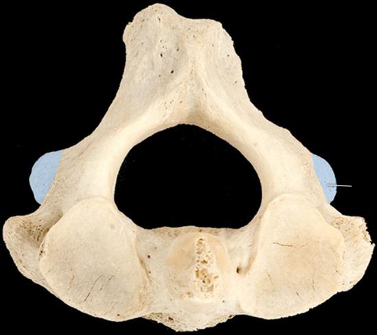 Which structure is highlighted? | back 83 inferior articular process |
front 84 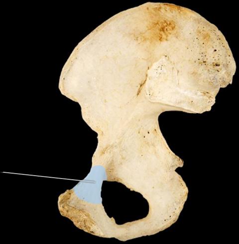 Which structure is highlighted? | back 84 superior ramus of pubis |
front 85  Which structure is highlighted? | back 85 median sacral crest |
front 86 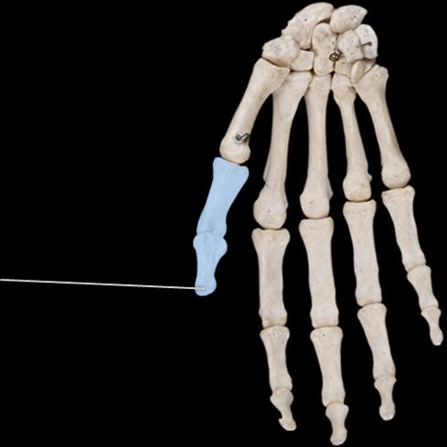 How would you classify the group of highlighted bones? | back 86 long |
front 87 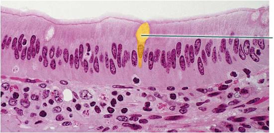 What is secreted by the highlighted cell? | back 87 mucin |
front 88 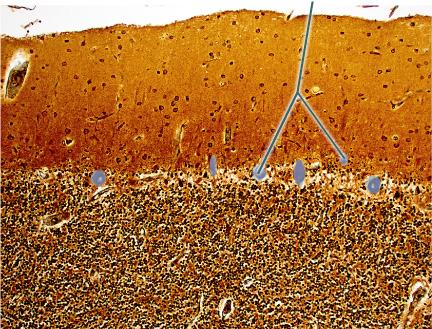 Which structures are highlighted? | back 88 cell bodies of Purkinje cells |
front 89 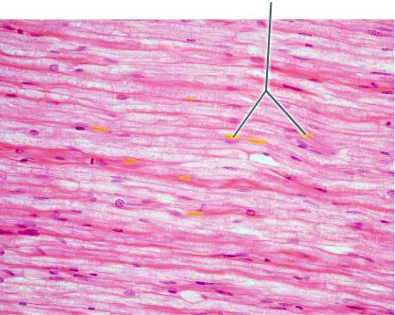 Which structures are highlighted? | back 89 Schwann cell nuclei |
front 90 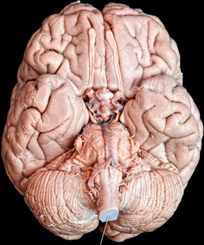 Which structure is highlighted? | back 90 brainstem |
front 91  When does the highlighted curvature develop? | back 91 at the time that a child is able to sit up |
front 92 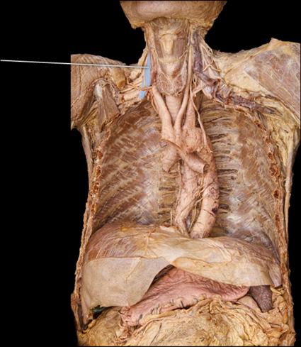 Which muscle is highlighted? | back 92 anterior scalene |
front 93 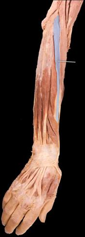 Which nerve innervates the highlighted muscle? | back 93 radial |
front 94 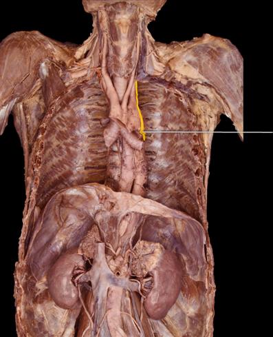 Which nerve is highlighted? | back 94 left vagus |
front 95 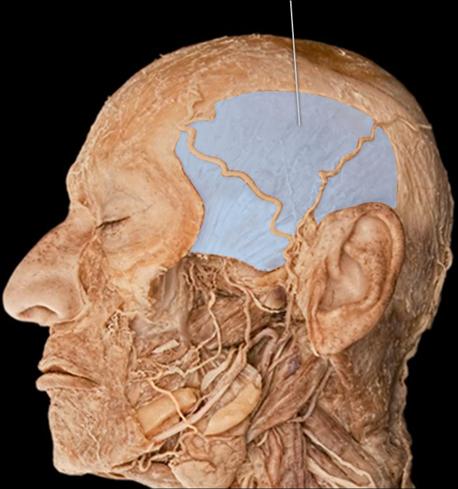 The highlighted muscle originates from which of the following structures of the cranium? | back 95 temporal fossa |
front 96 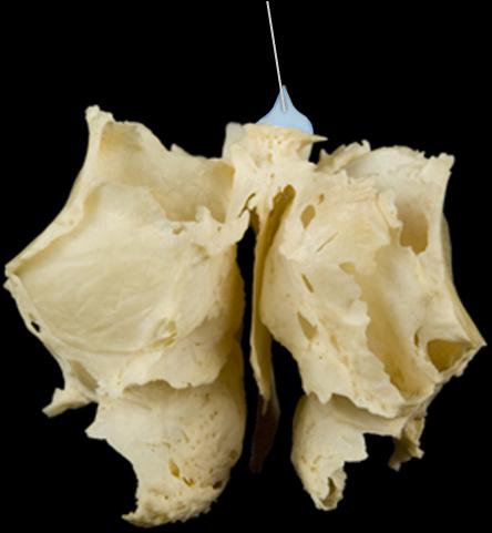 Which structure is highlighted? | back 96 crista galli |
front 97 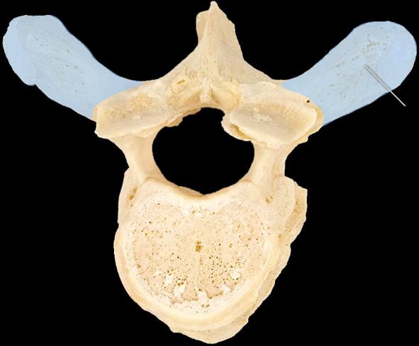 Which structure is highlighted? | back 97 transverse process |
front 98 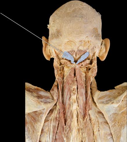 Which muscles are highlighted? | back 98 rectus capitis posterior major |
front 99 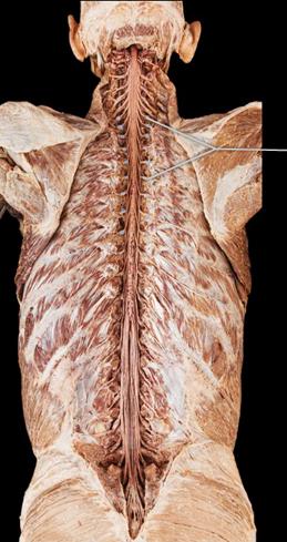 Which structures are highlighted? | back 99 dorsal root ganglia |
front 100 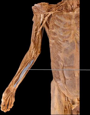 The highlighted muscle flexes the interphalangeal joints of digits ________. | back 100 2-5 |
front 101 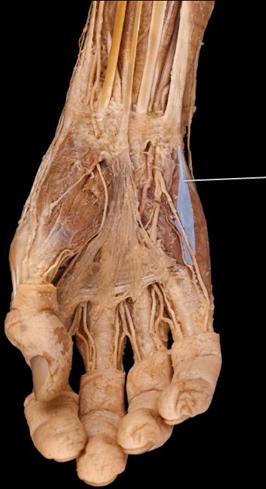 Which muscle is highlighted? | back 101 Flexor digiti minimi brevis |
front 102 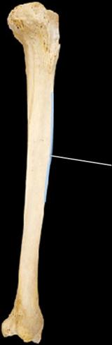 Which structure is highlighted? | back 102 anterior crest |
front 103 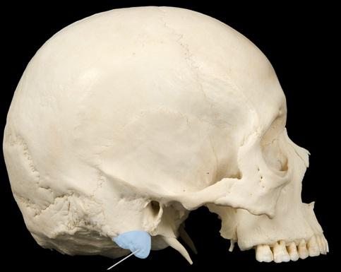 Which structure is highlighted? | back 103 mastoid process |
front 104 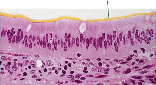 Which structures are highlighted? | back 104 microvilli |
front 105 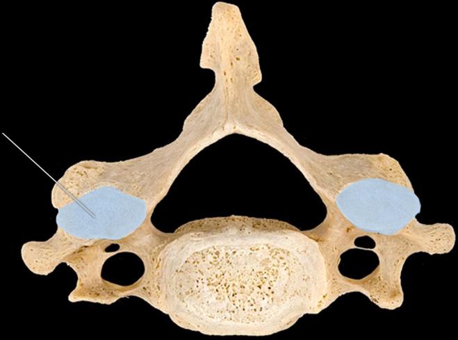 Which structures are highlighted? | back 105 superior articular facet |
front 106 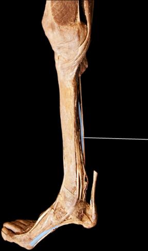 Which muscle is highlighted? | back 106 flexor hallucis longus |
front 107 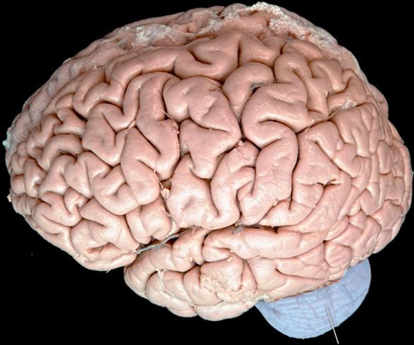 What is a primary function of the highlighted structure? | back 107 coordination of movement |
front 108 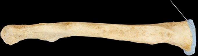 Which structure is highlighted? | back 108 sternal end |
front 109 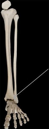 Which joint is highlighted? | back 109 distal tibiofibular |
front 110 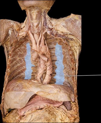 Which muscle is highlighted? | back 110 subcostalis |
front 111 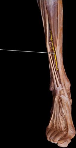 Which nerve is highlighted? | back 111 deep fibular |
front 112 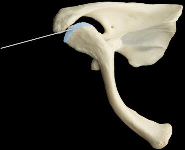 Which joint is highlighted? | back 112 acromioclavicular |
front 113  Which bone is in this image? | back 113 sternum |
front 114 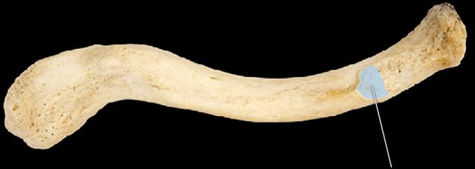 Which structure is highlighted? | back 114 costal tubercle |
front 115 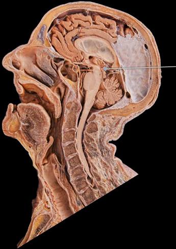 Which structure is highlighted? | back 115 pineal gland |
front 116 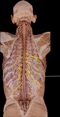 The highlighted structures innervate which of the following? | back 116 deep muscles of thorax |
front 117 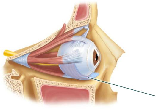 What is the action of the highlighted structure? | back 117 elevates eye and turns it laterally |
front 118 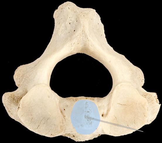 Which of the following best describes the function of the highlighted structure? | back 118 allows lateral rotation of the head |
front 119 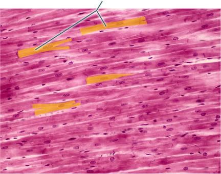 What structure connects the highlighted muscle cells to one another? | back 119 intercalated discs |
front 120 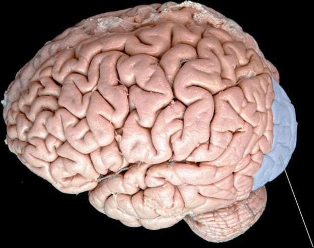 Which lobe is highlighted? | back 120 occipital |
front 121 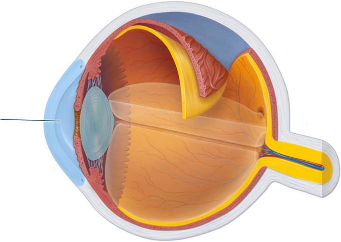 Which structure is highlighted? (124) | back 121 cornea |
front 122  Which structure is highlighted? | back 122 sclera |
front 123 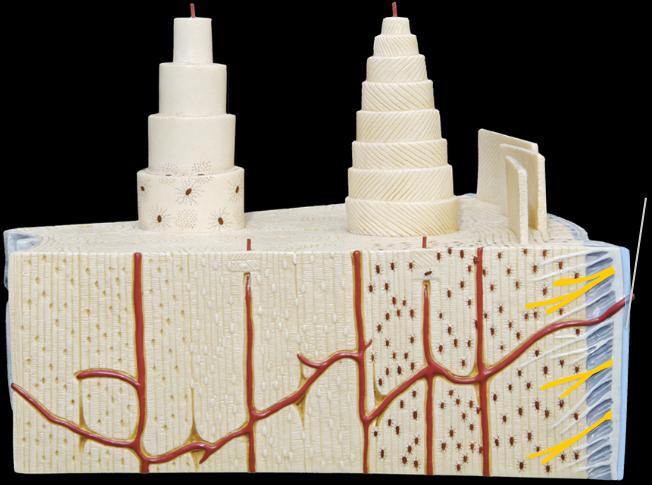 True or false? Fibroblasts can be found within the highlighted structure. | back 123 True |
front 124 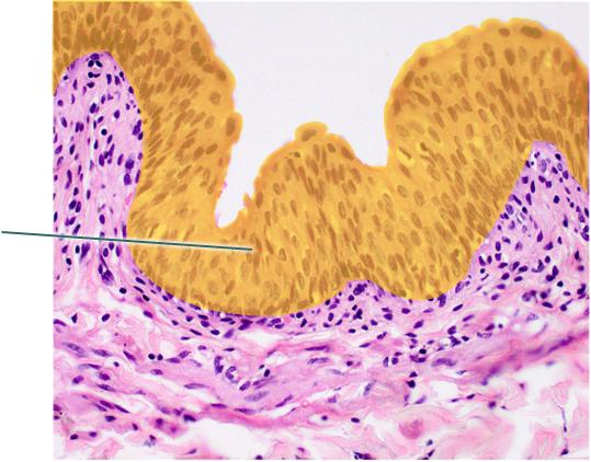 Which epithelial type is highlighted? | back 124 transitional epithelium |
front 125 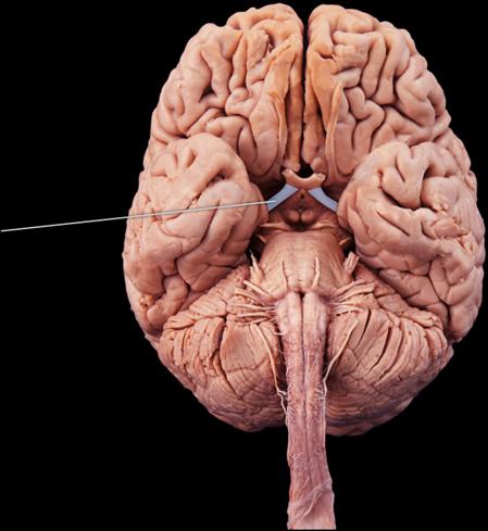 Which structures are highlighted? | back 125 optic tracts |
front 126 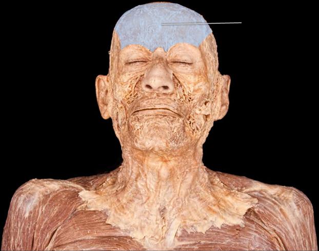 What is the action of the highlighted muscle? | back 126 raises eyebrows |
front 127  Which structure is highlighted? | back 127 lateral epicondyle |