Instructions for Side by Side Printing
- Print the notecards
- Fold each page in half along the solid vertical line
- Cut out the notecards by cutting along each horizontal dotted line
- Optional: Glue, tape or staple the ends of each notecard together
A&P I Lab Final Pictures
front 1 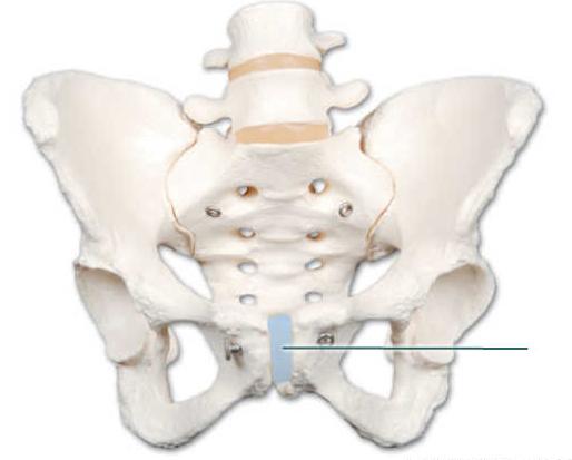 | back 1 Pubic Symphsis |
front 2 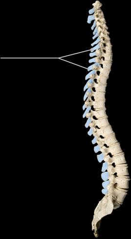 _____ is a condition characterized by the exaggerated lateral bending of the highlighted structure. | back 2 Scoliosis |
front 3 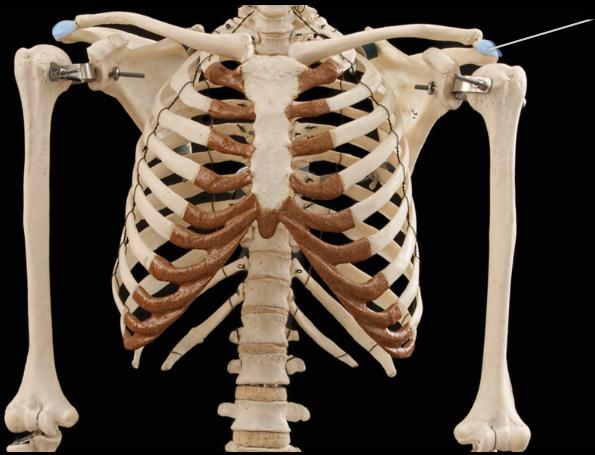 The highlighted features are found on which bone? | back 3 Scapula |
front 4 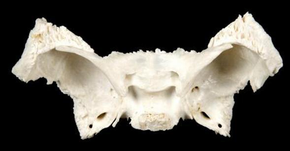 Which bone is in this image? | back 4 Sphenoid |
front 5  What structure is highlighted? | back 5 Axillary Border |
front 6 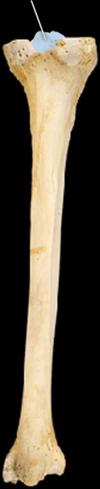 What structure is highlighted? | back 6 Intercondylar eminence |
front 7 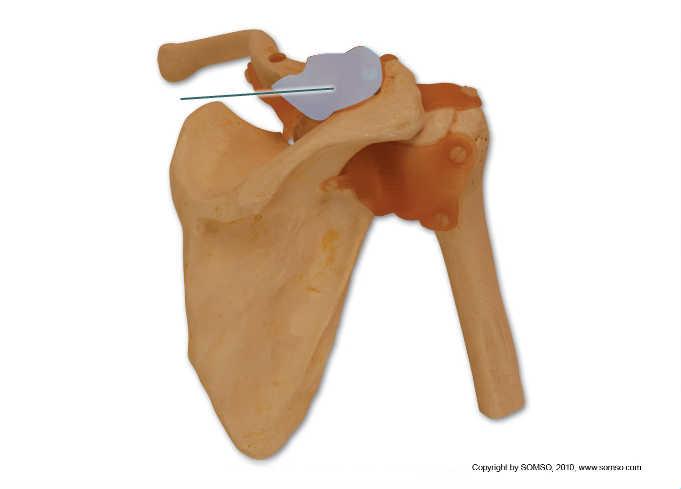 Which structure is highlighted? | back 7 Acromioclavicular Ligament |
front 8 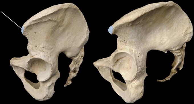 What structure is highlighted? | back 8 Anterior superior illiac spine |
front 9 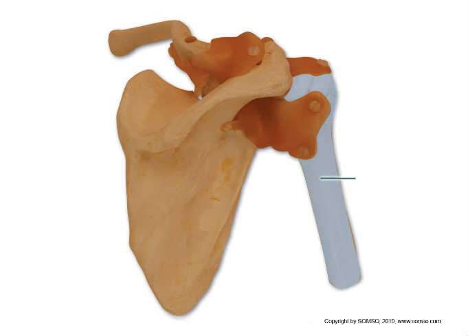 The proximal end of the highlighted bone participates in which joint? | back 9 Glenohumeral |
front 10 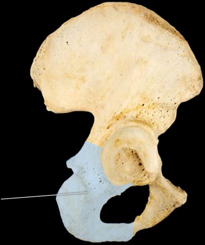 Which bone is highlighted? | back 10 Ischium |
front 11 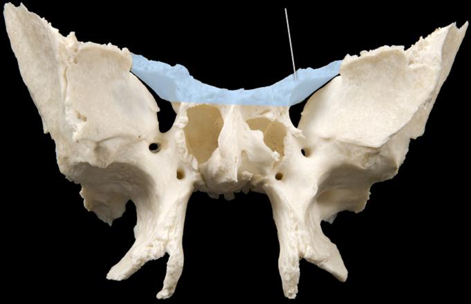 Which structure is highlighted? | back 11 Lesser wings |
front 12 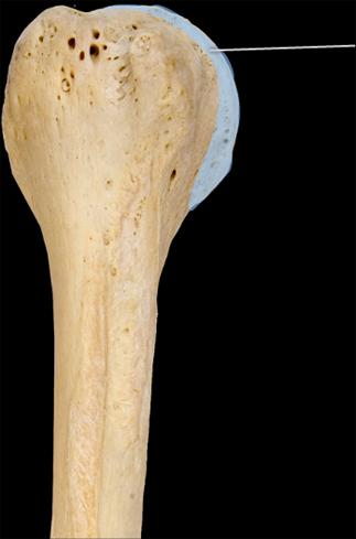 Which structure is highlighted? | back 12 Head |
front 13 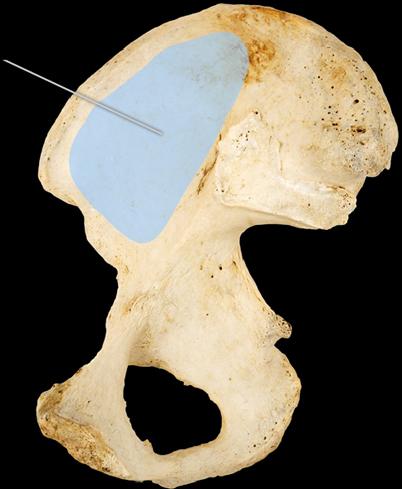 Which structure is highlighted? | back 13 illiac Fossa |
front 14  Which structure is highlighted? | back 14 Acromin |
front 15 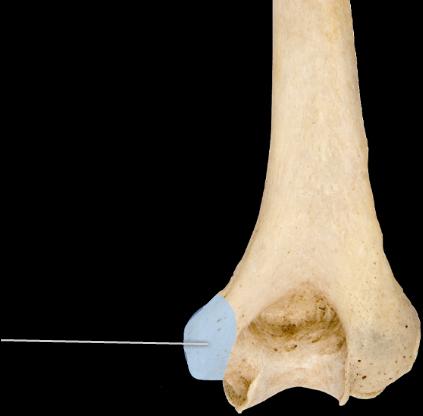 Which structure is highlighted? | back 15 Medial epicondyle |
front 16 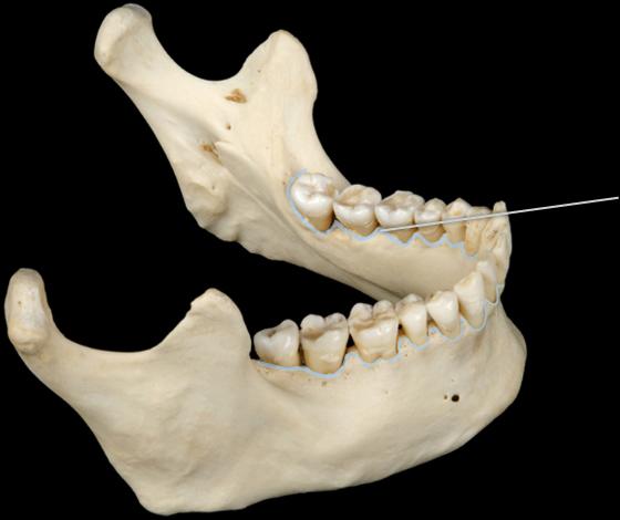 Which structure is highlighted? | back 16 Alveolar Margin |
front 17 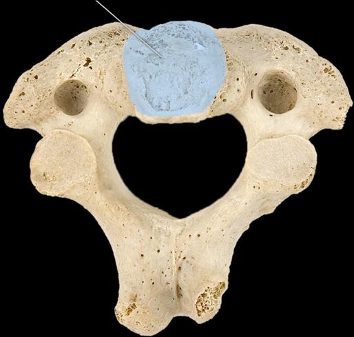 This highlighted structure articulates with which of the following? | back 17 Cervical Vertebra |
front 18 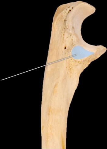 Which structure is highlighted? | back 18 Radial Notch |
front 19 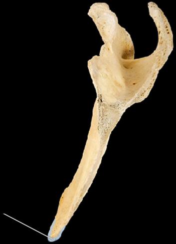 Which structure is highlighted? | back 19 Inferior angle |
front 20 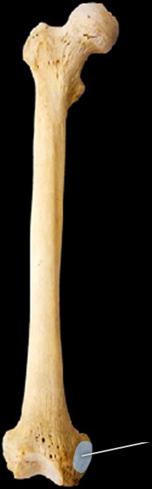 Which of the following ligaments attaches to the structure is highlighted? | back 20 Medical Collateral |
front 21 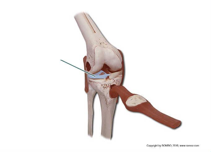 What type of tissue comprises the highlighted structure? | back 21 Fibrocartilage |
front 22 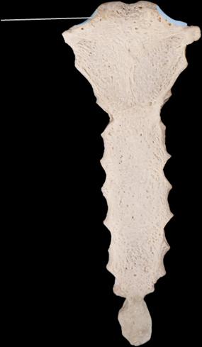 Which structure is highlighted? | back 22 Clavicular Notches |
front 23 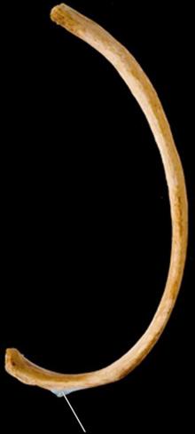 What surface is highlighted? | back 23 articular tubercal and facet |
front 24 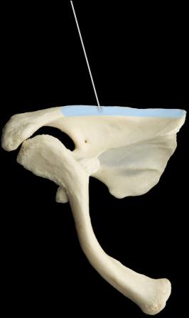 Which structure is highlighted? | back 24 Spine |
front 25 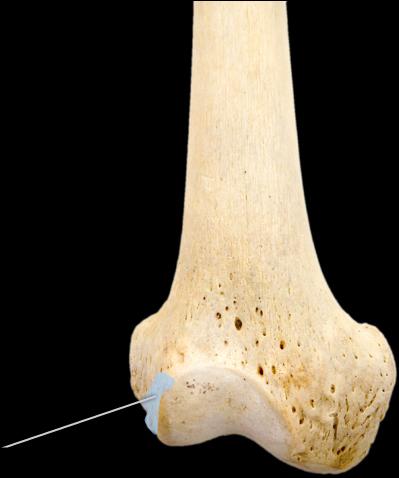 Which structure is highlighted? | back 25 Medial epicondyle |
front 26 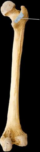 Which structure is highlighted? | back 26 Intertrochanteric Crest |
front 27 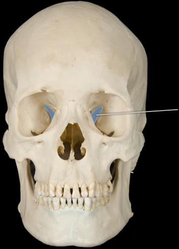 Which openings are highlighted? | back 27 Superior Orbital Fissures |
front 28 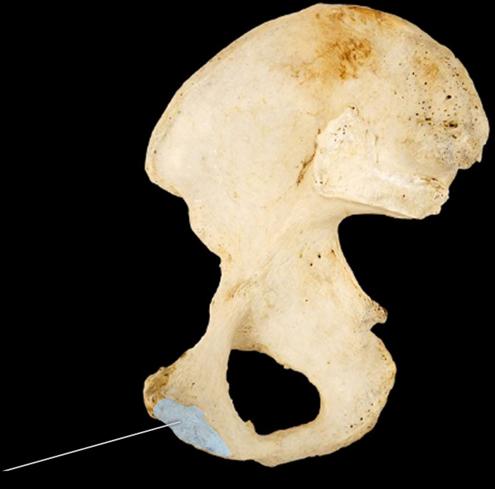 Which structure is highlighted? | back 28 Pubic Symphyseal Fossa |
front 29 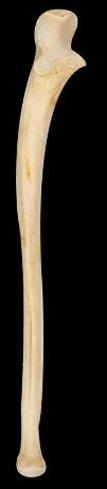 Which bone is in this image? | back 29 Ulna |
front 30 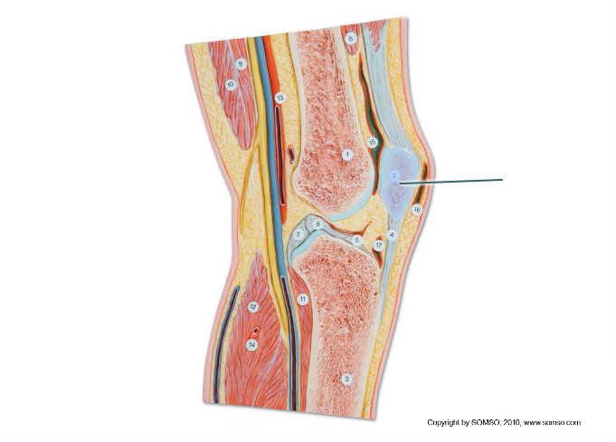 What type of bone is highlighted? | back 30 Seasmoid Long Flat |
front 31 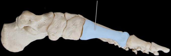 What bone is highlighted | back 31 1st Metatarshal |
front 32 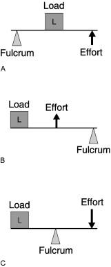 First Class Lever. | back 32 C |
front 33 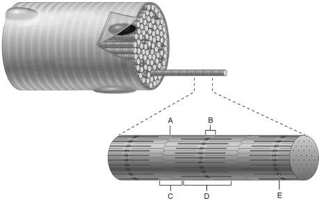 H Zone. | back 33 B |
front 34 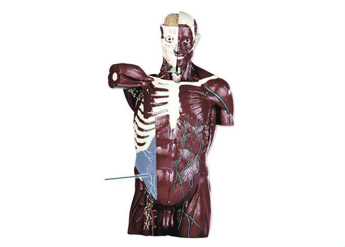 Which Muscle is highlighted? | back 34 External Oblique |
front 35 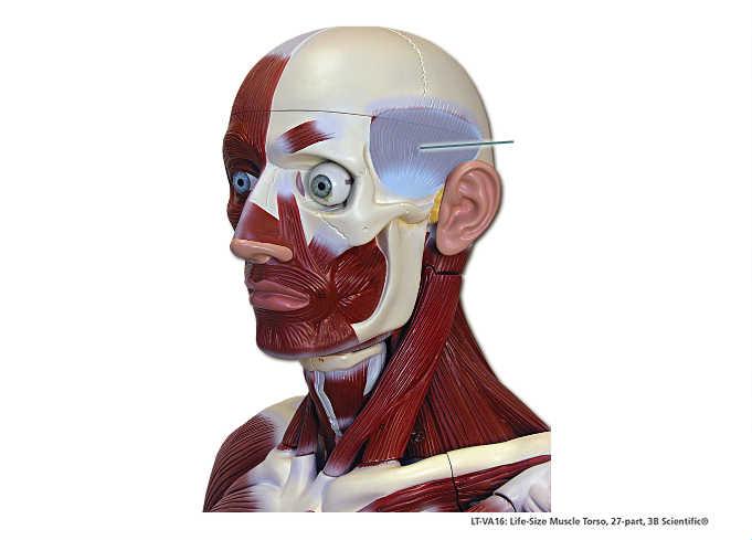 Which Muscle is highlighted? | back 35 Temporalis |
front 36 A Band. | back 36 D |
front 37 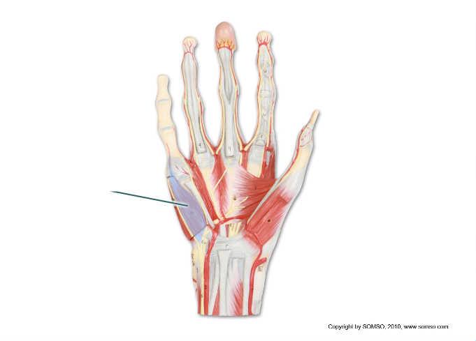 Which Muscle is highlighted? | back 37 Flexor Digiti minimi brevis |
front 38 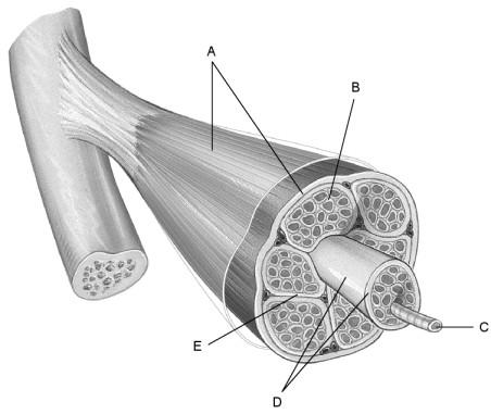 Connective tissue covering the exterior of a muscle organ? | back 38 A |
front 39 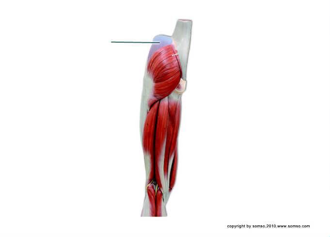 Which Muscle is highlighted? | back 39 Gluteus Medius |
front 40 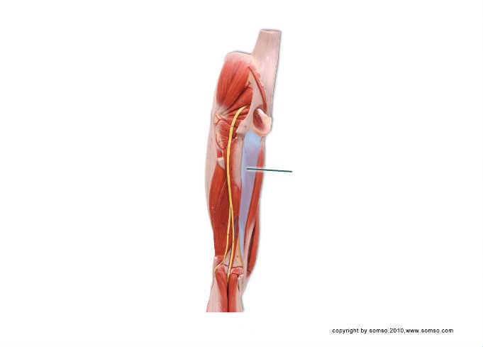 Which Muscle is highlighted? | back 40 vastus lateralis |
front 41 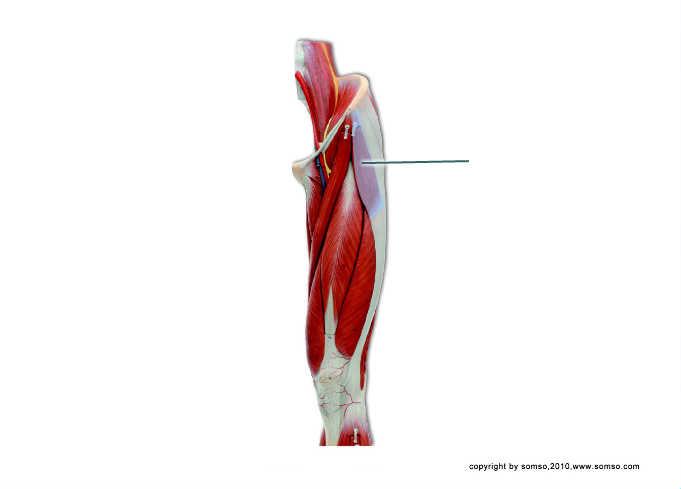 Which Muscle is highlighted? | back 41 Tensor Fasciae latae |
front 42 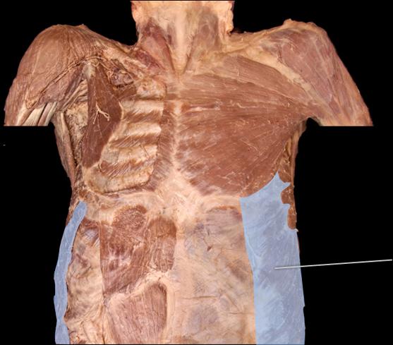 Which Muscle is highlighted? | back 42 External Oblique |
front 43 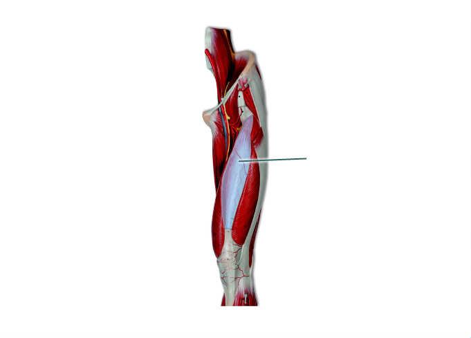 Which Muscle is highlighted? | back 43 Vastus Intermedius |
front 44 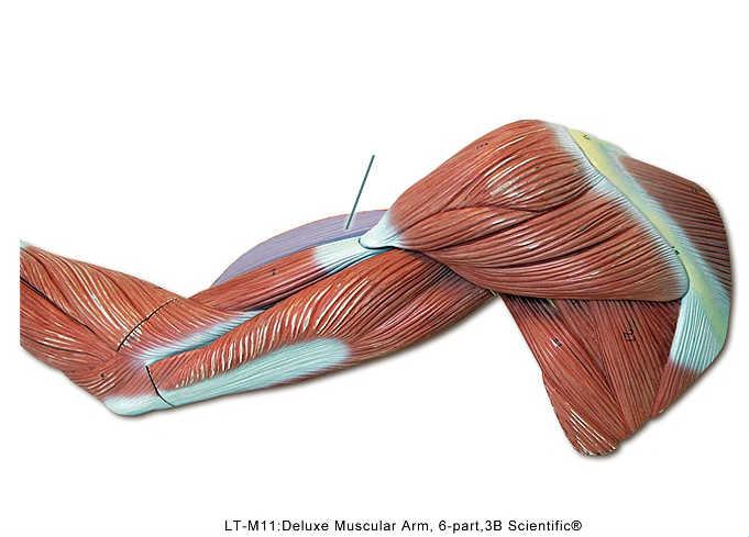 Which Muscle is highlighted? | back 44 Biceps brachii |
front 45 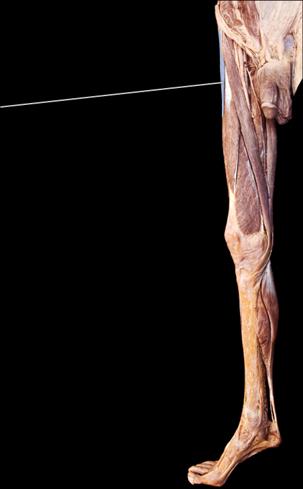 Which Muscle is highlighted? | back 45 Tensor Fasciae latae |
front 46 M Line. | back 46 E |
front 47 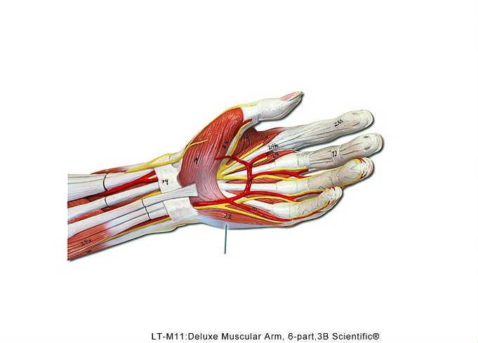 | back 47 abductor digiti minimi |
front 48 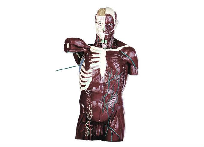 Which Muscle is highlighted? | back 48 serratus anterior |
front 49 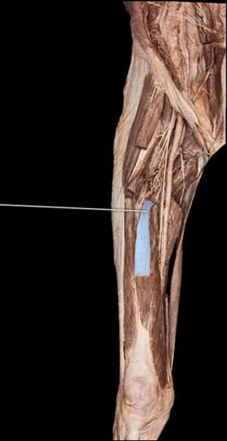 Which Muscle is highlighted? | back 49 Vastus intermedius |
front 50  Which Muscle is highlighted? | back 50 teres major |
front 51 I Band. | back 51 C |
front 52 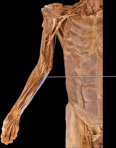 Which Muscle is highlighted? | back 52 Flexor Carpi Ulnaris |
front 53  Which Muscle is highlighted? | back 53 Coracobrachialis |
front 54 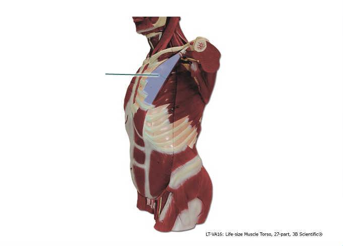 Which Muscle is highlighted? | back 54 Pectoralis Minor |
front 55  Which Muscle is highlighted? | back 55 Mentalis |
front 56 Atlanto-Occiptal Joint. | back 56 C |
front 57 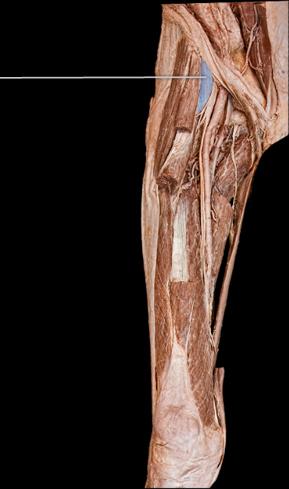 Which Muscle is highlighted? | back 57 Iliopsoas |
front 58 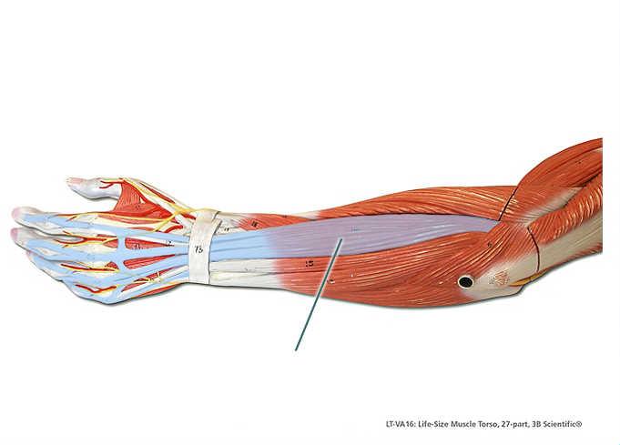 Which Muscle is highlighted? | back 58 Extensor Digitorum |
front 59 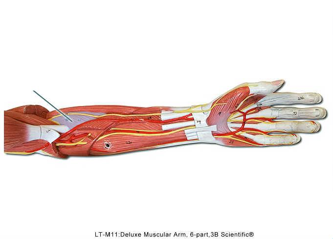 Which Muscle is highlighted? | back 59 Pronator teres |
front 60 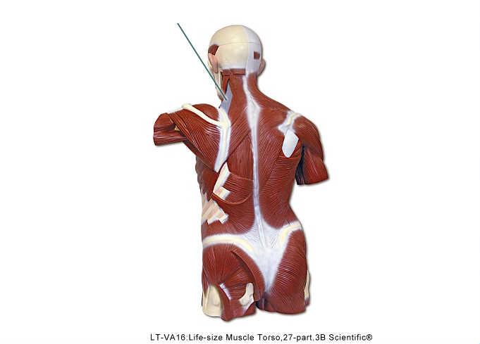 Which Muscle is highlighted? | back 60 Levator Scapulae |
front 61 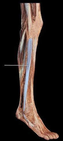 Which Muscle is highlighted? | back 61 Fibularious longus |
front 62 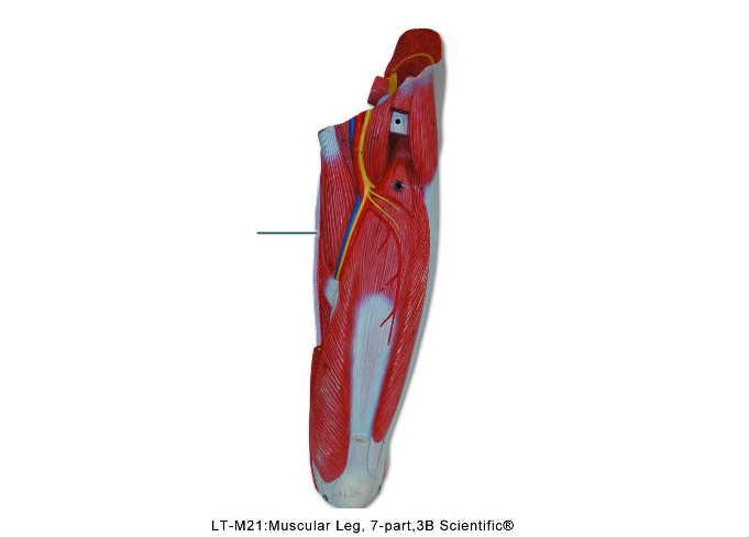 Which Muscle is highlighted? | back 62 Gracilis |
front 63 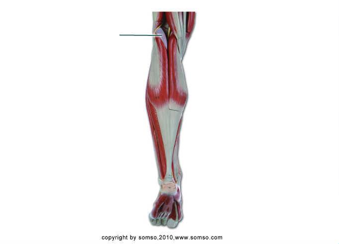 Which Muscle is highlighted? | back 63 plantaris |
front 64 Z Disc. | back 64 A |
front 65 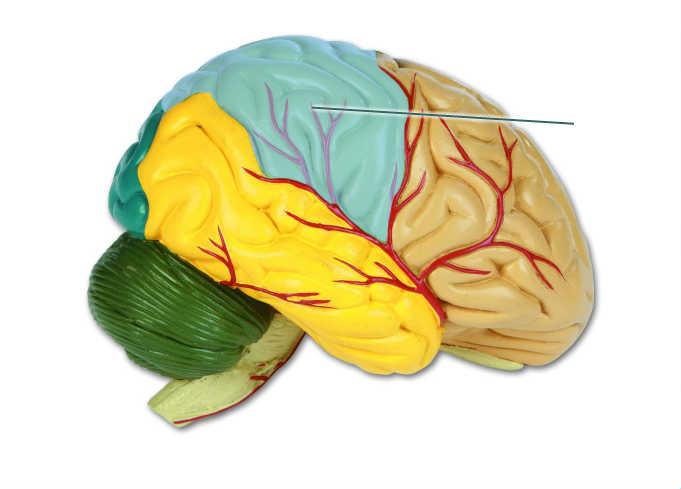 What structure is highlighted? | back 65 Parietal Lobe |
front 66 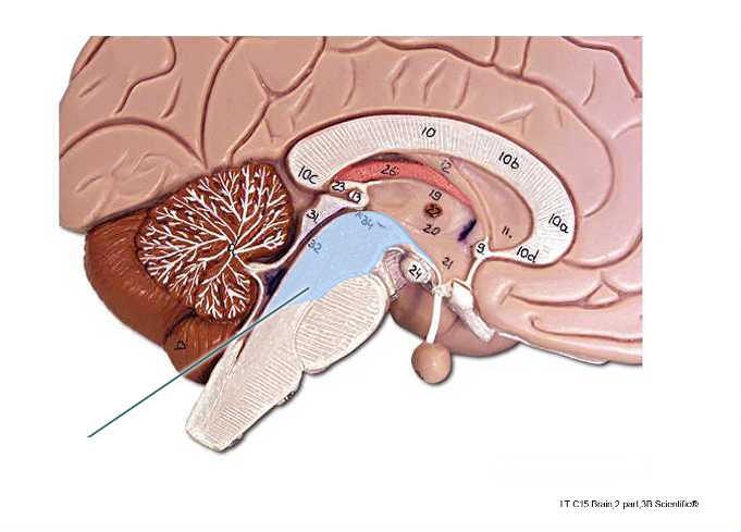 Th central space of the highlighted structure is the ____. | back 66 cerebral aqueduct, |
front 67 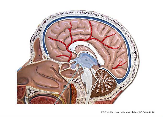 The structure highlighted? | back 67 hypothalmus |
front 68 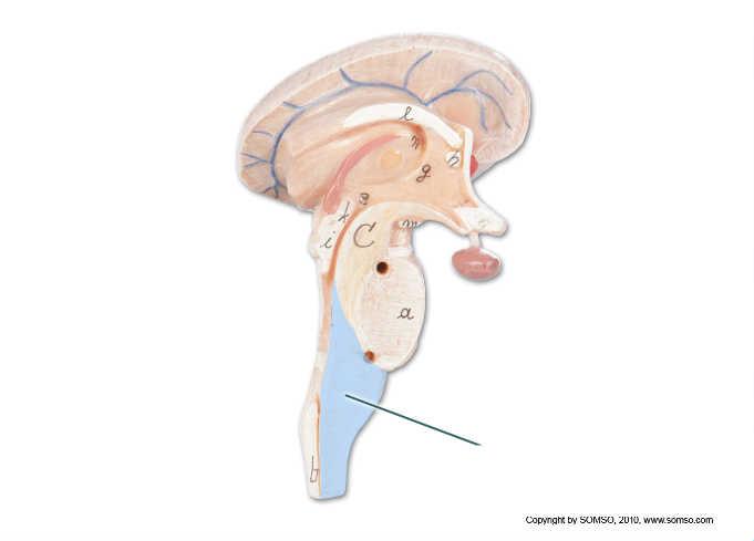 The structure highlighted? | back 68 medulla oblongata |
front 69 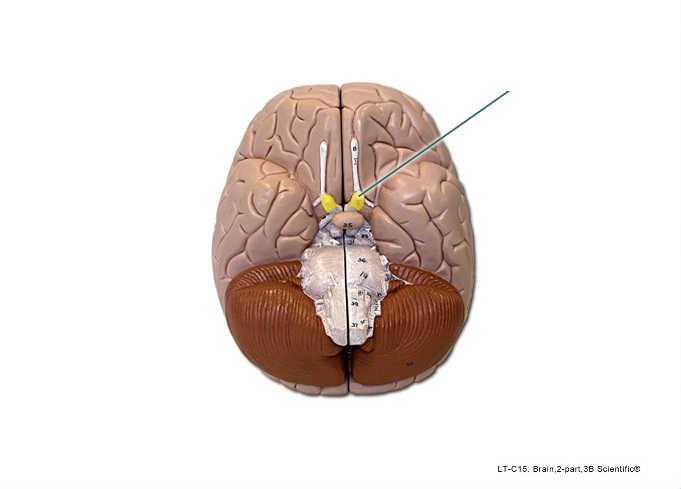 The structure highlighted? | back 69 Optic Nerve (II) |
front 70 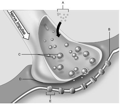 Ion Channel. | back 70 E |
front 71 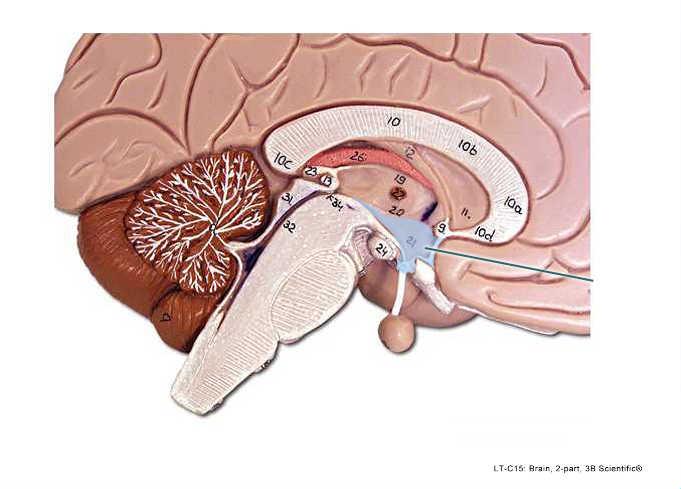 The structure highlighted? | back 71 Hypothalamus |
front 72 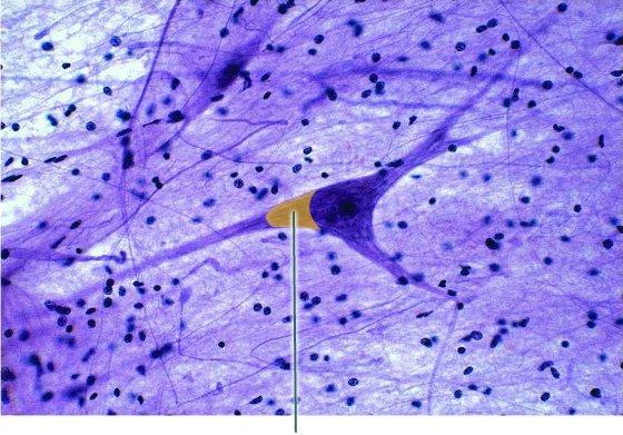 The structure highlighted? | back 72 Axon hillock |
front 73 Synaptic Cleft. | back 73 D |
front 74 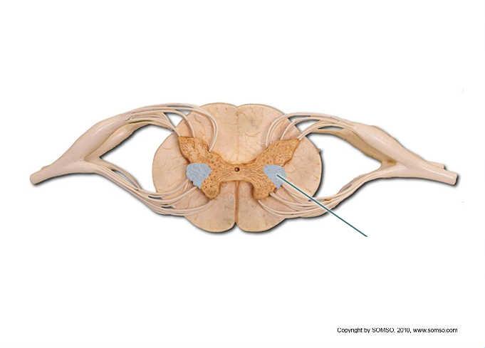 The structure highlighted? | back 74 lateral Horn |
front 75 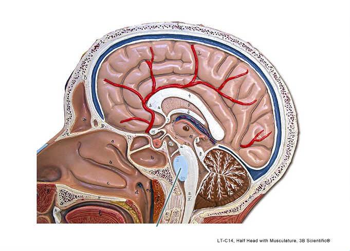 The structure highlighted? | back 75 Pons |
front 76 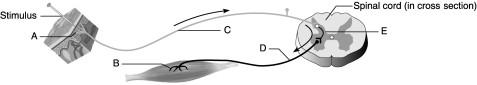 Motor Neuron. | back 76 D |
front 77 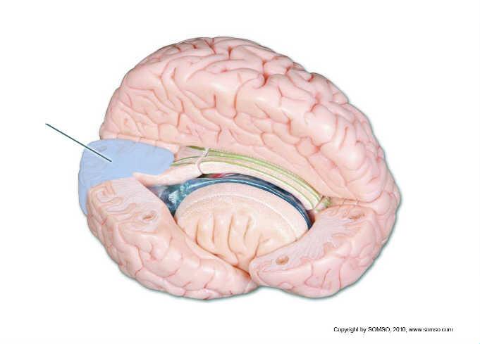 The structure highlighted? | back 77 Occipital Lobe |
front 78 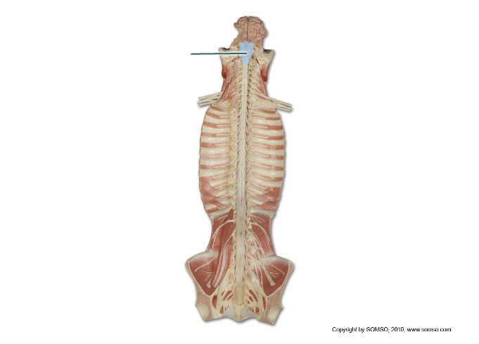 The structure highlighted? | back 78 Brain Stem |
front 79 Receptor. | back 79 A |
front 80 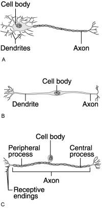 Which neuron would connect to a muscle? | back 80 A |
front 81 Which neuron is a sensory neuron found in the relfex arch? | back 81 C |
front 82 Synapthic Vesicles. | back 82 C |
front 83 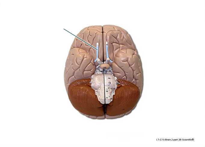 The structure highlighted? | back 83 olfactory tract |
front 84 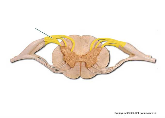 What structures comprise the highlighted structure? | back 84 cell bodies of sensory neurons |
front 85 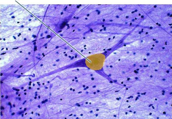 Conduction velocity in the highlighted structure is dependent on which of the following features? | back 85 axon length axon terminus axon diameter |
front 86 The structure highlighted? | back 86 dorsal root |
front 87 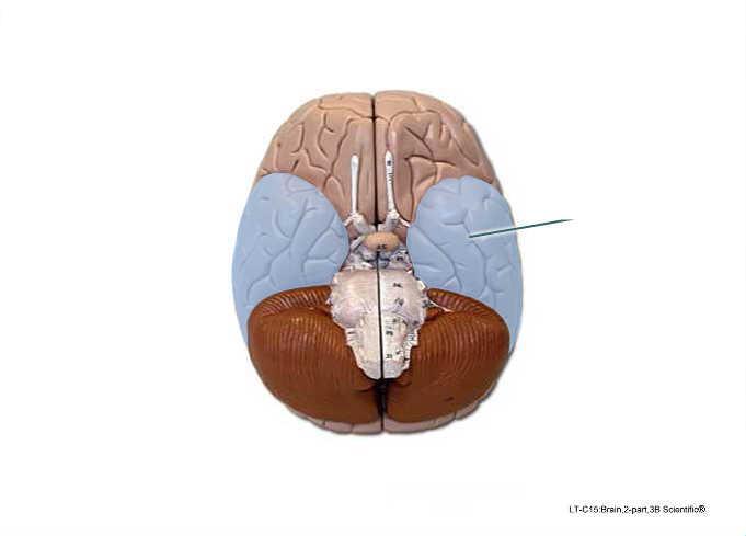 The structure highlighted? | back 87 temporal lobe |
front 88 Integration center. | back 88 E |
front 89 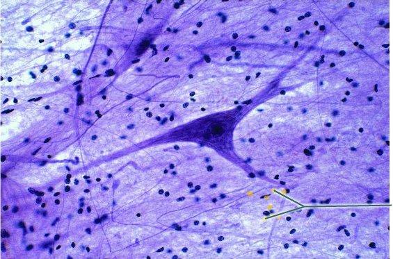 Which cell type is highlighted? | back 89 satellite cell ependymal cell neuroglial cell ?? |
front 90 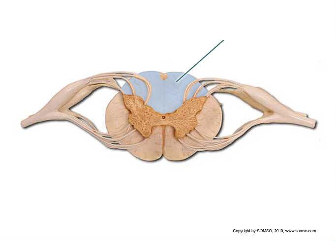 The structure highlighted? | back 90 Anterior Funiculus |
front 91 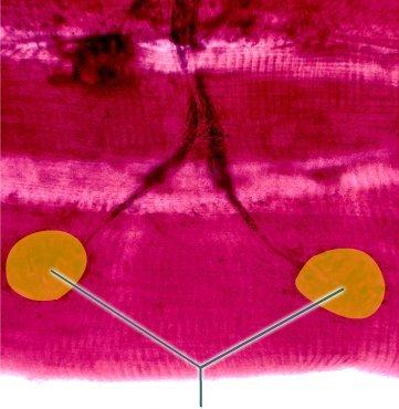 The structure highlighted? | back 91 motor end plates |
front 92 Effector | back 92 B |
front 93 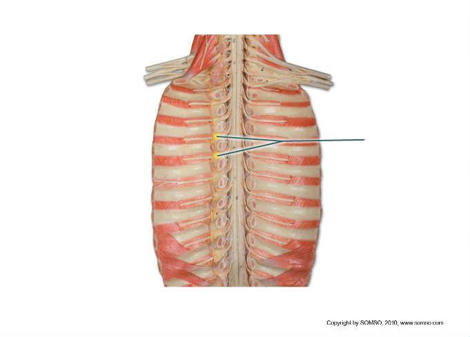 The structure highlighted? | back 93 Spinal Nerves |
front 94  The structure highlighted? | back 94 Pineal gland |
front 95 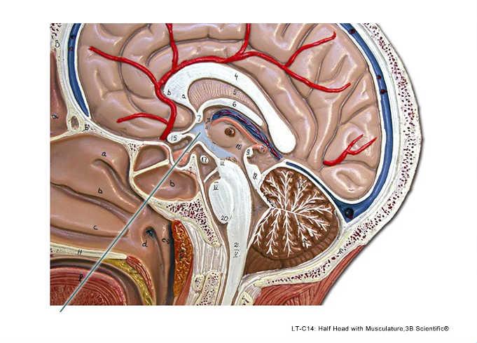 The structure highlighted? | back 95 hypothalmus |
front 96 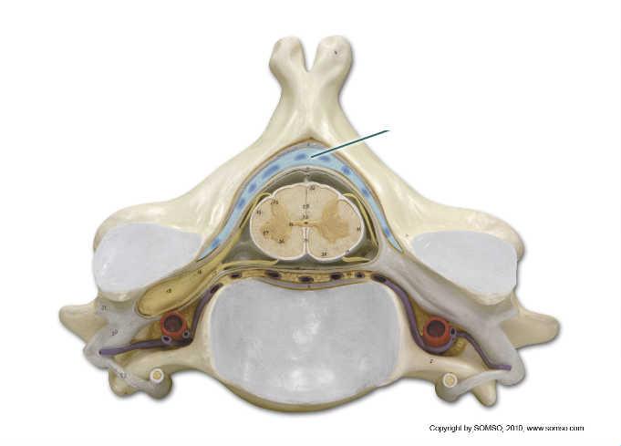 The structure highlighted? | back 96 Dura Mater |
front 97 Sensory neuron | back 97 C |
front 98 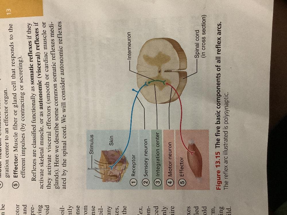 | back 98 no data |