Print Options

Card layout: ?

← Back to notecard set|Easy Notecards home page

Instructions for Side by Side Printing
  1. Print the notecards
  2. Fold each page in half along the solid vertical line
  3. Cut out the notecards by cutting along each horizontal dotted line
  4. Optional: Glue, tape or staple the ends of each notecard together
  1. Verify Front of pages is selected for Viewing and print the front of the notecards
  2. Select Back of pages for Viewing and print the back of the notecards
    NOTE: Since the back of the pages are printed in reverse order (last page is printed first), keep the pages in the same order as they were after Step 1. Also, be sure to feed the pages in the same direction as you did in Step 1.
  3. Cut out the notecards by cutting along each horizontal and vertical dotted line
To print: Ctrl+PPrint as a list

100 notecards = 25 pages (4 cards per page)

Viewing:

Chapter 9: Allergies and Immunologic Diseases

front 1

Clinical Features

  • localized variant
    • involve one to several fungiform papillae
    • mildly painful, elevated, red-yellow papules
    • occur on anterior dorsal surface of tongue
  • generalized variant
    • involves many fungiform papillae
    • sensitive, enlarged, erythematous
    • occur on tip and lateral dorsal surface of tongue
  • papulokeratotic variant
    • diffuse involvement of fungiform papillae
    • asymptomatic, parakeratotic white-yellow papules
    • occur on tip and lateral dorsal surface of tongue

Histopathologic Features

  • focal areas of exocytosis or ulceration
  • proliferation of small vascular channels
  • mixed inflammatory cellular infiltrate
  • hyperparakeratosis with bacterial colonization

back 1

transient lingual papillitis

front 2

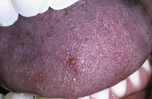
Tender, yellow-pink papule on the dorsum of the tongue.

back 2

transient lingual papillitis, localized

front 3

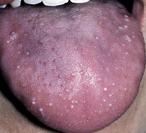
Multiple painful white papules on the lateral dorsum and tip of tongue.

back 3

transient lingual papillitis, generalized

front 4

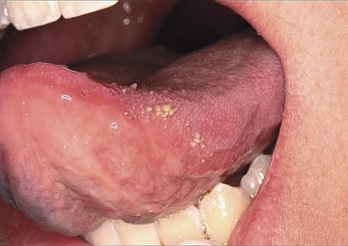
Clusters of asymptomatic, elevated, yellow papules on the dorsolateral surface of the tongue.

back 4

transient lingual papillitis, papulokeratotic

front 5

What is the treatment and prognosis for transient lingual papillitis?

back 5

  • resolves without therapy
  • topical corticosteroids or anesthetics may be used to reduce pain or duration

front 6

Clinical Features

  • one of the most common oral mucosal pathoses (20%)
  • T cell–mediated immunologic reaction with production TNF-α
  • appear to be initiated by some antigenic stimulus
  • may be associated with systemic disorders
  • three clinical variations: mild, moderate, herpetaform
  • occur most in children and young adults (80% before 30 years)

Histopathological Features

  • central zone of ulceration covered by fibrinopurulent membrane
  • increased vascularity of connective tissue
  • mixed inflammatory cellular infiltrate

back 6

recurrent aphthous stomatitis

front 7

Clinical Features

  • fewest recurrences and shortest duration
  • arise on nonkeratinized mucosa, especially buccal and labial
  • yellow-white, fibrinopurulent membrane with erythematous halo
  • between 3 and 10 mm in diameter
  • heal in 7 to 14 days without scarring
  • one to five lesions at a time
  • disproportionately painful

back 7

minor aphthous ulceration

front 8

Clinical Features

  • largest lesions and longest duration
  • measure 1 to 3 cm in diameter
  • yellow-white ulceration with red halo
  • take from 2 to 6 weeks to heal and may cause scarring
  • occur most on labial mucosa, soft palate, tonsillar fauces
  • onset after puberty

back 8

major aphthous ulceration

front 9

Clinical Features

  • greatest number of lesions and most frequent recurrences
  • measure 1 to 3 mm in diameter
  • up to a hundred present at once
  • superficial resemblance to primary HSV infection
  • may coalesce into larger irregular ulcerations
  • heal within 7 to 10 days
  • occur on nonkeratinized, movable mucosa
  • female predominance, with onset in adulthood

back 9

herpetiform aphthous ulceration

front 10

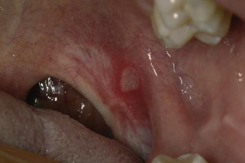
Erythematous halo encircling a yellowish ulceration of the soft palate on the left side.

back 10

minor aphthous ulceration

front 11

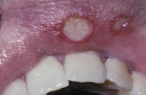
Two ulcerations of different sizes located on the maxillary labial mucosa.

back 11

minor aphthous ulceration

front 12

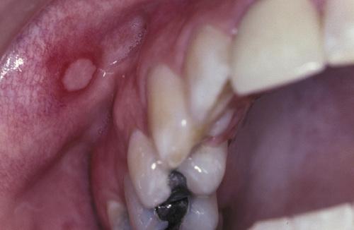
Single ulceration of the anterior buccal mucosa.

back 12

minor aphthous ulceration

front 13

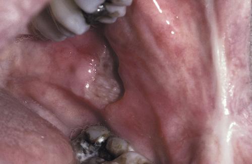
Large, deep, and irregular ulceration of the posterior buccal mucosa. Note extensive scarring of the anterior buccal mucosa from previous ulcerations.

back 13

major aphthous ulceration

front 14

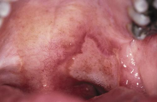
Large, irregular ulceration of the soft palate.

back 14

major aphthous ulceration

front 15

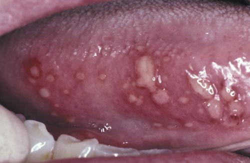
Numerous pinhead ulcerations of the ventral surface of the tongue, several of which have coalesced into larger, more irregular areas of ulceration.

back 15

herpetiform aphthous ulceration

front 16

Aphthous ulcerations that heal within 1 to 2 weeks and recur infrequently.

back 16

simple aphthosis

front 17

Aphthous ulcerations that occur in multiples and develop continuously.

back 17

complex aphthosis

front 18

How is recurrent aphthous stomatitis diagnosed?

back 18

No laboratory procedure provides definitive diagnosis; the diagnosis is made from the clinical presentation and from exclusion of other diseases.

front 19

What is the treatment and prognosis for recurrent aphthous stomatitis?

back 19

  • topical corticosteroids (e.g. dexamethasone, fluocinonide) for mild disease
  • potent corticosteroids (e.g. triamcinolone, clobetasol) for major disease
  • in resistant cases, systemic corticosteroids may be required for control
  • complex aphthosis demands thorough evaluation for systemic disease

front 20

  1. Large ulceration of the left anterior buccal mucosa.
  2. Same lesion after 5 days of therapy with betamethasone syrup used in a swish-and-swallow method. The patient was free of pain by the second day of therapy. The ulceration healed completely during the next week.

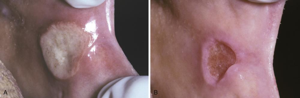
back 20

major aphthous ulceration

front 21

Clinical Features

  • chronic ocular inflammation and orogenital ulcerations
  • systemic vasculitis caused by an abnormal immune process
  • usually arises in the third and fourth decades
  • increased prevalence and worse clinical course in men
  • oral lesions similar to aphthous ulcers often herald onset
  • genital lesions (papules, vesicles, pustules, etc.) in 75% of cases
  • positive skin “pathergy”
  • self-limiting and nondeforming arthritis
  • ocular involvement (uveitis, conjunctivitis, etc.) in 70% of cases
  • vascular disease often presents as thrombophlebitis
  • variable gastrointestinal disease (pain, anorexia, diarrhea, etc.)
  • CNS involvement (paralysis, dementia) in 10% to 25% of cases

Histopathological Features

  • leukocytoclastic vasculitis
  • similar to aphthous stomatitis

back 21

Behçet syndrome

front 22

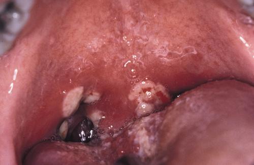
Diffuse erythema surrounding numerous irregular ulcerations of the soft palate. Oral ulcerations often herald the onset of this disease.

back 22

Behçet syndrome

front 23

Numerous irregular ulcerations of the labia majora and perineum as part of a disease characterized by chronic ocular inflammation and orogenital ulcerations.

back 23

Behçet syndrome

front 24

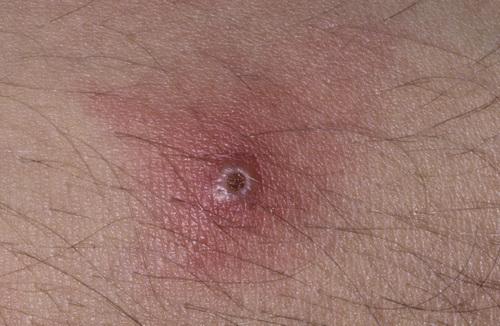
Sterile pustule of the skin that developed 1 day after injection of saline. This reaction is termed cutaneous pathergy.

back 24

Behçet syndrome

front 25

From a diagnostic standpoint, one of the most important skin manifestations in Behçet syndrome is the presence of positive “pathergy.” What does this refer to?

back 25

One or 2 days after the oblique insertion of a needle under sterile conditions, a tuberculin-like skin reaction or sterile pustule develops.

front 26

No laboratory finding is diagnostic of Behçet syndrome. What In an attempt to standardize diagnoses, five definitive criteria have been developed. What are they?

back 26

  1. recurrent oral ulceration, plus and two of the following:
  2. recurrent genital ulcerations
  3. eye lesions
  4. skin lesions
  5. positive pathergy test

front 27

What is the treatment and prognosis for Behçet syndrome?

back 27

  • most treatment is symptomatic until the disease goes into remission
  • old or female patients have a better prognosis than male or young
  • ocular lesions or CNS involvement mandate more aggressive therapy
  • systemic medications include azathioprine, colchicine, corticosteroids
  • orogenital ulcerations respond to topical or intralesional corticosteroids
  • relapsing and remitting pattern, with attacks lessening after 5 to 7 years

front 28

Clinical Features

  • arises prior to age 50, with predilection for females and blacks
  • acute cases exhibit fever, fatigue, anorexia, or weight loss
  • chronic cases exhibit pulmonary symptoms
  • affects lungs, lymph nodes, skin, eyes, and salivary glands
  • chronic, violaceous, indurated skin lesions ("lupus pernio")
  • scattered, tender erythematous nodules ("erythema nodosum")
  • ocular involvement (25%) often appears as anterior uveitis
  • salivary gland involvement results in enlargement and xerostomia
  • submucosal masses, isolated papules, granularity, or ulcerations
  • occur on buccal mucosa, gingiva, lips, tongue, and palate

Histopathological Features

  • granulomatous inflammation
  • aggregates of epithelioid histiocytes
  • surrounding rim of lymphocytes
  • scattered Langhans' or foreign body type giant cells
  • laminated, basophilic, calcified lysosomes ("Schaumann bodies")
  • stellate inclusions of trapped collegen ("asteroid bodies")

back 28

sarcoidosis

front 29

Violaceous indurated plaques of the right malar area and bridge of nose.

back 29

lupus pernio (sarcoidosis)

front 30

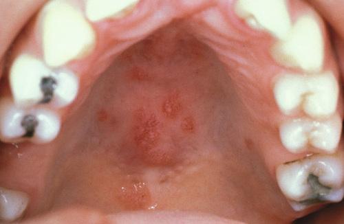
Multiple erythematous macules of the hard palate. Oral lesions are uncommon in this disease, but may be normal in color, brown-red, violaceous, or hyperkeratotic.

back 30

sarcoidosis

front 31

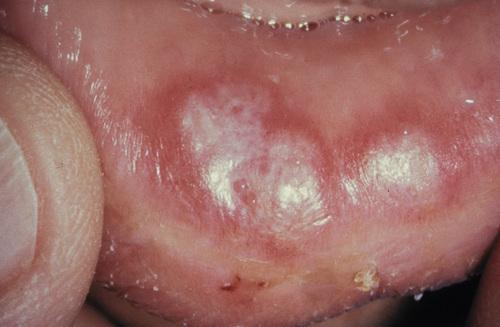
Erythematous macules with central hyperkeratosis of the lower labial mucosa. Oral lesions are uncommon in this disease.

back 31

sarcoidosis

front 32

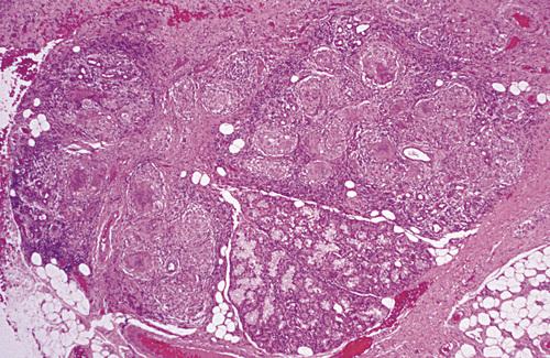
Labial minor salivary gland demonstrating granulomatous inflammation characterized by circumscribed collections of histiocytes, lymphocytes, and multinucleated giant cells.

back 32

sarcoidosis

front 33

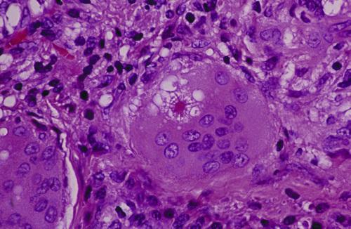
Multinucleated giant cell with intracytoplasmic asteroid body, a stellate inclusion consisting of entrapped fragments of collagen.

back 33

sarcoidosis

front 34

A clinical syndrome associated with acute sarcoidosis consisting of erythema nodosum, bilateral hilar lymphadenopathy, and arthralgia.

back 34

Löfgren syndrome

front 35

A clinical syndrome associated with acute sarcoidosis consisting of parotid enlargement, anterior uveitis of the eye, facial paralysis, and fever.

back 35

Heerfordt syndrome (uveoparotid fever)

front 36

How is sarcoidosis diagnosed?

back 36

  1. The diagnosis is established by clinical and radiographic presentations, the histopathologic appearance, and negative tests for casative organisms.
  2. Elevated serum angiotensin-converting enzyme (ACE) levels supports the diagnosis, but is reported in only 60% of patients with the disease.
  3. In the past, the Kveim test, was performed by intradermal injection of a sterilized suspension of human sarcoid tissue.

front 37

What is the treatment and prognosis for sarcoidosis?

back 37

  • symptoms may resolve spontaneously within 2 years (60%)
  • intervention is recommended for progressive disease
  • corticosteroids remain first-line therapy
  • resistance and relapses are common
  • resolution may not occur even with treatment (10% to 20%)
  • patients may die of pulmonary, cardiac, or CNS complications (4% to 10%)

front 38

Clinical Features

  • primarily affects adults, but can occur at any age
  • nontender, persistent swelling involving lips
  • may be combined with facial paralysis and fissured tongue
  • intraoral lesions including edema, ulcers, and papules
  • tongue may develop fissures, edema, paresthesia, or erosions
  • gingiva can develop swelling, erythema, or pain
  • buccal mucosa often exhibits a cobblestone
  • linear hyperplastic folds may occur in the sulcus

Histopathological Features

  • edema in superficial lamina propria
  • dilation of lymphatic vessels
  • scattered lymphocytes
  • fibrosis in long-term lesions
  • noncaseating granulomatous inflammation
  • granulomas clustered around scattered vessels

back 38

orofacial granulomatosis

front 39

The variant of orofacial granulomatosis in which there is nontender, persistent swelling of the lips combined with facial paralysis and a fissured tongue.

back 39

Melkersson-Rosenthal syndrome

front 40

The variant of orofacial granulomatosis in which there is nontender, persistent swelling of the lips in isolation.

back 40

cheilitis granulomatosa (of Miescher)

front 41

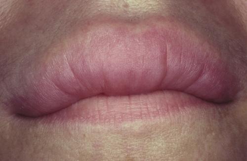
Nontender, persistent enlargement of the upper lip. This patient did not present with facial paralysis nor a fissured tongue.

back 41

cheilitis granulomatosa (orofacial granulomatosis)

front 42

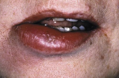
Persistent enlargement of the lower lip. This patient also presented with numerous furrows on the dorsal surface of the tongue.

back 42

Melkersson-Rosenthal syndrome (orofacial granulomatosis)

front 43

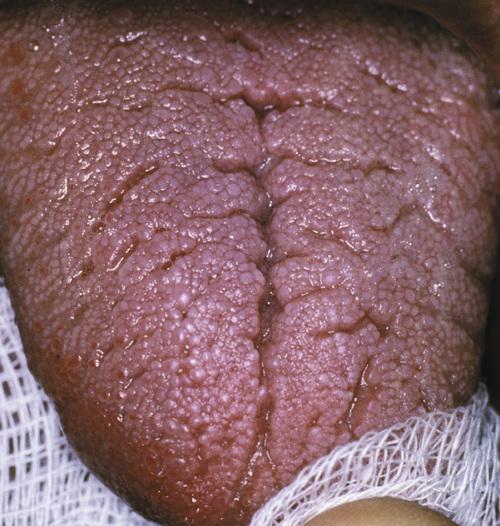
Note numerous furrows on the dorsal surface of the tongue. This patient also presented with persistent enlargement of the lower lip.

back 43

Melkersson-Rosenthal syndrome (orofacial granulomatosis)

front 44

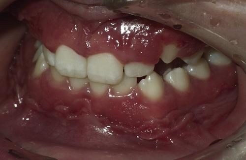
Hyperplastic mucosa noted bilaterally in the mandibular mucobuccal fold with elongated ulcerations in the base of these folds.

back 44

orofacial granulomatosis

front 45

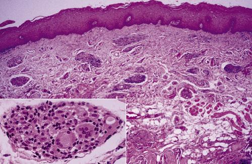
Clusters of granulomatous inflammation around scattered vessels. The inset illustrates the histiocytes and multinucleated giant cells within the granulomas.

back 45

orofacial granulomatosis

front 46

How is orofacial granulomatosis diagnosed?

back 46

Based on histopathologic demonstration of granulomatous inflammation that is associated with negative special stains for organisms and no foreign material.

front 47

What is the treatment and prognosis for orofacial granulomatosis?

back 47

  • first goal of management is discovery of initiating cause
  • intralesional delayed-release high-concentrate triamcinolone
  • prognosis is highly variable; some lesions resolve spontaneously

front 48

  1. Diffuse enlargement of the upper lip.
  2. Same patient after intralesional triamcinolone injections.

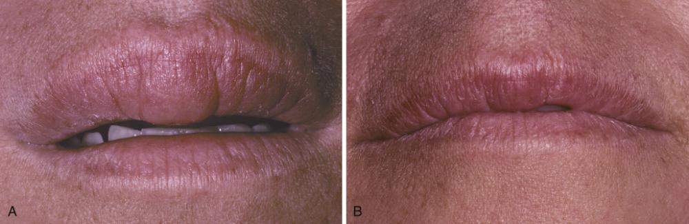
back 48

orofacial granulomatosis

front 49

  1. Clinical appearance before local therapy.
  2. Significant resolution after intralesional corticosteroid therapy.

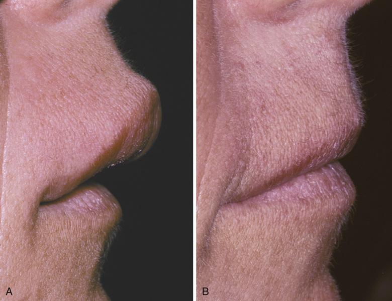
back 49

orofacial granulomatosis

front 50

Clinical Features

  • wide age range (mean, 41 years), 90% in Caucasians
  • necrotizing granulomatous lesions of respiratory tract (limited)
  • may also exhibit necrotizing glomerulonephritis (generalized)
  • may exhibit lesions of the skin and mucosa (superficial)
  • nasal drainage, sinus pain, nasal ulceration (URT involvement)
  • dry cough, hemoptysis, dyspnea, chest pain (LRT involvement)
  • proteinuria and red blood cell casts (renal involvement)
  • florid and granular ginvival hyperplasia ("strawberry gingivitis")
  • oral ulcerations (usually diagnosed at a later stage)
  • salivary gland enlargement

Histopathological Features

  • mixed inflammation centered around blood vessels
  • heavy neutrophilic infiltration, necrosis, and nuclear dust
  • inflammatory cellular infiltrate
  • pseudoepitheliomatous hyperplasia and subepithelial abscesses
  • prominent vascularity with extensive red blood cell extravasation

back 50

Wegener granulomatosis

front 51

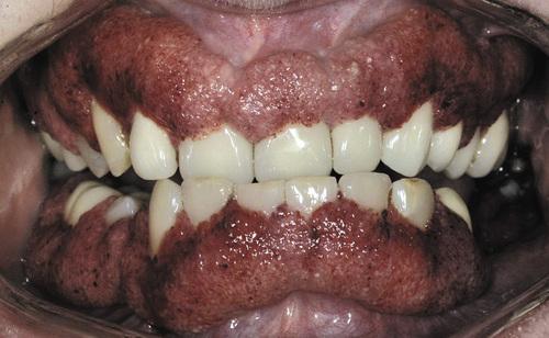
Florid and granular gingival hyperplasia with hemorrhagic and friable bulbous projections ("strawberry gingivitis").

back 51

Wegener granulomatosis

front 52

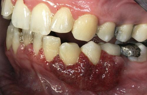
Hyperplastic and hemorrhagic mucosa of the facial mandibular gingiva on the left side. This red, bumpy surface is responsible for the strawberry-like appearance

back 52

Wegener granulomatosis

front 53

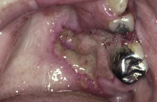
Deep, irregular ulceration of the hard palate on the left side. These represent late-stage disease; more than 60% of patients will have renal involvement.

back 53

Wegener granulomatosis

front 54

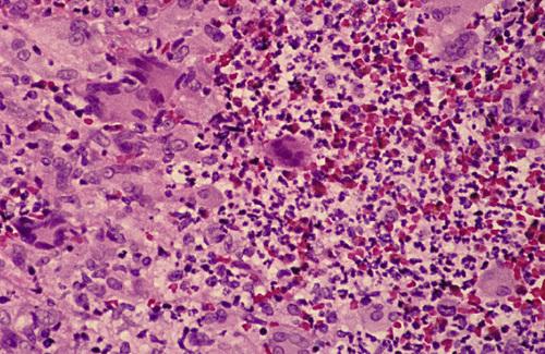
Connective tissue containing proliferation of numerous vascular channels and a heavy inflammatory infiltrate consisting of lymphocytes, neutrophils, eosinophils, and multinucleated giant cells.

back 54

Wegener granulomatosis

front 55

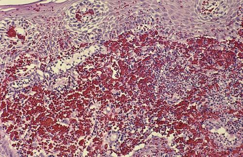
Gingival biopsy specimen showing a mixed inflammatory cellular infiltrate obscured by extensive extravasation of red blood cells.

back 55

Wegener granulomatosis

front 56

The American College of Rheumatology proposed four diagnostic criteria with a minimum of two required for a diagnosis of Wegener granulomatosis. What are these criteria?

back 56

  1. oral ulcerations or nasal discharge
  2. nodules, fixed infiltrates, or cavities on chest radiograph
  3. abnormal urinary sediment (e.g. RBCs or casts
  4. granulomatous inflammation upon biopsy

front 57

What marker is the most useful in the diagnosis of Wegener granulomatosis, seen in 90% to 95% of generalized cases and 60% of the early or localized cases?

back 57

antibodies against proteinase-3 (PR3-ANCA)

front 58

What is the treatment and prognosis for Wegener granulomatosis?

back 58

  • mean survival of untreated patients with generalized disease is 5 months
  • 80% of the patients are dead at 1 year and 90% within 2 years
  • prognosis is better for the limited and superficial forms
  • first line therapy is oral prednisone and cyclophosphamide
  • prolonged remission is noted in up to 75% of affected patients
  • for maintenance, cyclophosphamide may be replaced with methotrexate
  • relapse rate up to 30%, maintenance therapy is necessary in many patients

front 59

The risk of an adverse reaction is approximately ___% with the use of two medications, ___% with five drugs, and almost ___% with eight or more.

back 59

6%, 50%, 100%

front 60

What is a Type A drug reaction?

back 60

An exaggerated but otherwise expected pharmacologic action of the prescribed medication (e.g. bleeding associated with warfarin).

front 61

What is a Type B drug reaction?

back 61

An idiosyncratic reaction that is not expected, usually arising from immune-mediated effects (e.g. hypersensitivity reactions).

front 62

A reaction of the oral mucosa to the systemic administration of a medication.

back 62

stomatitis medicamentosa

front 63

Several different patterns of stomatitis medicamentosa exist. What are the seven classes of mucosal reactions to systemic drug administration?

back 63

  1. anaphylactic stomatitis
  2. intraoral fixed drug eruptions
  3. lichenoid drug reactions
  4. pemphigoid-like drug reactions
  5. pemphigus-like drug reactions
  6. lupus erythematosus–like eruptions
  7. nonspecific erosive, ulcerative, or aphthous-like lesions

front 64

Clinical Features

  • various patterns of mucosal alterations
  • erythema or aphthous-like ulcerations
  • localized areas of erythema and edema
  • vesiculoerosive lesions of the labial mucosa
  • lupus-, pemphigoid-, or pemphigus-like lesions
  • bilateral and symmetrical lesions are common

Histopathological Features

  • nonspecific pattern of subacute mucositis
  • mixed inflammatory cellular infiltrate
  • spongiosis and exocytosis of the epithelium
  • lupus-, pemphigoid-, or pemphigus-like findings
  • DIF for IgG shows annular pattern along basal cell layer ("sting of pearls")
  • may detect circulating antibody ("basal cell cytoplasmic antibody")

back 64

mucosal reactions to systemic drug administration

front 65

  1. Bilateral erosions of lower labial mucosa with intermixed striae. Biopsy revealed lichenoid pattern of mucositis but with numerous plasma cells intermixed with the lymphocytes.
  2. Same patient depicted in A after discontinuation of simvastatin.

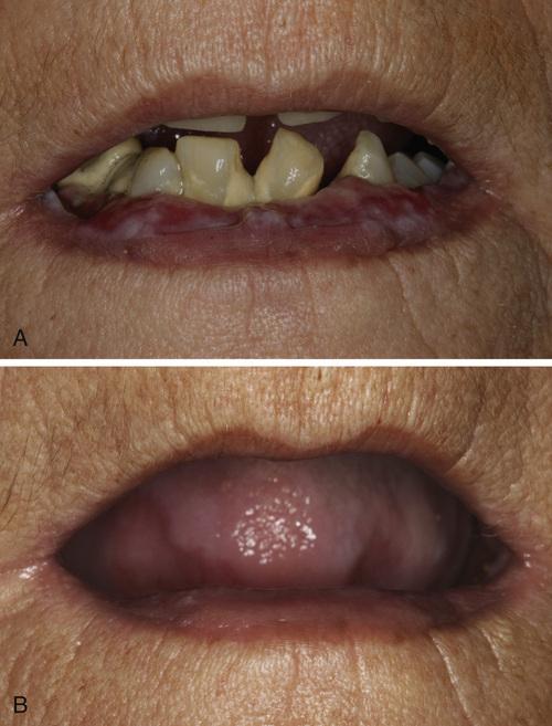
back 65

allergic mucosal reaction to systemic drug administration

front 66

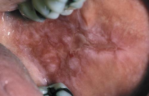
Irregular area of superficial erosion of the left buccal mucosa. Lesions were also present on the contralateral buccal mucosa and bilaterally on the lateral borders of the tongue. Lesions disappeared after discontinuing allopurinol.

back 66

lichenoid drug reaction

front 67

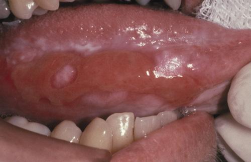
Large irregular erosion of the right ventral surface of the tongue. The lesion arose secondary to use of oxaprozin, a nonsteroidal antiinflammatory drug (NSAID).

back 67

allergic mucosal reaction to systemic drug administration

front 68

How are mucosal reactions to systemic drug administration diagnosed?

back 68

  1. identify potentially offending medication through detailed medical history and establish a temporal relationship with the mucosal alteration
    1. if more than one suspected, start with most recently added
    2. if last-introduced drug is not responsible, do serial elimination
  2. definitive diagnosis can be made if mucosal alterations resolve after discontinuation of medication and recur on reintroduction
  3. presumptive diagnosis usually is sufficient and justified when the mucosal alterations clear after cessation of the offending medication
  4. for lupus-like reactions, consider serum evaluation for ANAs dsDNA Ab

front 69

What is the treatment for mucosal reactions to systemic drug administration?

back 69

  1. discontinue and replace causative drug and if possible
  2. lesions can be resolved with topical corticosteroids
  3. If discontinuation is contraindicated, palliative care can be provided

front 70

Clinical Features

  • can be acute or chronic
  • distinct female predominance
  • can be localized or diffuse
  • burning is the most frequent symptom
  • mucosa may be barely red to brilliantly erythematous
  • superficial ulcerations occasionally arise
  • muscosa may be white and hyperkeratotic
  • desquamation of the superficial layers of the epithelium
  • dryness, scaling, fissuring, or cracking of the vermilion border

back 70

allergic contact stomatitis

front 71

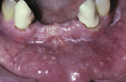
Mucosal erythema and vesicles of the lower labial mucosa caused by use of aluminum chloride on gingival retraction cord.

back 71

allergic contact stomatitis

front 72

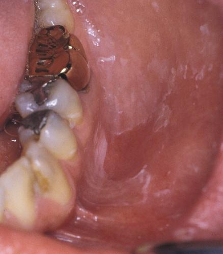
Erythematous mucosa with superficial epithelial desquamation. This patient reported recently changing brands of toothepaste.

back 72

allergic contact stomatitis

front 73

How is allergic contact stomatitis diagnosed?

back 73

  • diagnosis of acute contact stomatitis is straightforward due to temporal relationship between agent and resultant eruption
  • diagnosis of chronic contact stomatitis is more difficult, requirring good oral health and elimination of all other possible causes

front 74

What is the treatment and prognosis for allergic contact stomatitis?

back 74

  • for mild acute cases, simply removal of suspected allergen
  • for severe acute cases, antihistamines and topical anesthetics
  • for chronic cases, topical corticosteroid gel or oral suspension
  • use of plain baking soda or flavor-free toothpaste
  • avoidance of mouthwash, gum, mints, chocolate, cinnamon products

front 75

Clinical Features

  • associated with use of potent corticosteroids on face
  • worsened by exposure to UV light, heat, and wind
  • persistent erythematous papules, vesicles, and pustules
  • occur on skin surrounding the vermilion border
  • may involve perinasal skin (40%)
  • zone of spared skin adjacent to vermilion border
  • most occurs in women between ages of 20 to 45 years

Histopathologic Findings

  • chronic lymphohistiocytic dermatitis with spongiosis of hair follicles
  • rosacea-like pattern with perifollicular granulomatous inflammation
  • may be misdiagnosed as sarcoidosis

back 75

perioral dermatitis

front 76

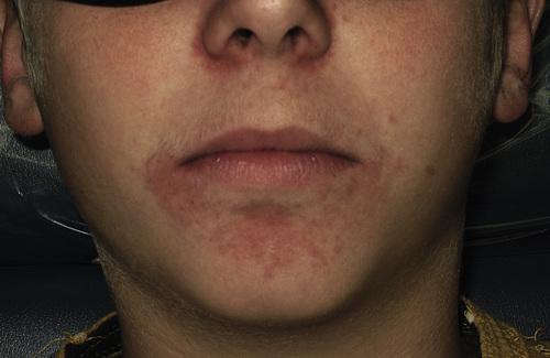
Multiple erythematous papules of the skin surrounding the vermilion border of the lips. Note similar involvement around the nasal orifices.

back 76

perioral dermatitis

front 77

What is the treatment for perioral dermatitis?

back 77

  • discontinue corticosteroids, cosmetics, facial creams (“zero therapy”)
  • oral tetracycline is "gold standard," except in childhood and pregnancy
  • responds well to topical pimecrolimus or topical erythromycin
  • improvement within weeks, resolution in months, recurrence uncommon

front 78

Clinical Features

  • associated with candy, chewing gum, and toothpaste
  • pain and burning are common in all cases
  • affects gingiva, buccal mucosa, lateral borders of tongue
  • may resemble plasma cell gingivitis
  • enlargement, edema, and erythema
  • hyperkeratosis of surface epithelium

Histopathologic Findings

  • acanthotic, often with elongated rete ridges
  • thinning of the suprapapillary plates
  • hyperkeratosis and extensive neutrophilic exocytosis
  • heavy inflammatory cell infiltrate

back 78

contact stomatitis from cinnamon flavoring

front 79

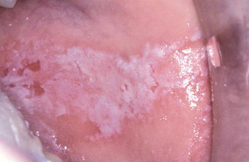
Oblong area of sensitive erythema with overlying shaggy hyperkeratosis. Buccal mucosal lesions often are aligned along the occlusal plane.

back 79

contact stomatitis from cinnamon flavoring

front 80

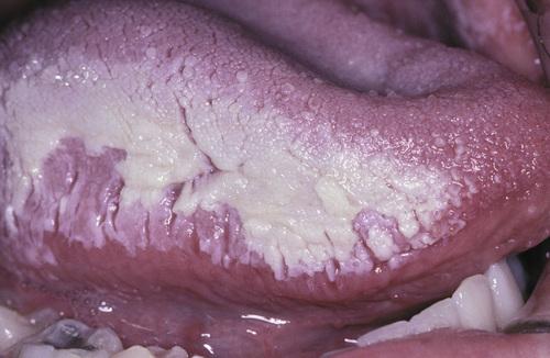
Sensitive and thickened hyperkeratosis of the lateral and dorsal surface of the tongue on the right side.

back 80

contact stomatitis from cinnamon flavoring

front 81

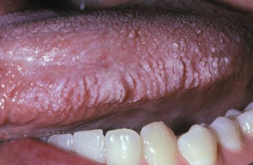
Left lateral border of the tongue demonstrating linear rows of hyperkeratosis that resemble oral hairy leukoplakia (OHL).

back 81

contact stomatitis from cinnamon flavoring

front 82

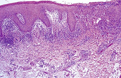
Oral mucosa demonstrating significant interface mucositis and deeper perivascular inflammation. Heavy inflammatory cell infiltrate often obscures the epithelium and connective tissue interface.

back 82

contact stomatitis from cinnamon flavoring

front 83

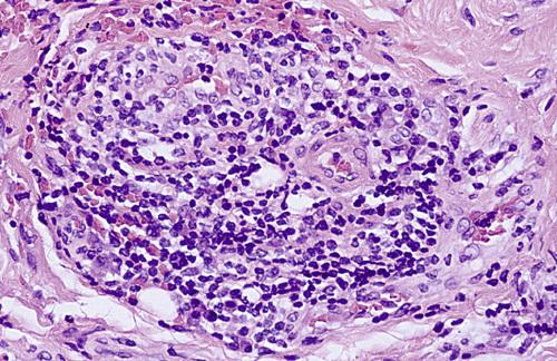
Perivascular inflammatory infiltrate consisting predominantly of lymphocytes and plasma cells, a characteristic feature in localized cases.

back 83

contact stomatitis from cinnamon flavoring

front 84

How is contact stomatitis from cinnamon flavoring diagnosed?

back 84

Based on the clinical appearance and the history of cinnamon use; often biopsies are performed for atypical or extensive cases.

front 85

What is the treatment and prognosis for contact stomatitis from cinnamon flavoring?

back 85

  • disappears within 1 week of discontinuation of cinnamon product
  • patient may benefit from short-term use of a topical corticosteroid

front 86

The vast majority of hypersensitivity reactions to dental restorative materials are to dental amalgam. What component is largely responsible?

back 86

mercury

front 87

Although rare acute reactions to mercury may be seen following placement of amalgam, the vast majority of adverse alterations are associated with older and corroded amalgams. What type of hypersensitivity reactions are these?

back 87

type IV hypersensitivity reaction

front 88

Clinical Features

  • associated with dental restorations
  • can be acute (rare) or chronic
  • buccal mucosa and ventrolateral tongue
  • white or erythematous, with or without peripheral striae
  • no symptoms, but periodic erosion may be noted

Histopathologic Findings

  • numerous features of lichen planus
  • hyperkeratotic, atrophic, or ulcerated surface epithelium
  • hydropic degeneration of basal cell layer
  • dense bandlike chronic inflammatory cellular infiltrate

back 88

lichenoid contact reaction to dental restorative material

front 89

  1. Hyperkeratotic lesion with a peripheral radiating pattern on the lateral border of the tongue on the right side; the altered mucosa contacted the amalgams of the adjacent mandibular molar teeth.
  2. Appearance of previously altered area of the tongue 14 days after removal of adjacent amalgams. Note total resolution of the mucosal alterations.

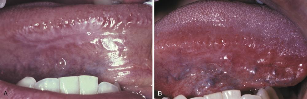
back 89

lichenoid contact reaction to dental amalgam

front 90

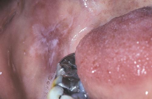
Radiating pattern of hyperkeratotic striae on the posterior buccal mucosa that contacts a large distobuccal amalgam of the permanent mandibular second molar.

back 90

lichenoid contact reaction to dental amalgam

front 91

How is lichenoid contact reaction to dental amalgam diagnosed?

back 91

Based on (a) the clinical appearance of the lesion, (b) the lack of lesional migration, and (c) the correlation to adjacent dental metal.

front 92

What is the treatment and prognosis for lichenoid contact reaction to dental amalgam?

back 92

  • acute reactions are self-limiting and resolve within 2 to 3 days
  • for chronic reactions, local measures should be attempted first
  • if unsuccessful, the amalgam in question should be replaced

front 93

A diffuse edematous swelling of the soft tissues, which has also been referred to historically as Quincke disease.

back 93

angioedema

front 94

Angioedema is a diffuse edematous swelling of the soft tissues. What are the potential causes of angioedema?

back 94

  1. allergic IgE-mediated hypersensitivity reactions
  2. angiotensin-converting enzyme (ACE) inhibitors
  3. hereditary—type I (C1-INH deficiency) or type II (C1-INH dysfunction)
  4. acquired—lymphoproliferative or autoimmune

front 95

An unusual pattern of drug reaction that can produce severe forms of angioedema that are not mediated by IgE is the type associated with ACE inhibitors. What is thought to be the cause of this reaction?

back 95

excess bradykinin (ACE degrades bradykinin)

front 96

Clinical Features

  • rapid onset of soft, nontender tissue swelling
  • may be solitary or multiple
  • extremities most common cutaneous sites
  • edema of larynx, pharynx, uvula, or soft palate
  • deeper voice, hoarseness, aphonia, and dyspnea
  • pain is unusual, but itching is common
  • typically resolves over 24 to 72 hours
  • drug-associated form more common in blacks

back 96

angioedema

front 97

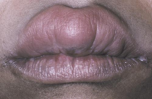
Diffuse upper lip swelling that arose rapidly.

back 97

angioedema

front 98

  1. A, Soft, nontender tissue swelling of the face arose relatively rapidly after dental treatment.
  2. Facial appearance after resolution of edematous facial enlargement.

back 98

angioedema

front 99

How is angioedema diagnosed?

back 99

  1. for allergic causes, diagnosis is based on clinical presentation in conjunction with the known antigenic stimulus
  2. case unrelated to antigenic exposure or medications should be evaluated for the presence of adequate functional C1-INH:
    1. type I hereditary—normal C1, low C1-INH
    2. type II hereditary—normal C1, normal C1-INH
    3. lymphoproliferative—low C1, low C1-INH
    4. autoimmune—low C1, normal C1-INH

front 100

What is the treatment and prognosis for angioedema?

back 100

  • oral antihistamines, IM epinephrine, IV corticosteroids
  • discontinue ACE inhibitors