Instructions for Side by Side Printing
- Print the notecards
- Fold each page in half along the solid vertical line
- Cut out the notecards by cutting along each horizontal dotted line
- Optional: Glue, tape or staple the ends of each notecard together
Chapter 4: Mechanics of Mandibular Movement
front 1 What two types of movement occur in the TMJ? | back 1 rotational and translational |
front 2 Rotational moment of mandible the occurs within the (...) cavity of the joint between the superior surface of the (...) and the inferior surface of the (...). | back 2 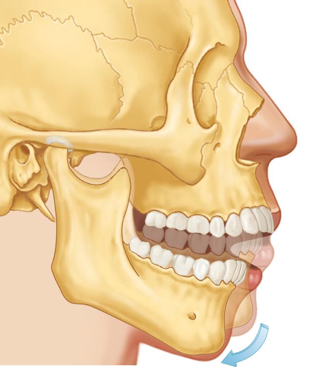 inferior; condyle; articular disc |
front 3 What three planes can rotational movement of the mandible can occur in? | back 3 horizontal, frontal, and sagittal |
front 4 In each the horizontal, frontal, and sagittal planes, rotational movement of the mandible occurs around a point, called the (...). | back 4 axis |
front 5 Mandibular movement around the horizontal axis is referred to as a (...) movement, and the axis around which it occurs is referred to as the (...). | back 5 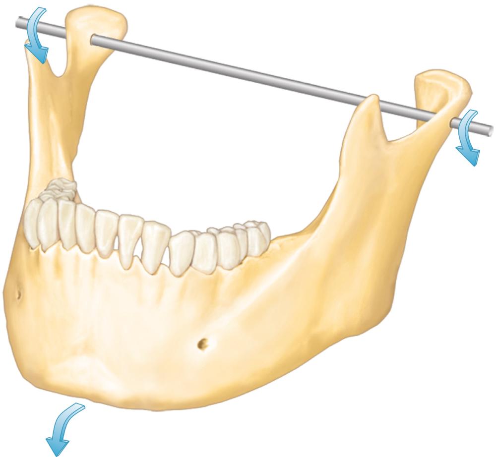 hinge; hinge axis |
front 6 When the condyles are in their most superior position in the articular fossae and the movement of the mandible is purely rotatational, the axis around which movement occurs is called the (...). | back 6 terminal hinge axis |
front 7 Mandibular movement around the frontal axis occurs when one condyle moves (...), while the vertical axis of the opposite condyle remains in the (...). | back 7 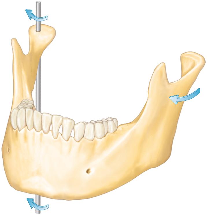 anteriorly; terminal hinge position |
front 8 Mandibular movement around the sagittal axis occurs when one condyle moves (...) while the vertical axis of the opposite condyle remains in the (...). | back 8 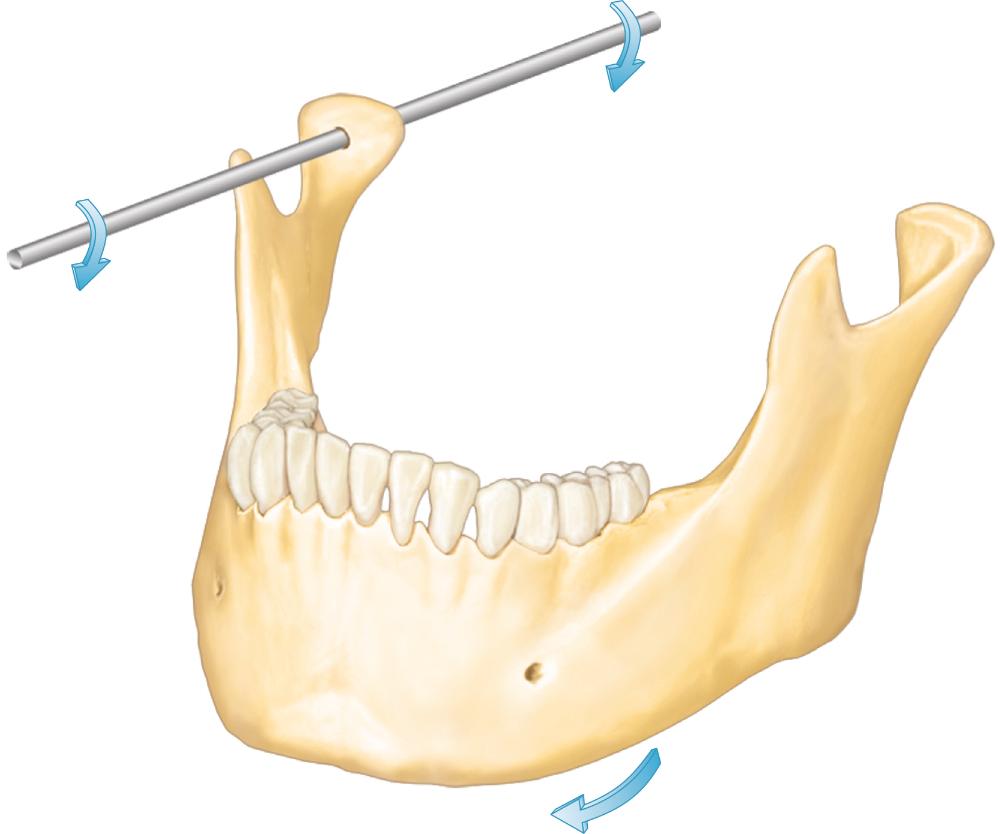 inferiorly; terminal hinge position |
front 9 Translational movement of mandible the occurs within the (...) cavity of the joint between the superior surface of the (...) and the inferior surface of the (...). | back 9 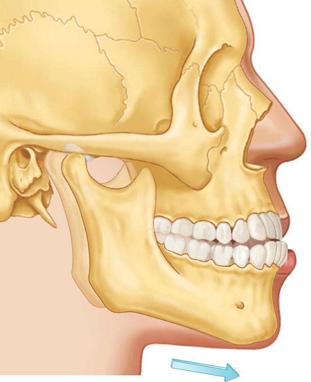 inferior; articular disc; articular fossa |
front 10 When the mandible moves through the outer range of motion, reproducible describable limits result, which are called (...). | back 10 border movements |
front 11 What are the four components of mandibular motion in the sagittal plane? | back 11 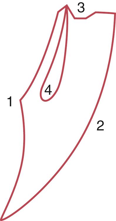
|
front 12 The range of posterior and anterior border movements is determined primarily by (...); the superior border movements are determined by the (...). | back 12 ligaments; occlusal surfaces of the teeth |
front 13 Posterior opening border movements in the sagittal plane occur as two-stage hinging movements. In the first stage, the condyles are in centric relation, and (...) movement occurs until the anterior teeth are (...) mm mm apart. | back 13 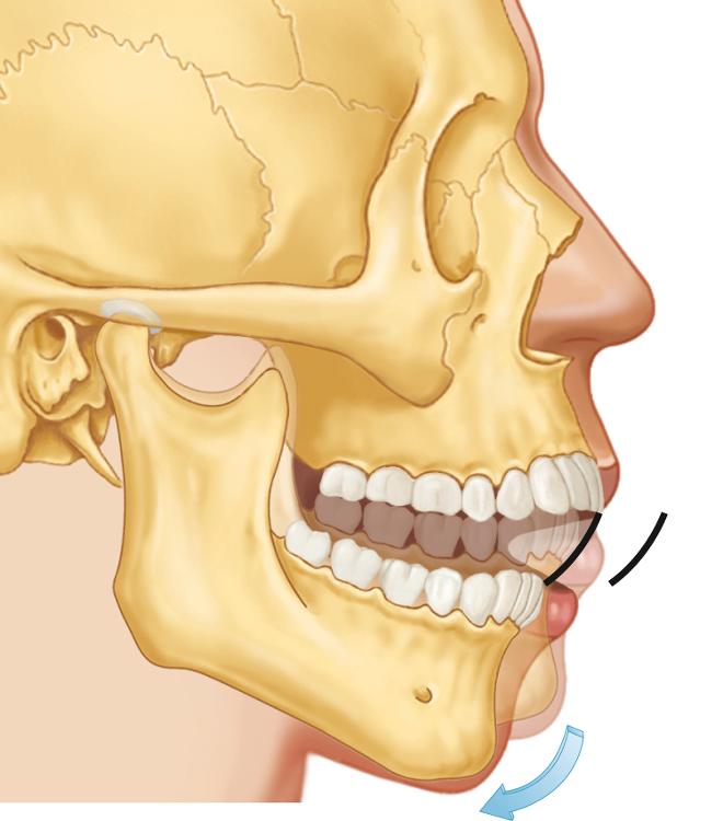 rotational; 20 to 25 mm |
front 14 Which ligament limit rotational movement of the mandible? | back 14 TM ligament |
front 15 Posterior opening border movements in the sagittal plane occur as two-stage hinging movements. In the second stage, the TM ligaments tighten, and the condyle is (...) as the mouth rotates open to its maximum limit of (...) mm. | back 15 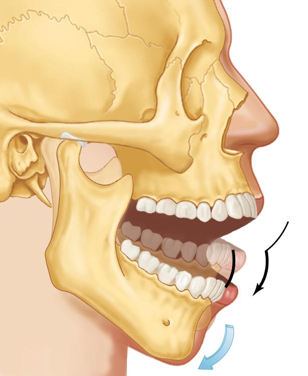 translated; 40 to 60 mm |
front 16 Which ligament limits maximum opening of the mandible? | back 16 capsular ligament |
front 17 With the mandible maximally opened, closure accompanied by contraction of the (...) will generate the anterior closing border movement and tightening of the (...) produces a posterior movement of the condyles. | back 17 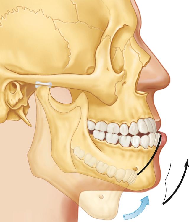 inferior lateral pterygoids; stylomandibular ligaments |
front 18 Which ligament limits the posterior border movement? | back 18 stylomandibular ligament |
front 19 Whereas the border movements previously discussed are limited by (...), the superior contact border movement is determined by the characteristics of the (...). | back 19 ligaments; occluding surfaces of the teeth |
front 20 The precise delineation of the superior contact border depends on the:
| back 20
|
front 21 The initial tooth contact in (...) occurs between the mesial inclines of a maxillary tooth and the distal inclines of a mandibular tooth. | back 21 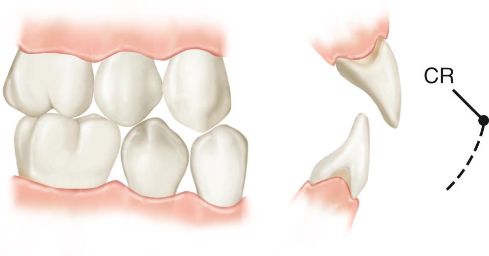 terminal hinge closure (CR) |
front 22 If muscular force is applied to the mandible in terminal hinge closure, a superoanterior movement or shift will result until the (...) is reached. | back 22 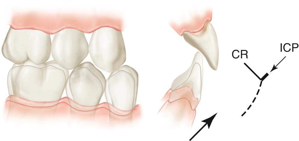 intercuspal position (ICP) |
front 23 The slide from CR to ICP is present in approximately 90% of the population, and the average distance is (...) mm. | back 23 1 to 1.25 mm |
front 24 When the mandible is protruded from maximum intercuspation, contact between the incisal edges of the mandibular anterior teeth and the lingual inclines of the maxillary anterior teeth results in an (...) movement of the mandible. | back 24 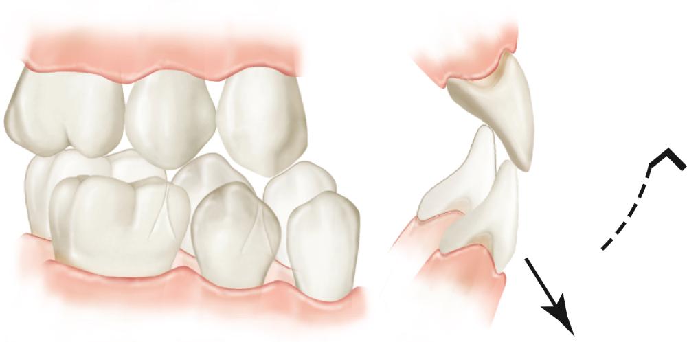 anteroinferior |
front 25 As maxillary and mandibular anterior teeth enter an edge-to-edge relationship in protrusion, the the mandible follows a (...) pathway of movement, which continues until the mandibular incisal edges pass beyond the maxillary incisal edges. | back 25 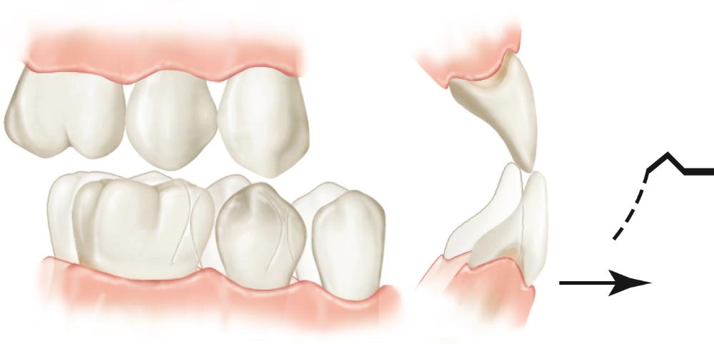 horizontal |
front 26 Once the mandibular incisal edges pass beyond the maxillary incisal edges in protrusion, the mandible moves in a (...) direction until the posterior teeth contact. | back 26 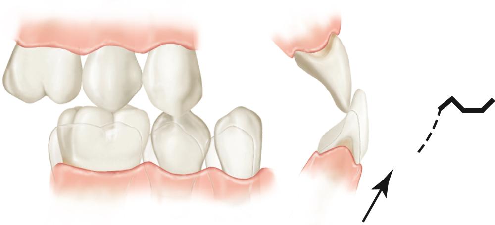 superior |
front 27 Once the posterior teeth contact in protrusion, their occlusal surfaces dictate the remaining pathway to the maximum protrusive movement, which joins with the most superior position of the (...) border movement. | back 27 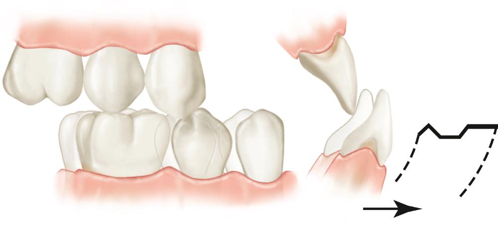 anterior |
front 28 How is the description of the superior contact border movement is altered when a person has no discrepancy between CR and maximum intercuspation? | back 28 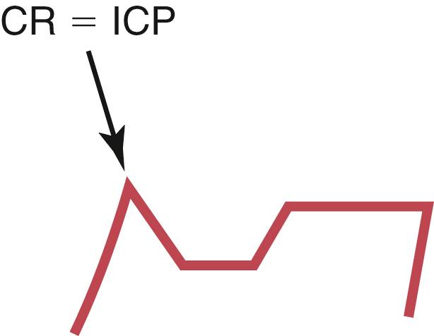 There is no superior slide from CR to ICP. |
front 29 (...) movements occur during activity of the mandible. They usually take place within the border movements and therefore are considered free movements. | back 29 functional |
front 30 Most functional activities require maximum intercuspation and therefore typically begin at and below the ICP. When the mandible is at rest, it is found to be located approximately (...) mm below ICP. This position has been called the (...). | back 30 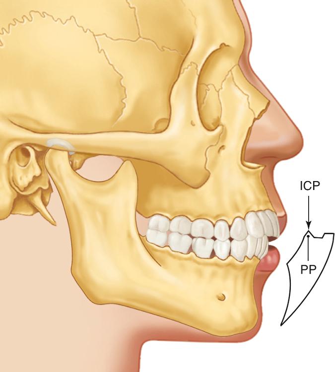 2 to 4 mm; clinical rest position (or postural position) |
front 31 If the chewing stroke is examined in the sagittal plane, the movement will be seen to begin and end at the (...) potision. | back 31 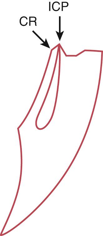 intercuspal (ICP) |
front 32 With the head upright the teeth are elevated directly into (...) from the postural position. | back 32 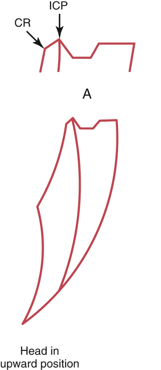 maximum intercuspation |
front 33 With the head raised 45°, the postural position of the mandible is shifted (...), thus when the teeth occlude, contacts occur (...) to the intercuspal position. | back 33 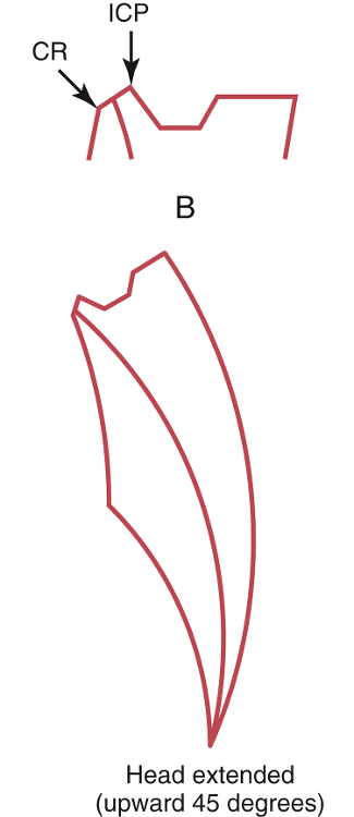 posteriorly; posterior |
front 34 With the head lowered 30°, the postural position of the mandible is shifted (...), thus when the teeth occlude, contacts occur (...) to the intercuspal position. | back 34 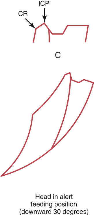 anteriorly; anterior |
front 35 It has been stated that the normal head position during eating is with the face directed downward 30°. This is referred to as the (...). | back 35 alert feeding position |
front 36 A (...) is used to record the mandibular border movements in the horizontal plane. As the mandible moves, the stylus attached to the mandibular teeth generates a pathway on the recording table attached to the maxillary teeth. | back 36 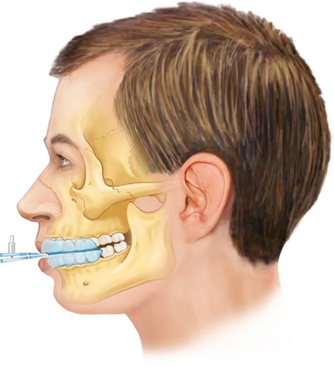 Gothic arch tracer |
front 37 What are the four components of mandibular motion in the horizontal plane? | back 37 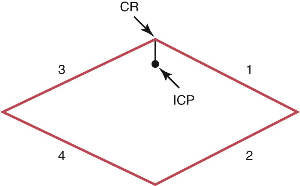
|
front 38 With the condyles in the CR position, contraction of the right inferior lateral pterygoid will cause the right condyle to move (...). If the left condyle remains situated in CR, this will result in the (...) border movement. | back 38 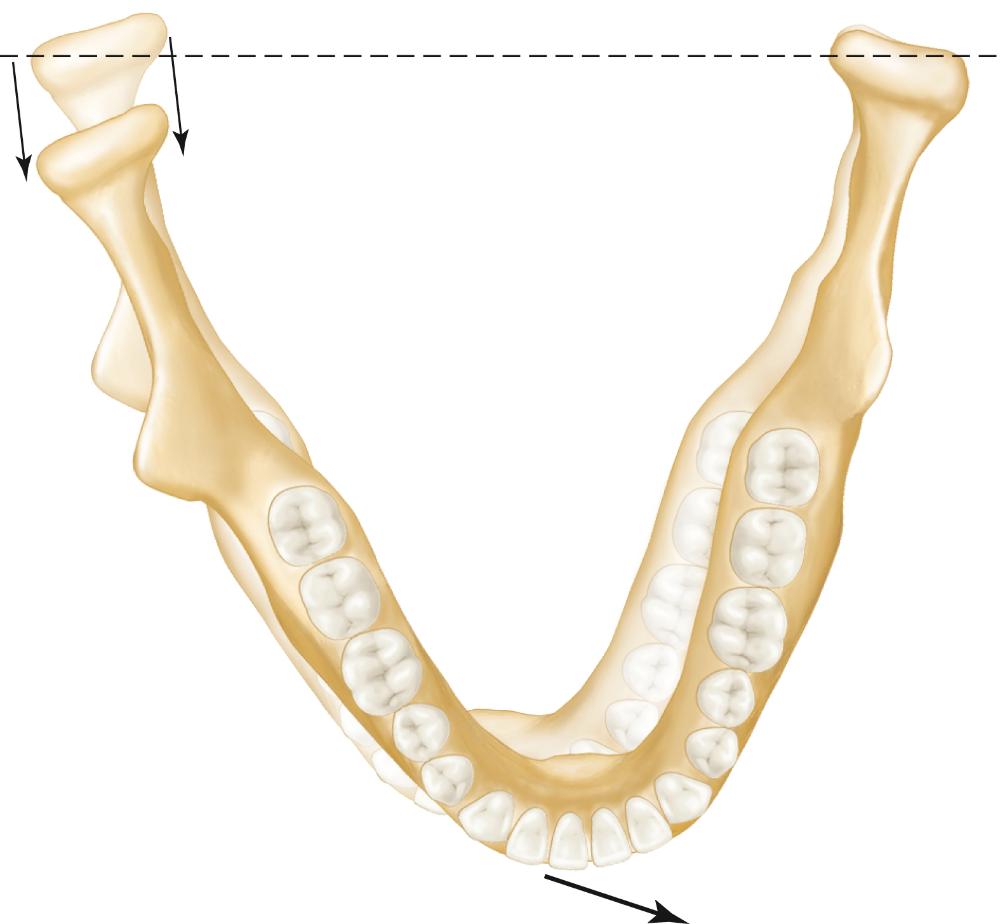 anteromedially; left lateral |
front 39 During the left border movement, the right condyle is called the (...) condyle and the left condyle is called the (...) condyle. | back 39 orbiting; rotating |
front 40 With the mandible in the left lateral border position, contraction of the left inferior lateral pterygoid muscle along with continued contraction of the right inferior lateral pterygoid muscle will cause the left condyle to move (...), producing the (...) border movement. | back 40  anteromedially; continued left lateral |
front 41 With the condyles in the CR position, contraction of the left inferior lateral pterygoid will cause the left condyle to move (...). If the right condyle remains situated in CR, this will result in the (...) border movement. | back 41 anteromedially; right lateral |
front 42 During the right border movement, the right condyle is called the (...) condyle and the left condyle is called the (...) condyle. | back 42 rotating; orbiting |
front 43 With the mandible in the right lateral border position, contraction of the right inferior lateral pterygoid muscle along with continued contraction of the left inferior lateral pterygoid muscle will cause the right condyle to move (...), producing the (...) border movement. | back 43 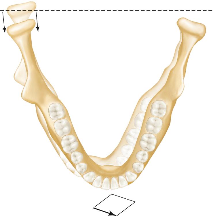 anteromedially; continued right lateral |
front 44 As mandibular opening increases, the size of the tracings generated by horizontal border movements become successively (...). | back 44 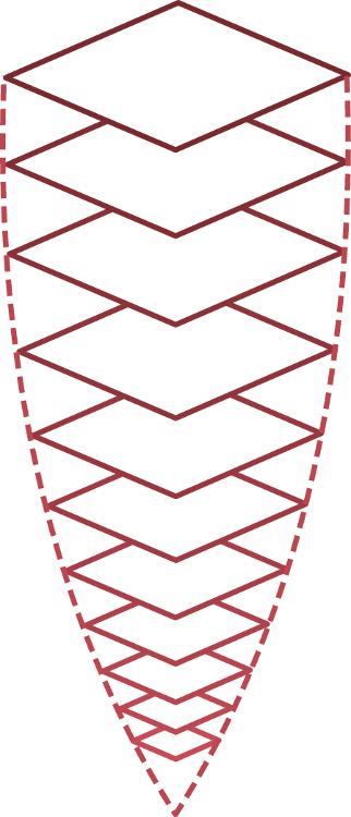 smaller |
front 45 The exact position of the mandible during chewing is dictated by the existing (...). | back 45 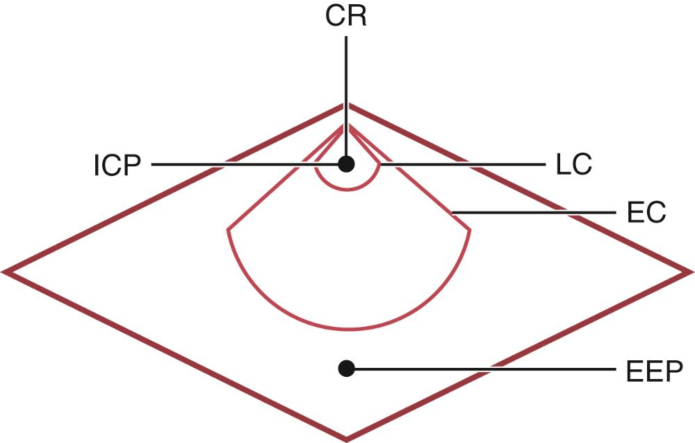 occlusal configuration |
front 46 What are the four components of mandibular motion in the frontal plane? | back 46 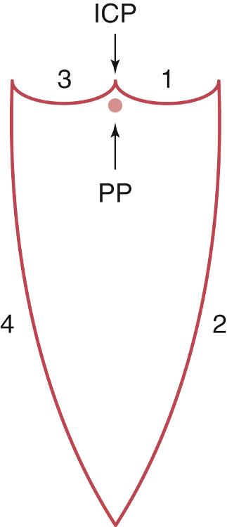
|
front 47 With the mandible in maximum intercuspation, a lateral movement is made to the left. A recording device will disclose an (...) path being generated, which forms the (...) border movement. | back 47 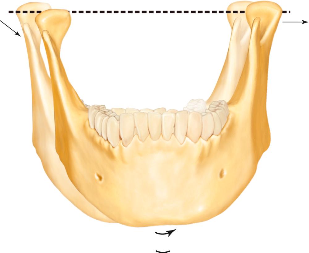 inferiorly concave; left lateral |
front 48 The precise nature of the left and right superior border movements in the frontal plane are primarily determined by (...); of secondary influence are the (...). | back 48 tooth contacts; condyle-disc-fossa relationship |
front 49 From the maximum left lateral superior border position, an opening movement of the mandible produces a (...) path. As maximum opening is approached, ligaments tighten and produce a (...) directed movement. | back 49 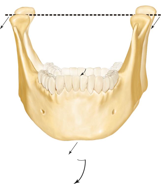 laterally convex; medially |
front 50 With the mandible in maximum intercuspation, a lateral movement is made to the left. A recording device will disclose an (...) path being generated, which forms the (...) border movement. | back 50 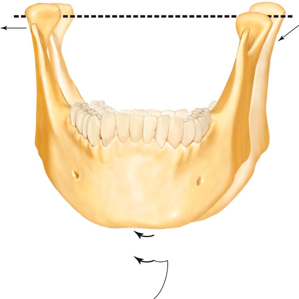 inferiorly concave; right lateral |
front 51 From the maximum right lateral superior border position, an opening movement of the mandible produces a (...) path. As maximum opening is approached, ligaments tighten and produce a (...) directed movement. | back 51 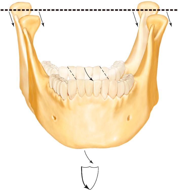 laterally convex; medially |
front 52 As in the other planes, functional movements in the frontal plane begin and end at the (...) position. | back 52 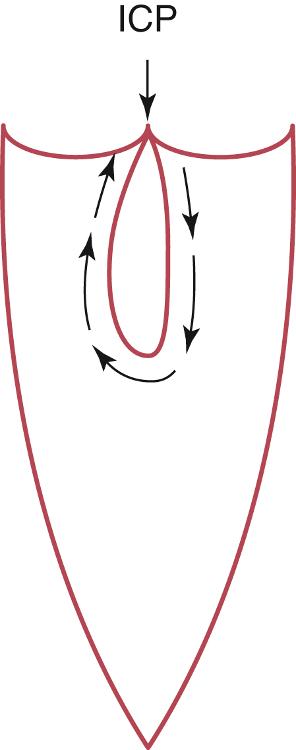 intercuspal (ICP) |
front 53 By combining mandibular border movements in the three planes (sagittal, horizontal, and frontal), a three-dimensional (...) is produced, that represents the maximum range of movement of the mandible | back 53  envelope of motion |