Instructions for Side by Side Printing
- Print the notecards
- Fold each page in half along the solid vertical line
- Cut out the notecards by cutting along each horizontal dotted line
- Optional: Glue, tape or staple the ends of each notecard together
Ocular Anatomy
front 1 Cornea | back 1 The cornea is the transparent structure on the front surface of the globe. It has 40 to 46 diopters of refractive power, an index of refraction of 1.37, and is the major refractive body of the eye. The cornea consists of 5 layers and is the structure that a contact lens rests upon. The average corneal diameter is 10.5 to 12.5mm. As a person ages, the cornea becomes less sensitive and less elastic. |
front 2 Eyelids | back 2 The eyelids or palpebra are folds of skin that serve as protective coverings for the globe and cornea. The eyelids contain Meibomian glands that help moisten the eye. The eyelids also regulate light entry. The palpebral fissure (opening of the eye) is approximately 10mm vertically and 30 mm horizontally. |
front 3 Conjunctiva | back 3 The conjunctiva is a clear, protective membrane that covers the white-part of the eye and the inside of the eyelids. The bulbar conjunctiva covers the sclera, while the palpebral conjunctiva line the inner surface of the eyelids. |
front 4 Iris and Pupil | back 4 The iris is a “shutter-like” muscle that forms the colored part of the eye. The pupil is the opening in the shutter that controls the amount of light entering the eye. It dilates, becomes larger, in dim light and constricts, becomes smaller, in bright light. These reactions are involuntary and can not be controlled without the presence of drugs. |
front 5 Aqueous Humor | back 5 The aqueous humor, with an index of refraction of 1.336, is a watery substance that fills the anterior and posterior chambers. The function of the aqueous humor is to maintain the shape of these chambers, and regulate the eye’s intraocular pressure. The aqueous humor also supplies the crystalline lens and cornea with nutrients. The aqueous humor is constantly being replaced and drains through the Canal of Schlem. |
front 6 Ciliary Muscle | back 6 The ciliary muscle controls the focusing or accommodation of the eye by changing the shape of the crystalline lens. |
front 7 Crystalline Lens | back 7 The crystalline lens is a resilient, transparent structure in the eye that focuses light by changing its front surface curvature. It is located near the front of the eye, directly behind the pupil. It is biconvex in shape and has an index of refraction of 1.42. |
front 8 Retina | back 8 The retina is a tissue that lines the inside of the back of the eye. It contains 10 layers of nerve endings, as well as, rods and cones. |
front 9 Rods and Cones | back 9 The rods enable the eye to see in dimly lit conditions and at night. The cones are responsible for color perception. |
front 10 Optic Nerve (or Optic Disk) | back 10 The optic nerve sends impulses from the retina to the brain for interpretation of visual images. It also does not contain any photoreceptor cells and thus causes a scotoma (blind spot). |
front 11 Macula and Fovea | back 11 The macula is the central area of the retina. Within the macula is the fovea which is the location of the most acute vision. |
front 12 Choroid | back 12 The choroid is a layer of blood vessels in the back of the eye between the sclera and the retina. Its primary function is to nourish the retina. |
front 13 Vitreous Humor | back 13 The vitreous humor or body is a jelly-like substance found between the back of the crystalline lens and the retina. Its primary function is to maintain the eye’s proper shape and temperature. The index of refraction of the vitreous is 1.336. |
front 14 Presbyopia | back 14 "Presbyopia is a gradual loss of the focusing ability (accommodation) of the eye. This condition restricts the eye’s ability to focus for near vision. This lack of accommodation is first noticed in people around the age of 40. Presbyopia can be corrected with the use of a plus addition (or Add) for reading or multifocal glasses. There are two causes for the onset of presbyopia. 1. The layers of cells of the crystalline lens become rigid and therefore the lens can not reshape easily to its biconvex shape. 2. The ciliary muscles that are attached to the crystalline lens, via the suspensory ligaments, “weaken” and are no longer able to help the crystalline lens reshape." |
front 15 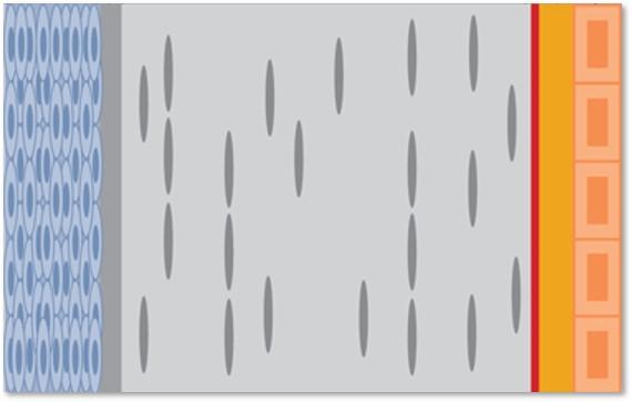 What are the 5 layers of the cornea anterior to posterior? | back 15 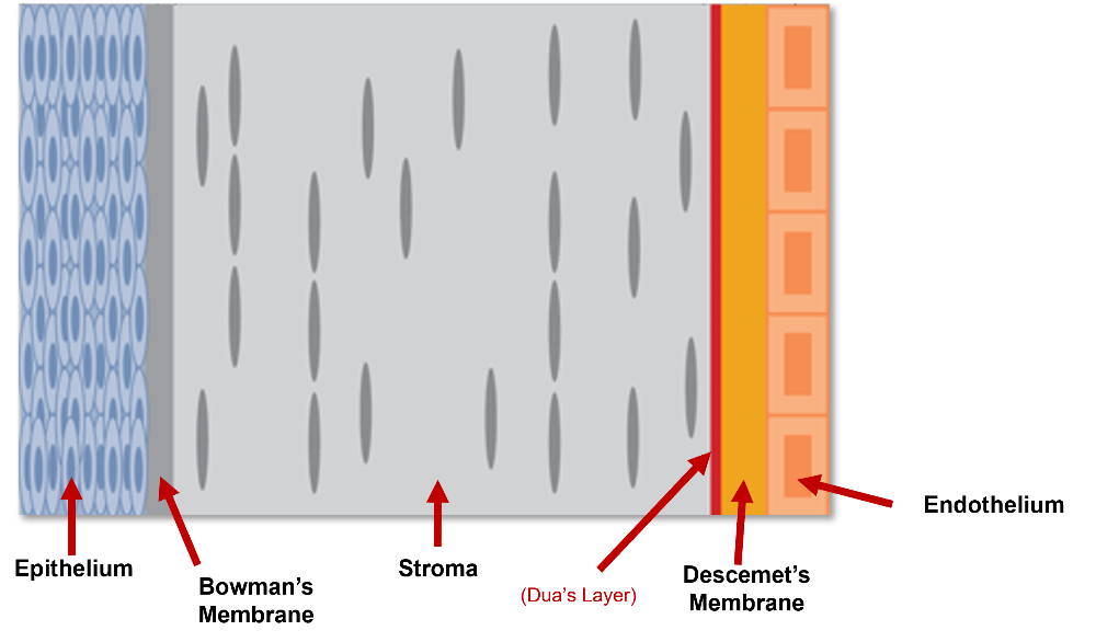 Epithelium, Bowman's membrane, Stroma, Descemet's membrane, Endothelium (Memorization help: Epi-bo-stro-desce-endo) |
front 16 Epithelium | back 16 Characteristics: - Outermost layer of the cornea - Smooth surface - Constantly regenerating Functions: - Blocks foreign material - Absorbs oxygen and nutrients - Distributes nutrients to the rest of the cornea |
front 17 Bowman's Membrane | back 17 Characteristics: - Acellular - No regenerative properties Function: Provides strength to the cornea |
front 18 Stroma | back 18 Characteristics: - 90 percent of the cornea's thickness - 78% water, 16 % collagen - A vascular Function: Gives cornea it's shape, elasticity, and form |
front 19 Descement's Membrane | back 19 Characteristics: - Thin, strong sheet of tissue - Composed of collagen fibers - Made by the endothelial cells that lie below it Function: Protective barrier against infection and injury |
front 20 Endothelium | back 20 Characteristics: - Innermost layer of the cornea - Single layer of flattened cells - Very susceptible to trauma and pathology - Very rarely regenerates Functions: - Deturgescene (transports fluids from stroma to anterior chamber) - "Leak" nutrients from the anterior chamber into the stroma and beyond - Maintains the cornea in a slightly dehydrated state |
front 21 Limbus | back 21 Borders the cornea and the sclera; about 1 mm in width. Functions: corneal nutrients pass through this area, helps drain extraneous aqueous humor from the anterior chamber |
front 22 Canal of Schlemm | back 22 A circular lymphatic-like vessel that helps drain extraneous aqueous humor from the posterior chamber. |
front 23 Meibomian glands | back 23 Located in the tarsal plate of both upper and lower eyelids. Produces the lipid layer of tears (Clogged meibomian gland = chalazion; Infected meibomian gland = stye) |
front 24 Lacrimal glands | back 24 These glands continually releases fluid which cleanses and protects the eye's surface as it lubricates and moistens it. Responsible for producing the aqueous layer of the tear film. |
front 25 What are the three layers of the pre-corneal film (or tear film) from anterior to posterior? | back 25 Lipid, aqueous, mucoid (or Mucin) |
front 26 Lipid Layer of the Tear Film | back 26 - Outermost (oil) layer - helps prevent evaporation - prevents irritation from the eyelids during blinking -created by the Meibomian glands |
front 27 Aqueous Layer of the Tear Film | back 27 - Center layer of the tear film - Largest volume - Contains some salt and proteins - Created by lacrimal glands |
front 28 Mucin (Mucoid) Layer of the Tear Film | back 28 - contains mucus -bonds tears to front surface of the cornea - prevents bacteria and debris from adhering to the eye - created by Goblet Cells |