The Stomach
The Stomach
How does the extra oblique layer of smooth muscle found in the stomach correlate with the stomach's churning movements? (Hint: 3 functions and what do they do? )
1. Churn food
2. Mix food
3. Pummel food
All above helps tot physically reduce the food to smaller fragments.
The chief cells produce what?
Produces pepsinogen
The parietal cells secret what?
HCl
Enteroendocrine cells release what?
Hormones
Gastroesophageal junction:
1. Describe the epithelium found in the esophagus and the beginning of the stomach (right after the gastroesophageal junction).
2. What is the functional importance of the epithelial differences seen in the esophagus and the stomach?
1. Stratified squamous epithelium is found in the esophagus, and simple columnar epithelium is found in the stomach (right after the gastroesophageal junction).
2. Stratified squamous epithelium=protection, Simple columnar epithelium=absorption
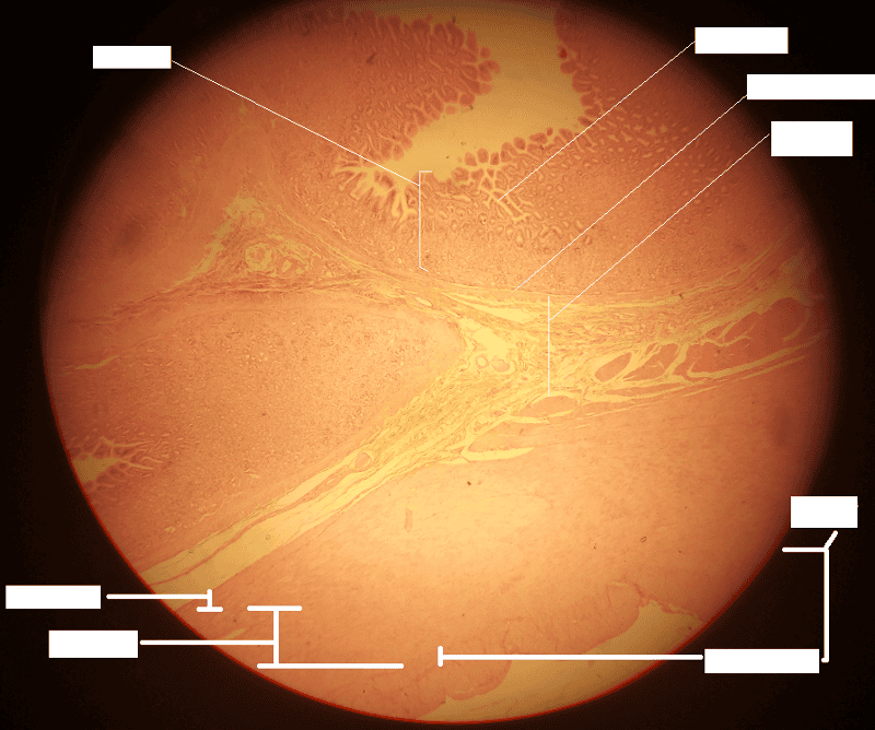
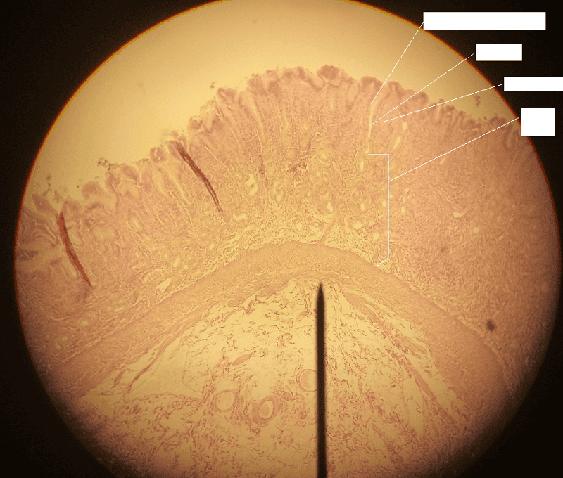
1. Identify the blanks.
2. What feature of the alimentary canal is shown in this histology slide? (Hint: organ)
1. Look at the picture.
2. Stomach wall
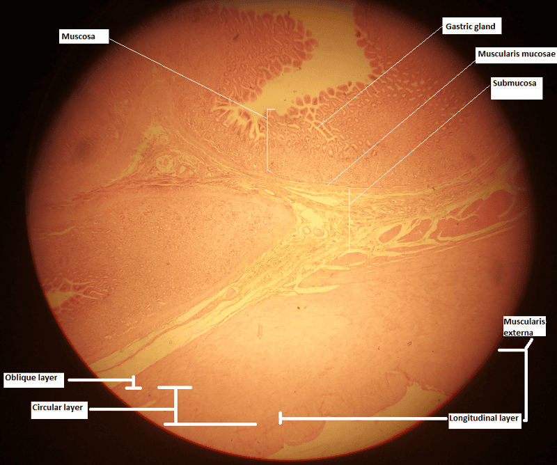
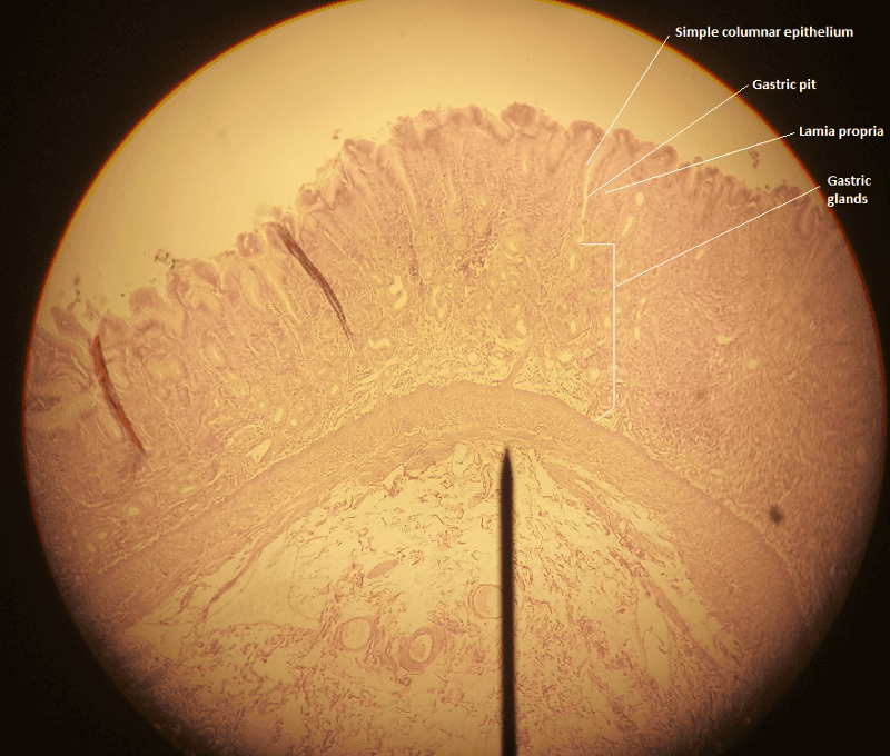
1. Identify the blanks.
2. What feature of the alimentary canal is shown in this histology slide? (Hint: organ)
1. Look at the picture.
2. Stomach wall
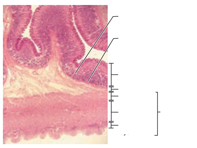
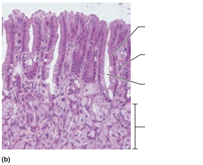
1. Identify the blanks.
2. What feature of the alimentary canal is shown in this histology slide? (Hint: organ)
1. Look at the picture.
2. Stomach wall
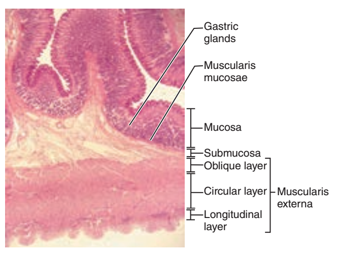
1. Identify the blanks.
2. What feature of the alimentary canal is shown in this histology slide? (Hint: organ)
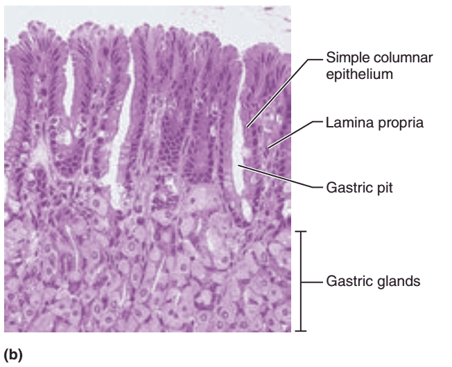
1. Look at the picture.
2. Stomach wall
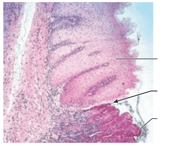
1. Identify the blanks.
2. What feature of the alimentary canal is shown in this histology slide? (Hint: junction)
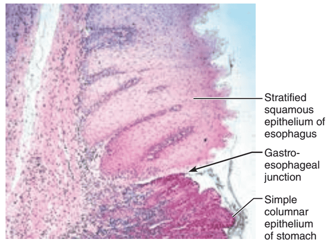
1. Look at the picture.
2. Gastroesophageal junction
The Small Intestine
The Small Intestine
The small intestine extends from what sphincter to what valve?
Extends from the pyloric sphincter to the ileocecal valve
The small intestine is suspended by what double-layered peritoneum and from what abdominal wall?
Suspended by the mesentery from the posterior abdominal wall
What are the 3 subdivisions of the small intestine?
1. Duodenum
2. Jejunum
3. ileum
The duodenum extends from what sphincter and curves around the head of what organ?
Extends from the pyloric sphincter and curves around the head of the pancreas
The jejunum is continuous with what subdivision of the small intestine?
Duodenum
Most of the jejunum occupies what region of the abdominal cavity?
The umbilical region of the abdominal cavity
The ileum makes up what portion of the small intestine?
Terminal portion of the small intestine
The ileum joins the large intestine at what valve?
Joins the large intestine at the ileocecal valve
The major portion of the ileum lies in what region of the abdominal cavity?
Hypogastric region of the abdominal cavity
What 2 enzymes complete digestion in the small intestine?
1. Brush border enzymes
2. Pancreatic enzymes
The brush border enzymes are what type of enzymes?
Hydrolytic enzymes
The brush border enzymes are bound to what structures of what epithelium?
Bound to the microvilli of the simple columnar epithelium of the stomach
How do pancreatic enzymes enter the stomach, and which specific subdivision of the small intestine do they enter through?
Enter by the main pancreatic duct and are ducted into the duodenum
Bile is formed in what organ?
Liver
How does bile enter the stomach, and which specific subdivision of the small intestine does it enter through?
Enter by the bile duct into the duodenum of the small intestine
At the duodenum, the pancreatic duct and the bile duct join to form what structure?
The hepato-pancreatic ampulla
The hepato-pancreatic ampulla empties into what lumen and through what structure?
Empties into the duodenal lumen through the major duodenal papilla
The major duodenal papilla is controlled by what valve?
The hepato-pancreatic sphincter
The hepato-pancreatic sphincter is what type of valve?
Muscle valve
Nearly all of what occurs in the small intestine?
Nutrient absorption
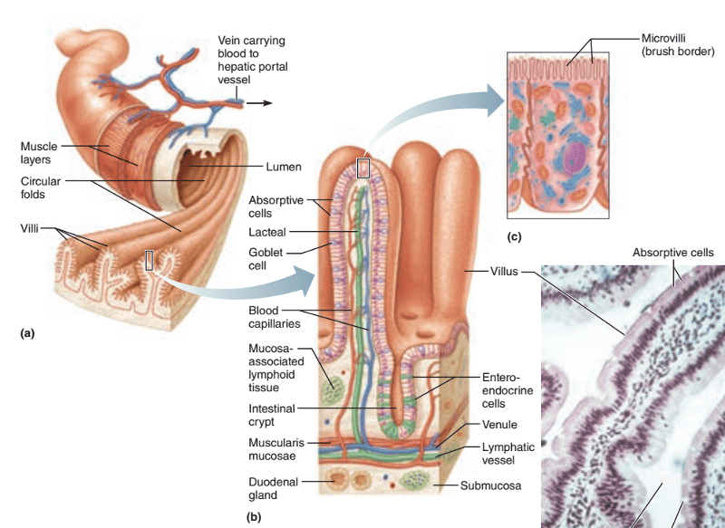
What 3 structural modifications increase the absorptive surface of the small intestine mucosa?
1. Microvilli
2. Villi
3. Circular folds
Microvilli are microscopic projections of the surface ____ of the ____.
Microscopic projections of the surface plasma membrane of the columnar epithelial cells of the mucosa.
Villi are ____ projections of what tunic?
Finger-like projections of the mucosa tunic
The circular folds are deep folds of what 2 layers?
1. Mucosa layer
2. Submucosa layer
What is the function of the circular folds of the small intestine?
Force chyme to spiral through the intestine, mixing it and slowing its progress
The 3 structural modifications that help with absorption (i.e. microvilli, villi, circular folds) of the small intestine decrease in frequency and size towards where?
Towards the end of the small intestine
What happens to any residue remaining undigested and unabsorbed food at the end of the small intestine? (Hint: enters what organ and through what valve?)
It enters the large intestine through the ileocecal valve
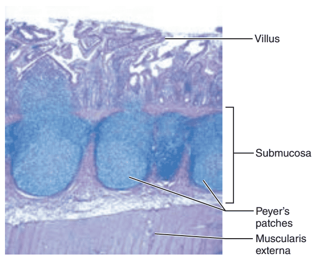
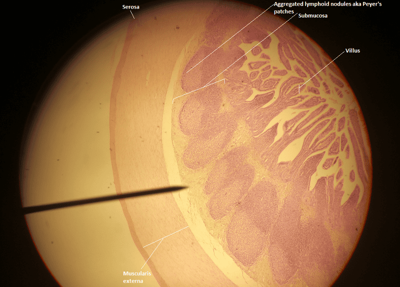
In contrast, while the 3 structural modifications that help with absorption decrease in frequency and size toward the end of the small intestine, what happens to the lymphoid tissue in the submucosa?
The lymphoid tissue in the submucosa of the small intestine increases along the length of the small intestine
The lymphoid tissue in the submucosa of the small intestine are also called what? (Hint: state common name as well)
Aggregated lymphoid nodules or Peyer's patches
The aggregated lymphoid nodules or Peyer's patches are very apparent in what subdivision of the small intestine?
The ileum
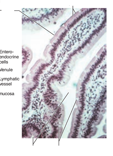
1. Identify the blanks.
2. What feature of the alimentary canal is shown in this histology slide?
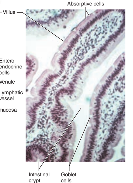
1. Look at the picture.
2. Small intestine
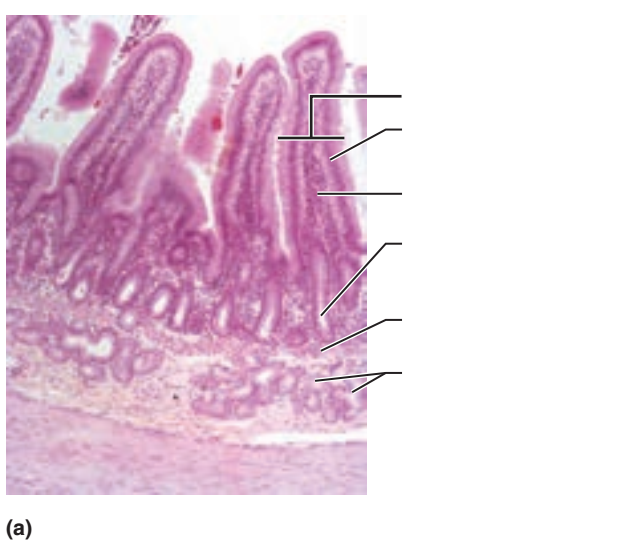
1. Identify the blanks.
2. What feature of the alimentary canal is shown in this histology slide? (Hint: What specific region of the organ?)
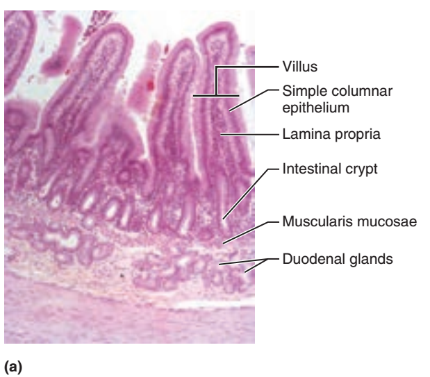
1. Look at the picture.
2. Dueodenum of small intestine
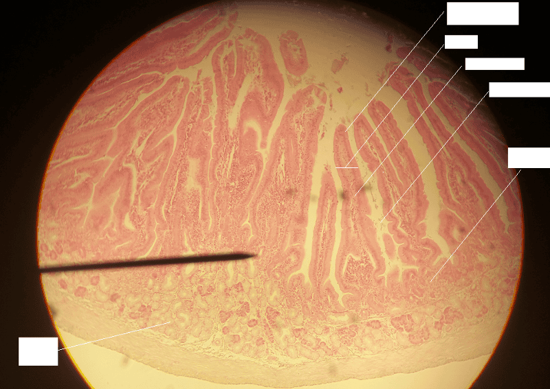
1. Identify the blanks.
2. What feature of the alimentary canal is shown in this histology slide? (Hint: What specific region of the organ?)
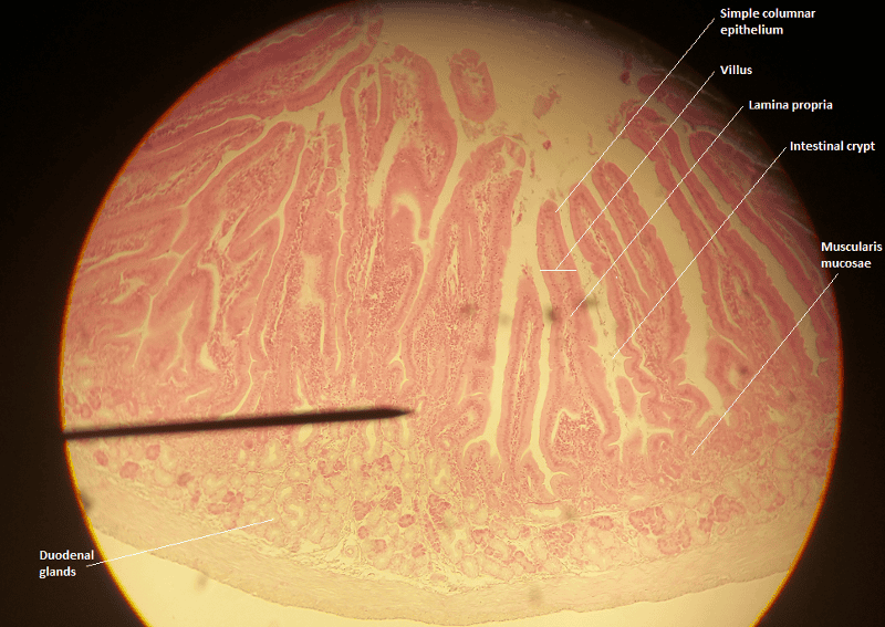
1. Look at the picture.
2. Duodenum of small intestine
1. Identify the blanks.
2. What feature of the alimentary canal is shown in this histology slide? (Hint: What specific region of the organ?)

1. Look at the picture.
2. Jejunum of small intestine
1. Identify the blanks.
2. What feature of the alimentary canal is shown in this histology slide? (Hint: What specific region of the organ?)
1. Look at the picture.
2. Ileum of small intestine
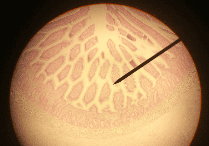
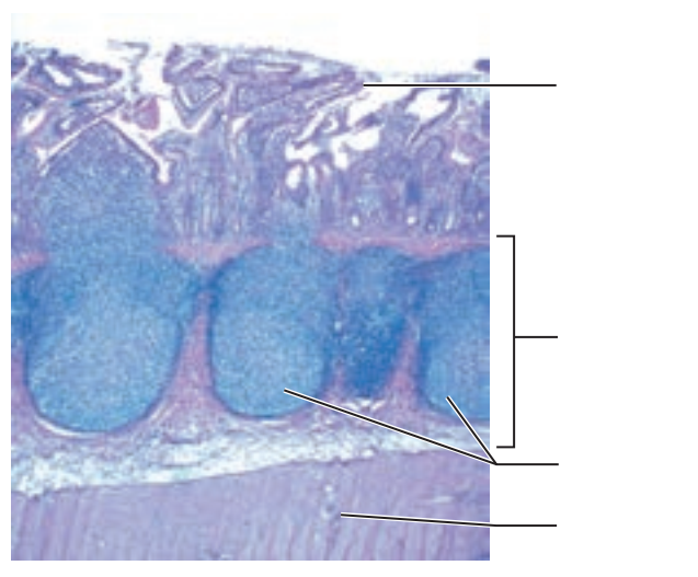
1. Identify the blanks.
2. What feature of the alimentary canal is shown in this histology slide? (Hint: What specific region of the organ?)
1. Look at the picture.
2. Ileum of the small intestine
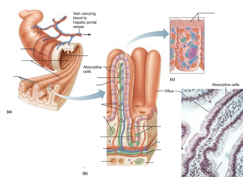
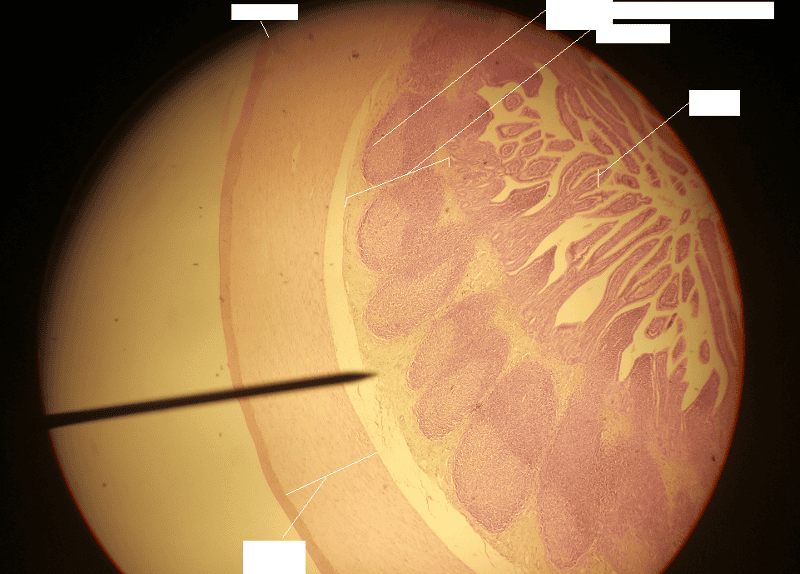
1. Identify the blanks.
2. What feature of the alimentary canal is shown in this histology slide?
1. Look at the picture.
2. Small intestine cutout