What is the major function of the cardiovascular system?
Transportation
What is used as the transport vehicle in the cardiovascular system?
Blood
Blood can carry what 4 essential substances?
1. Oxygen
2. Digested foods
3. Cell wastes
4. Electrolytes
What is the cardiovascular system's propulsive force?
Heart
The heart can be compared to what type of pump with what type of valves?
One-way pump with one-way valves
When the heart contracts, where does it force the blood?
It forces the blood into the blood vessels
The heart is about the size of what body part?
A closed fist
Where is the heart located?
Within the mediastinum of the thorax
The heart is flanked laterally by the ____, posteriorly by the ____, and anteriorly by the ____.
1. Lungs
2. Vertebral column
3. Sternum
Describe the apex of the heart. (Hint: How it looks like?)
It is sharp.
The apex slightly extends to which side of the body and rests on what organ?
Slightly extends to the left and rest on the diaphragm
The apex is approximately aligned at which intercostal space?
The 5th intercostal space
What emerges from the base of the heart?
The great blood vessels
The base of the heart lies beneath what rib and points toward what body feature ?
Lies beneath the second rib and points toward the right shoulder
The heart is enclosed within what structure?
Pericardium
What type of sac is the pericardium?
Double-walled sac
The pericardium has how many layers?
3
What are the 3 layers of pericardium covering the heart? (Hint: from most external to most internal)
1. Fibrous pericardium
2. Parietal pericardium
3. Visceral pericardium
The visceral pericardium is also called by what name?
Epicardium
How is the epicardium adhered to the heart muscle?
Closely adhered to the heart muscle
How loose is the parietal pericardium adhered to the heart muscle?
Loosely adhered
The parietal pericardium is attached at the heart apex to what structure?
Diaphragm
Which layer of the pericardium produces serous fluid?
Parietal pericardium
What is the function of the serous fluid produced by the parietal pericardium?
Allow the heart to beat without friction
The serous parietal pericardium lines what layer of the pericardium?
Fibrous pericardium
The fibrous pericardium is composed of what type of tissue?
dense connective tissue
How is the fibrous pericardium adhered to the heart muscle?
Loosely adhered
The walls of the actual heart are composed of how many layers?
3
What are the 3 walls of the actual heart (from outermost to innermost)?
1. Epicardium aka visceral pericardium
2. Myocardium
3. Endocardium
What wall of heart is the thickest layer?
Myocardium
The myocardium is composed mainly of what muscle type?
Cardiac muscle
The myocardium is then reinforced with what type of tissue?
Dense irregular connective tissue
The dense irregular connective tissue that lines the myocardium is also called what?
Cardiac skeleton
The cardiac skeleton (comprised of dense irregular connective tissue) that lines the myocardium is thicker around which 2 features of the heart?
1. Heart valves
2. Base of the great vessels
The endocardium covers what and is continuous with what?
Covers the heart valves and is continuous with the inner lining of the great vessels
The endocardium is composed of what epithelium?
Simple squamous epithelium
The endocardium rests on what type of tissue?
Areolar connective tissue
The heart is divided into how many chambers?
4
What are the 4 chambers of the heart?
2 atria and 2 ventricles
What structure divides the heart longitudinally?
Interventricular or interatrial septum
The interventricular septum divides what 2 chambers?
The 2 ventricles
The interatrial septum divides what 2 chambers?
The 2 atria
Functionally, the atria act as what type of chambers and are ineffective as what?
Act as receiving chambers and are ineffective as pumps
Which 2 chambers form the bulk of the heart?
The 2 ventricles
The 2 ventricles act as what type of chambers functionally?
Discharging chambers
What is the function of the ventricles in regards to blood?
They force blood out of the heart and into the large arteries that emerge from the base.
The heart has how many valves?
4
The 4 valves of the heart enforce what type of blood flow?
One-way blood flow
The atrioventricular (AV) valves are located between what chambers?
Between atria and ventricles
What are the 2 atrioventricular (AV) valves?
1. Tricuspid valve
2. Mitral or bicuspid valve
The semilunar (SL) valves are located between what 2 features of the heart?
1. Ventricles
2. Great vessels
What are the 2 semilunar (SL) valves?
1. Pulmonary (SL) valve
2. Aortic (SL) valve
What is the right atrioventricular (AV) valve?
Tricuspid valve
The tricuspid valve has how many flaplike cusps?
3
The 3 flaplike cusps of the tricuspid valve are anchored to what type of muscles?
Papillary muscles
Which chambers are papillary muscles found?
In the 2 ventricles
The tricuspid valve anchored to the papillary muscles by what structure?
By chordae tendineae
What is left atrioventricular (AV) valve called?
Mitral valve or bicuspid valve
The bicuspid valve has how many flaplike cusps?
2
The bicuspid valve anchored to the papillary muscles by what structure?
Chordae tendineae
When the AV valves are open and hang into the ventricles, and the ventricles are relaxed, blood is permitted to flow into which chambers?
The 2 atria and the 2 ventricles
When does the ventricles contract?
When they fill up with blood
When the ventricles contract, what happens to the atrioventricular (AV) valves?
They move superiorly to separate the atria and ventricles.
How does the papillary muscles and chordae tendineae prevent the backlow of blood?
The chordae tendineae are pulled tight by the contracting papillary muscles to anchor the cusps in the closed position. This prevents the backflow of blood from the ventricles back into the atria.
The pulmonary (SL) valve has how many cusps?
3 cusps
The pulmonary (SL) valve is located between what 2 heart structures?
1. Right ventricle
2. Pulmonary trunk
The aortic (SL) valve has how many cusps?
3
The aortic (SL) valve is located between what 2 heart structures?
1. Left ventricle
2. Aorta
When are the SL valves opened and flattened against the wall of the vessel?
During contraction of the ventricles that pushes blood into the great vessels
1. When do the SL valves close and their cusps fill up with blood?
2. How do the SL valves close?
1. When the ventricles relax
2.The flow of blood back toward the ventricle creates pressure that closes the cusps, closing the valves as well.
The closing of the SL valves prevents backflow of blood from what 2 heart features?
Prevents backflow of blood from the great vessels into the ventricles.

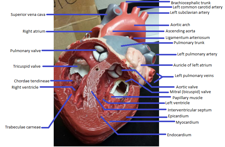
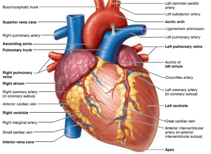
Name the missing parts.
Heart external anterior view

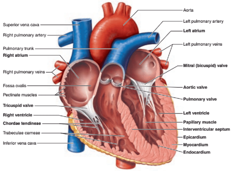
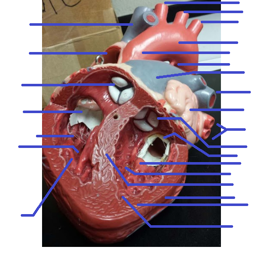
Name the missing parts.
Heart frontal section
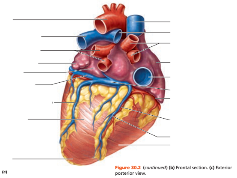
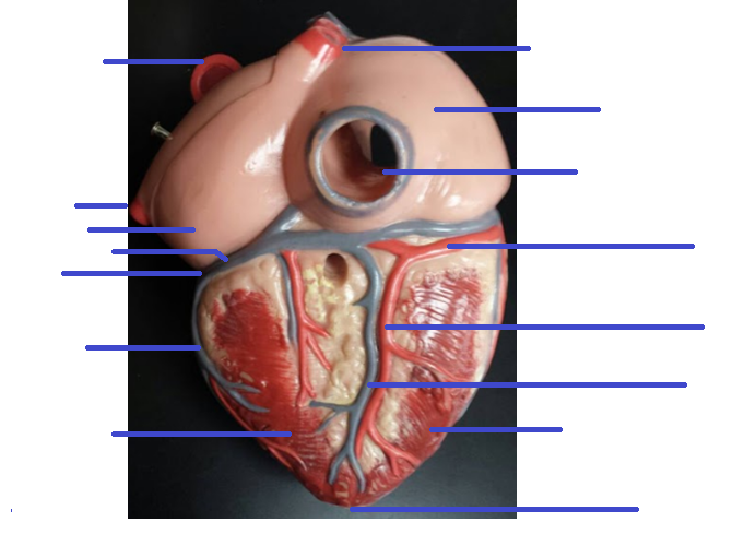
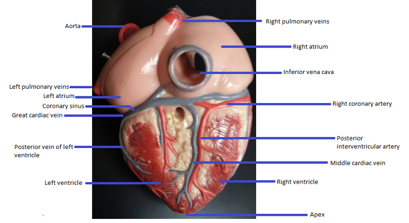
Name the missing parts.
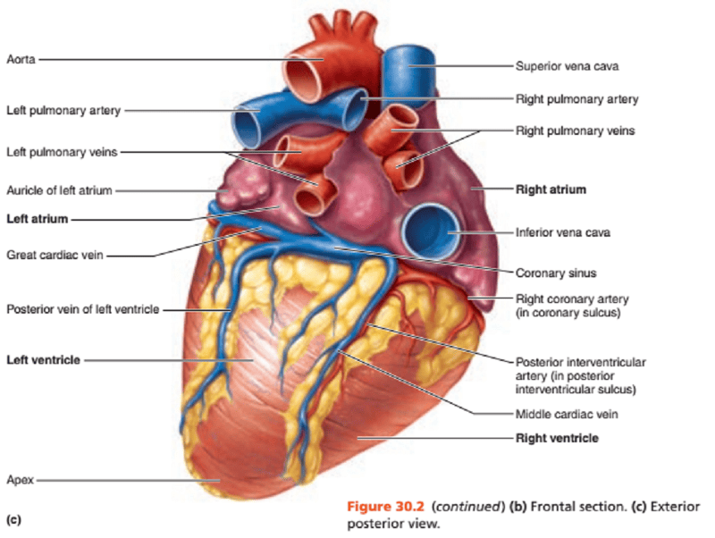
Heart external posterior view
Name the missing parts.
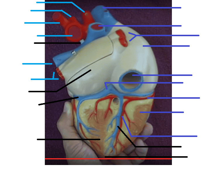
External posterior view

Name the missing parts.
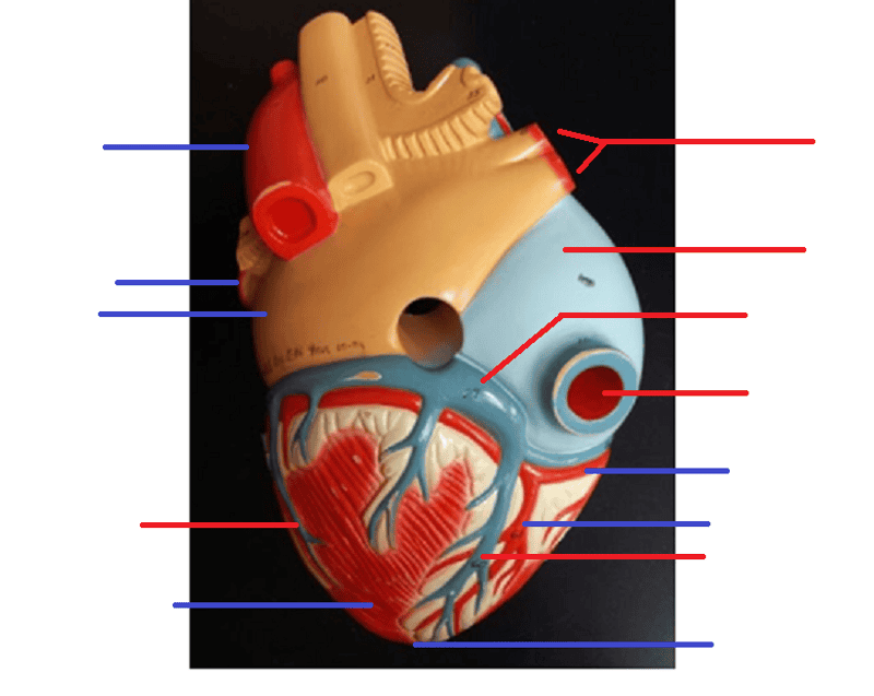
External posterior view
Name the missing parts.
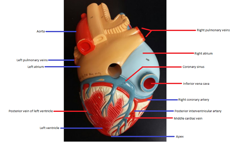
External posterior view
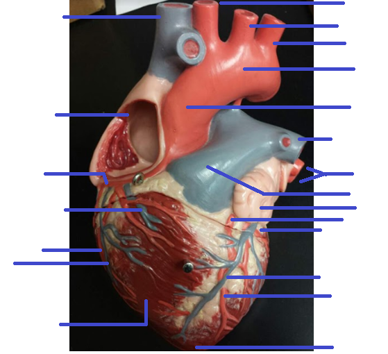
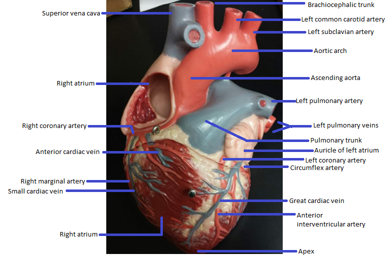
Name the missing parts.
External anterior view
Name the missing parts.
Frontal section