1.The shoulder consists of what 2 bones?
Scapula and clavical
2. The medial or sternal extremity articulates with the manubrium which is the upper part of the? The articulation is at what joint?
sternum, sternoclavicular joint
3. Whos clavicle is longer males or female?
males
4. Whos clavicle is usually thicker and more curved?
males
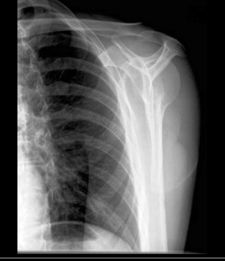
5. What exam is this and what is it checking for and how can you tell what it is?
Y view, you can tell because the vertebral border of scapula and it is checking for dislocation of humerus.
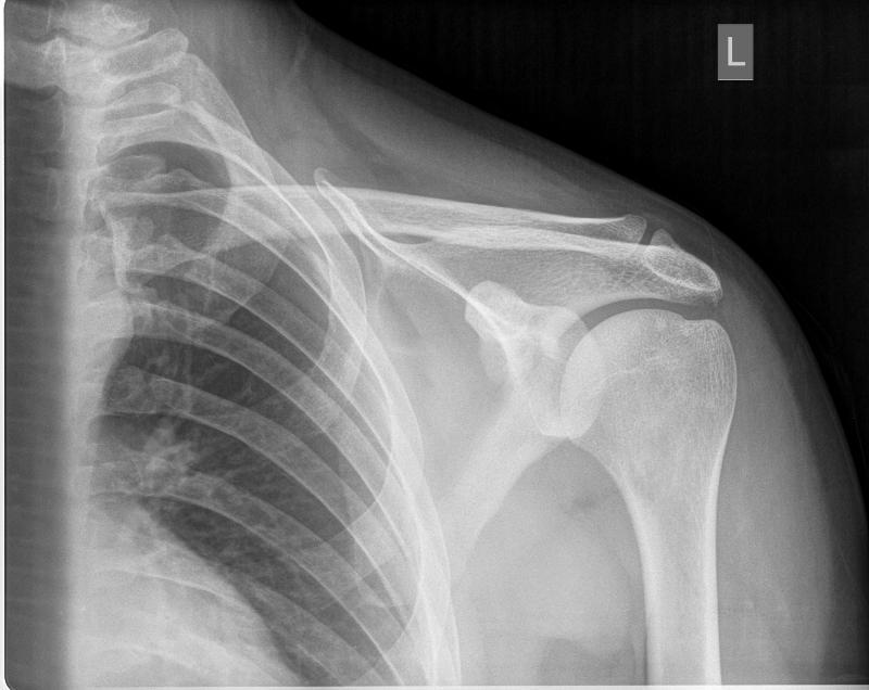
6. What shoulder exam is this and how can you tell
AP external rotation and you can see the greater tubercle
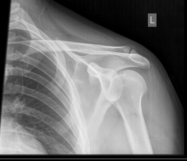
7. What view is this and how can you tell?
Internal rotation and the lesser tubercle is medial.

8. What is number 3 labeling
Acromion of the scapula
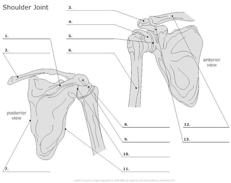
9. What is number 12 labeling
Body of clavicle
10. The three corners of the scapula are called angles, what are they?
Lateral, superior, inferior angles
11, The humeral head articulates with the glenoid cavity of the scapula to form what joint?
scapulohumeral joint
12. The three shoulder girdle joints are classified as what type of joints
synovial joint
13. What rotation is true AP projection
External rotation
14.The AP projection of the shoulder taken in the internal rotation is what position for the humerus
lateral position of the humerus
15. Shoulder and humerus kV range is?
70 - 80 with grid
65 - 70 without grid
16. Nuclear medicine bone scans are useful in demonstrating ____?
osteomyelitis, metastatic bone lesions, and cellulitis
17. Sonography is often used to check for ____ on the shoulder?
rotator cuff tears, bursa injuries, disruption and damage of the nerves
18. What clinical indication refers to an injury in which the distal clavicle usually is displaced superiorly. This is commonly caused by a fall.
Acromioclavicular dislocation
19. What clinical indication refers to trauma to the upper shoulder region resulting in a partial or complete tear of the AC or coracoclavicular ligament or both ligaments.
AC joint seperation
20. What clinical indication is an injury of the anterioinferior aspect of the glenoid labrum. This type of injury often is caused by anterior dislocation of the proximal humerus. Repeated dislocation may result in a small avulsion fracture in the anteriorinferior region of the glenoid rim.
Bankart lesion
21. What clinical indication is inflammation of the bursea, or fluid filled sacs enclosing the joints. It generally invloves the formation of calcification in associated tendons, causing pain and limitation of joint movement.
Bursitis
22. What clinical indication is a compression fracture of the articular surface of the posterolateral aspect of the humeral head that often is associated with an anterior dislocation of the humeral head.
Hill-Sachs defect
23. What clinical indication is a disability of the shoulder joint that is caused by chronic inflammation in and around the joint. It is characterised by pain and limitation of motion,
Idiopathic chronic adhesive capsulitis
24. What clinical indication is impingement of the greater tuberosity and soft tissues on the coracoacromial ligamentous and osseous arch, generally during abduction of the arm.
Impingement syndrome
25. What clinical indication is a noninflammatory joint disease characterized by gradual deterioration of the articular cartilage with hypertrophic bone formation? Usually happens in people over 50.
Osteoathritis
26. What clinical indication result in fractures due to a reduction in the quantity of bone or atrophy of skeletal tissue?
Osteoporosis
27. AP projection of the humerus CR?
Perpendicular to IR, directed to midpoint of humerus.
28. What landmarks can you see on a AP projection of the humerus
Acromion of scapula, Greater tubercle, Head of humerus, Lesser tubercle, Coracoid process of scapula, scapulohumeral joint and proximal humerus.
29. The use of a grid is not required for shoulder studies that measure less than 10 cm. true or false
true
30. The kV range for adult shoulder projections is between 80 and 90 kV for analog and 100 to 110 kV for digital systems. True or false
false
31. Low mA with short exposure time should be used for adult shoulder studies. true or false
false
31. Large focal spot setting should be selected for most adult shoulder studies. true or false
false
33. A high speed screen - IR system is recommended for analog shoulder studies when using a grid. true or false
true
34. A 72 inch SID is recommended for most shoulder girdle studies. true or false
false
35. CT arthography of the shoulder joint often requires the use of iodinated contrast media injected into the joint space. true or false
true
36. MRI is an excellent modality for demonstrating bony injuries of the shoulder girdle. true or false
false
37. How much is the CR angled for the inferosuperior axial projection (clements modification) if the patient cannot fully abduct the arm 90 degrees?
25 to 30
38. Which of nontrauma projections can be performed erect to provide a lateral view of the proximal humerus in relationship to the glenohumeral joint.
PA transaxillary projection (Hobbs modificaiton)
39. Which projections produces a tangential of the intertubercular groove?
Fisk modification
40. To best demonstrate a possible Hill - sachs defect, what additional positioning technique can be added to the inferosuperior axial projection?
Rotate affected arm externally appoximately 45 degree