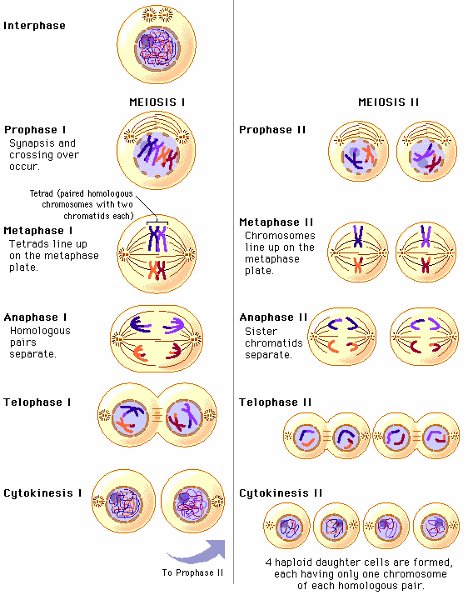
What’s the difference between mitosis and meiosis?
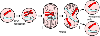
In mitosis two identical sets of daughter nuclei, -46
Meiosis number of chromosomes is divided into half- 23
What are the steps in oocyte production?
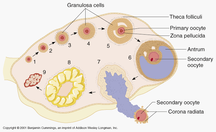
germ cells produce -->> primordial germ cell (PGC),-->>> mitosis, forming oogonia. oogenesis, the oogonia -->> primary oocytes.--->> SECONDARY
What are the steps in spermatocyte production?
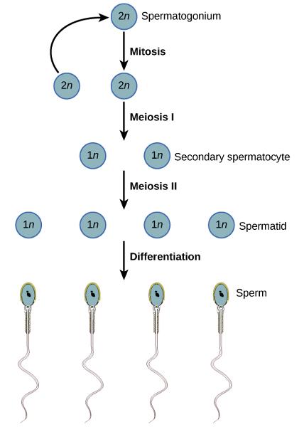
interphase before meiosis I,
synapsis before the first meiotic division.
meiosis II, the two daughter cells go through a second division to yield four cells containing a unique set of 23 single chromosomes into four sperm cells.
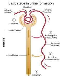
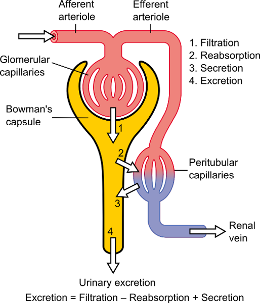
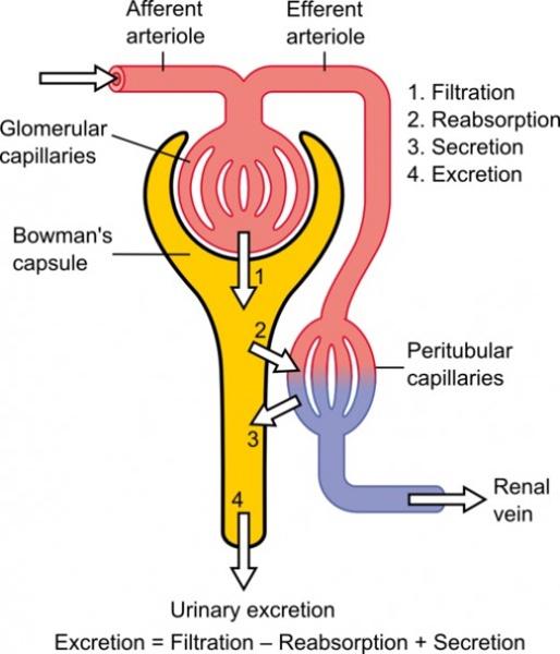
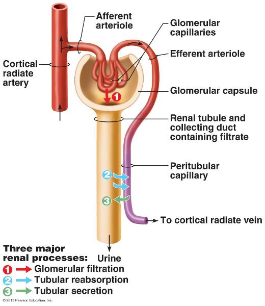
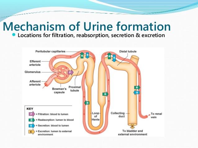
What are the three steps of urine formation and what is excreted and reabsorbed?

Filtration
As blood courses through the
glomeruli, soaks out of the blood through the membranes (by osmosis and
diffusion) filtered flows into the Bowman's capsule.
Reabsorption
( peritubular
copillaries). reabsorbed are water,
glucose and other nutrients, and sodium (Na+) ions
Secretion
secretion is reabsorption
in reverse. secretion moves substances out of
the blood and into the tubules converted into urine.
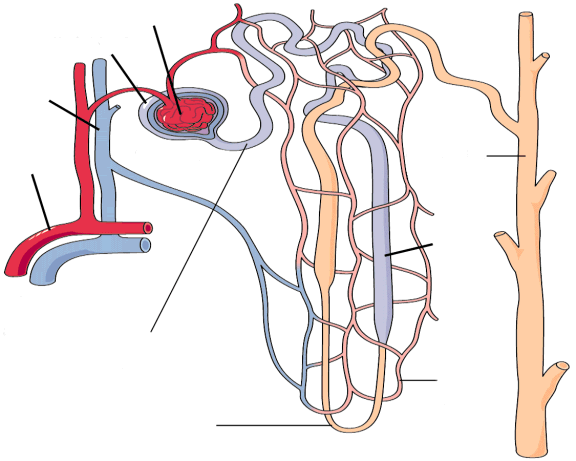
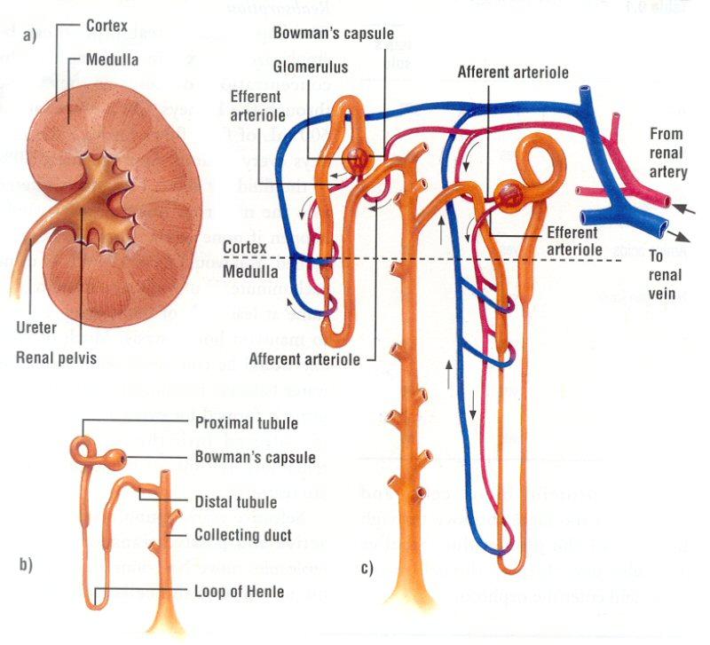
Trace the flow of blood and the flow of filtrate/urine through the kidney.
1-Glomerulus and
Bowman's capsule:
2-Proximal
tubule:
3-Descending
limb of loop of henle:
4-Ascending limb of
loop of henle:
5-Distal
tubule: blood into nephron
6-Collecting Duct:
What ions are predominant in extracellular fluid vs. intracellular fluid?
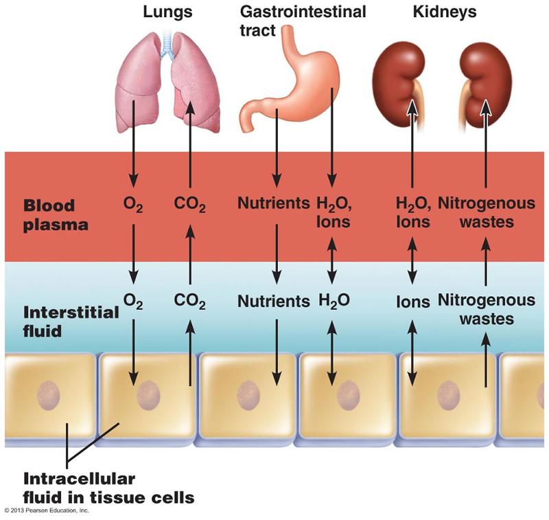
Extracellular fluids sodium, calcium, chloride and bicarbonate ions
Intracellular fluids potassium, magnesium,
phosphate, and sulfate ions
What are the different causes of Respiratory acidosis and alkalosis?
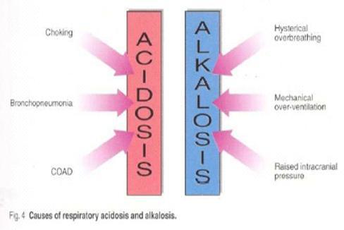
alkalosis pH above
acidosis pH below
Respiratory alkalosis isn’t enough carbon diox ide
- hyperventilation, which commonly occurs with anxiety
Respiratory Acidosis too much CO2
- chronic airway conditions, like asthma
- injury to the chest
What is normal body pH range?
7.35 to 7.45
What causes the flow of filtrate out of the glomeruli?
HYDROSTATIC PRESSURE
What is Boyle’s law?
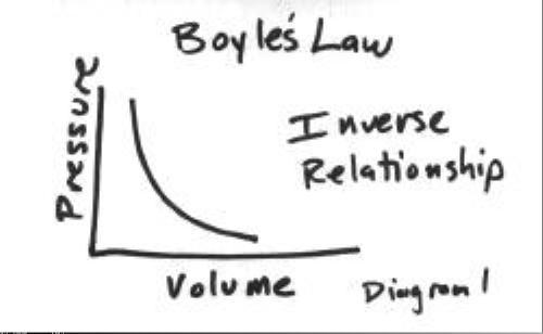
INVERSELY RELATED TO PRESSURE
What is Frank-Starling law?
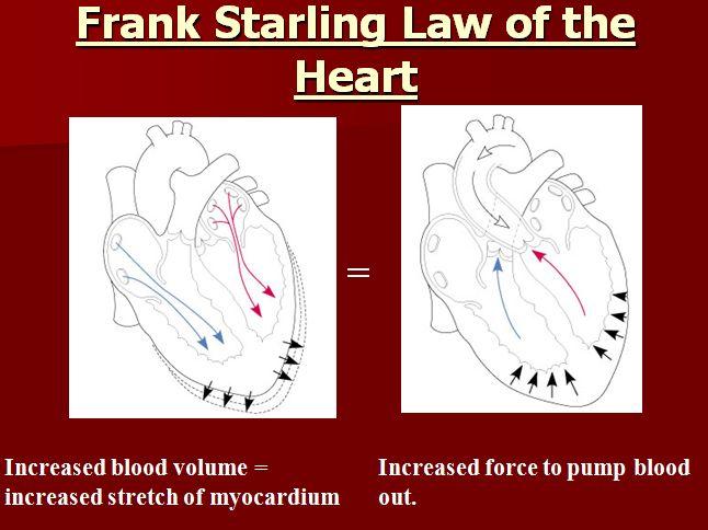
EQUAL IN / EQUAL OUT
What is Dalton’s law and how does it apply to respiration?
Total pressure=partial pressure gases
Production, activation and function of digestive enzymes and hormones including
gastrin, pepsinogen, pepsin, cholecystokinin, trypsin, trypsinogen, chymotrypsin, amylase.
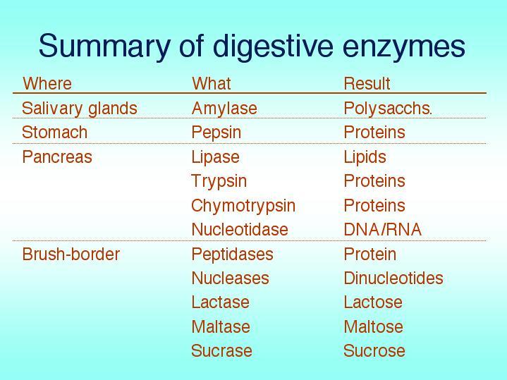
AMYLASE - a carbohydrase - an enzyme that breaks down starch into glucose
gastrin
is secreted by the stomach cells to regulate the production of gastric
juices.
PEPSIN Pepsin is an enzyme used to partially hydrolyze protein. Pepsin is released in an inactive form Pepsinogen.
pepsinogen reacts with HCl to form pepsin
cholecystokinin -a polypeptide hormone secreted in the small intestine,
stimulates gallbladder contraction and secretion of pancreatic enzymes.
Trypsin and Chymotrypsin are enzymes that break bonds next to specific amino acids
breaks proteins into amino acids in the DUODENUM
Summarize the correct sequence from the formation of a drop of urine to the elimination from the body.

Formation of urine Blood leaves the heart via the aorta and enters the renal artery where it flows into the interlobar arteries. From there it branches off to the arcuate artery, which curves along the outer edge of the pyramids. From the arcuate artery, blood flows to the cortical radiate arteries then into the afferent arteriole. From the afferent arteriole, it goes into the : glomerulus for filtering.
ADH- DCT DISTAL CONVELATED TUBE
all waste goes to the proximal convoluted tubule, while blood that will stay in the body goes to the efferent arteriole. More blood filtering takes place in the peritubular capillaries. Blood that will stay in the body exits through the cortical radiate veins, then goes into the arcuate vein, then into the interlobar vein, then the renal vein, then back to the heart via the inferior vena cava.
At the same time this is happening, the waste that was sent to the proximal convoluted tubule travels down the descending loop of henle, then up the ascending loop of henle to the distal convoluted tubule where it is dumped into the collecting duct. From the collecting duct, it goes into the ureters, travels to the bladder, and then leaves the body via the urethra.
What is aldosterone?
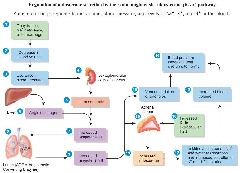
A corticosteroid hormone that stimulates absorption of sodium by the kidneys and so regulates water and salt balance (LH/ FSH)
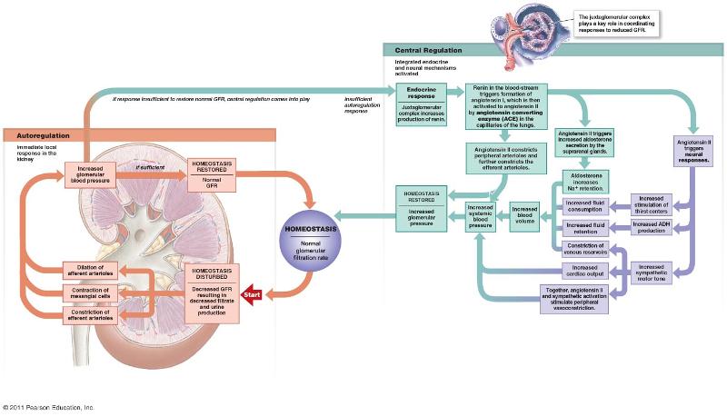
What is renin?
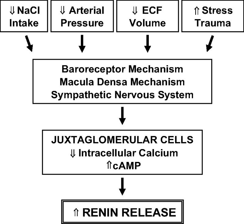
An enzyme secreted by and stored in the kidneys that promotes the production of the protein angiotensin (juxtaglomerular apparatus)
WHAT IS ANP ?
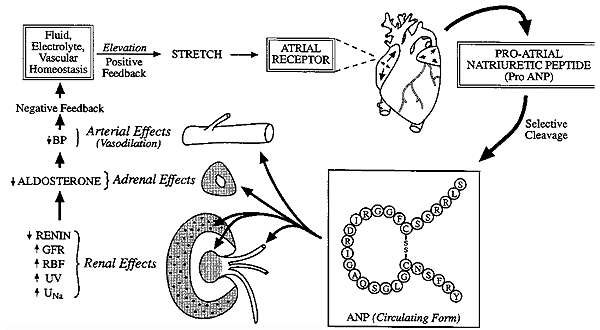
Atrial natriuretic peptide (ANP), (polypeptide) ANP acts to reduce the water, sodium and adipose loads on the circulatory system, thereby reducing blood pressure.
What is ADH?
water regulator in the body.
What are 2 ways you could check someone’s renal health?
Albuminuria-to-creatinine ratio (ACR).
Glomerular filtration rate (GFR)
If you wanted to create a more concentrated urine, what would your body do?
ADH causes the DCT and collecting ducts to be more permeable to water
What are the effects of angiotensin II?
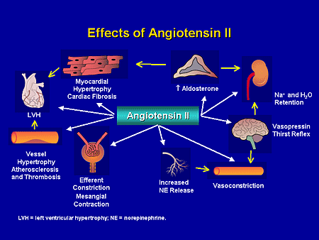
1. Constricts arteriolar smooth muscle, causing map to rise
2. stimulates reabsorption of Na+
Triggers adrenal cortex to release aldosterone
Aldosterone increases na+ reabsorption
3. Stimulates hypothalamus to release antidiuretic hormone (ADH) and activates thirst center ADH - causes water to be retained
4. Constricts efferent arterioles, decreasing capillar hydrostatic pressure and increasing fluid reabsorption
5. Causes glomerular cells to contract, decreasing surface area available for filtration
Explain the renin-angiotensin-aldosterone mechanism. What is its goal?

is a hormone system regulating blood pressure (BP) and fluid volume, a third major participant in this system called aldosterone. The level of activity of the renin-angiotensin system determines and is determined by the body’s BP
In the loop of Henle, how does the descending & ascending loop differ in their permeability to water & sodium?

descending (NO SALT): impermeable to Na+, but permeable to water/ h2o reabsorbed
ascending (NO H20) - permeable to na+ but impermeable to water : na+, k+, Cl-
What substances move through passive reabsorption?
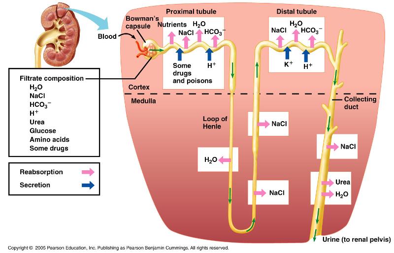
-H20 is highly permeable
-NaCl is highly permeable
-Glucose is highly permeable
In tubular reabsorption, substances are returned to the __________ from the ____________.
Distal convoluted tube
Proximal convoluted tubule (pct)
Why is pressure important in glomerular filtration?
The pressure helps to force liquid out of the blood.
What are the 3 steps in forming urine? Where does each occur?
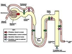
1. Glomerular filtration
2. tubular reabsorption : primarily in the proximal tubule
3. tubular secretion : distal tubules
What are the collecting ducts?
receive filtrate from many nephrons
principle cells of collecting ducts - help maintain water and salt balance
Name/describe the parts and processes of a nephron.

Glomerulus - tufts of capillaries; filtration
Bowman's capsule - enlarged, cup-shaped capsule surrounding glomerulus - collects filtrate
Proximal convoluted tubule (pct) - tubular reabsorption
Loop of henle - Sodium (Na+) and water balance
Distal convoluted tubule (DCT) - tubular secretion
What are mesangial cells?
Immunoreactive transformed smooth muscle cells that can contract in response to circulating vasoactive substances impeding glomerular blood flow and filtration.
What are the functions of the cells of JGA?
Granular cells (renin)
macula densa (monitor flow rate of filtrate.)
What are the layers of the glomerular filtration barrier?
Leaky endothelium (pores/fenestrae)
basement membrane (porous matrix of negatively charged glycoproteins)
podocytes (specialized epithelial cells with interdigitating pedicels separated by filtration slits).
What substances filter easily?
What substances don't filter?
What substances filter moderately?
Water, urea, glucose and inulin.
Albumin and hemoglobin.
Myoglobin.
The electrolytes of greatest importance to
sodium, potassium, calcium, magnesium, chloride, sulfate, phosphate, bicarbonate, and hydrogen ions
Posterior pituitary hormones and effect on the kidneys.

ADH antidiuretic hormone, which helps control body water balance through its effect on the kidneys and urine output
oxytocin, which triggers the contractions of the uterus that occur during labor.
Anterior pituitary hormones and effect on reproductive organs.
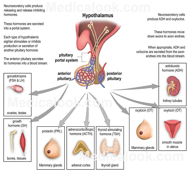
growth hormone, which stimulates the growth of bone and other body tissues and plays a role in the body's handling of nutrients and minerals
prolactin, which activates milk production in women who are breastfeeding
thyrotropin, which stimulates the thyroid gland to produce thyroid hormones
corticotropin, which stimulates the adrenal gland to produce certain hormones
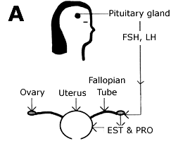
hormones that signal the ovaries and testes to make sex hormones. The pituitary gland also controls ovulation and the menstrual cycle in women.
How do the kidneys control blood pressure and pH?
buffers dissolved in the blood. remove excess chemicals remove H+ ions
How do the lungs control pH?
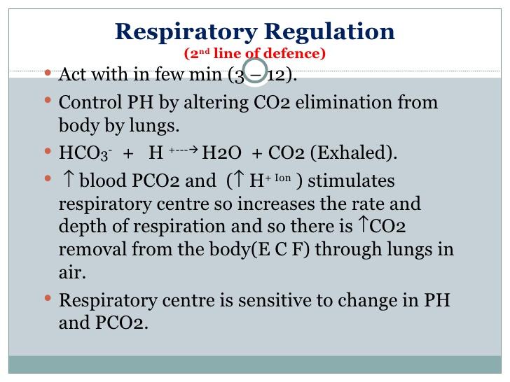
increased-breathing counteract the pH-lowering effects of exercise by removing CO 2, & pH buffer
Process of urine formation and micturition
- Urine is made in the kidneys
- Urine is stored in the bladder
- The sphincter muscles relax
- The bladder muscle (detrusor) contracts
- The bladder is emptied through the urethra and urine is removed from the body
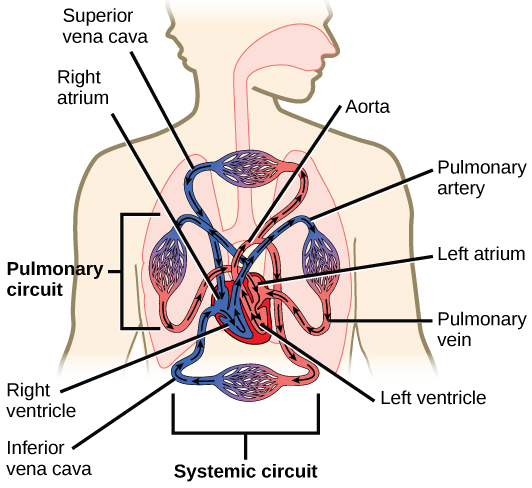
Flow of blood through the veins

superior and inferior vena cava, -->>>right atrium-->> tricuspid valve --> right ventricle -->>pulmonic valve-->> pulmonary artery-->> lungs
-->> pulmonary veins -->> left atrium -->>> mitral valve-->> left ventricle -->> aortic valve -->>> aorta-->>>> body
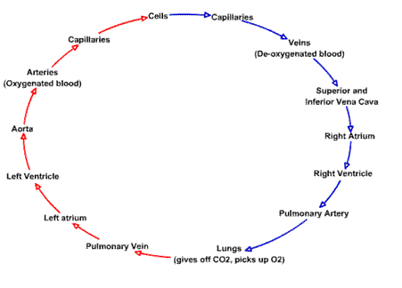
Flow of blood through the arteries
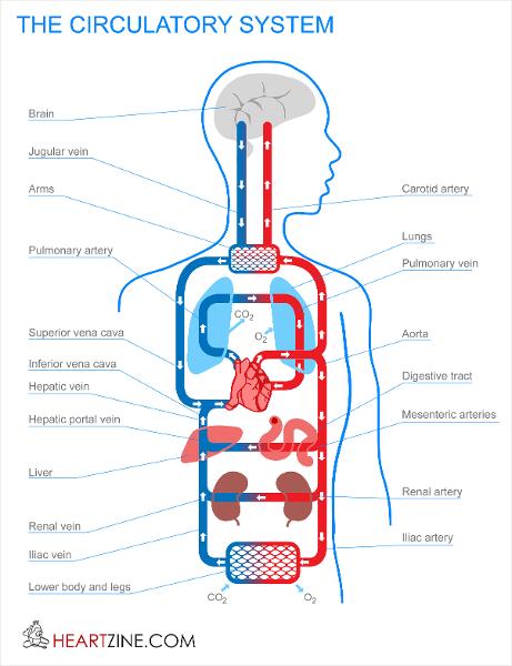
body >> vena cava >> right atrium of the heart -->> right atrium contracts -->>pumps the blood >>> tricuspid valve >>> right ventricle.>> pulmonary artery >>> lungs -->> tiny blood vessels called capillaries ( absorb carbon dioxide from the blood and replace it with oxygen) >>> pulmonary vein >>> left atrium>> mitral valve >>>> the left ventricle -->> left side of the heart >>>>> left ventricle >>>>> aortic arch -->> body -->> carotid artery and into the brain -->> auxiliary arteries -->> arms through the aorta -->> torso and legs -->> Blood >> arteries >> capillaries >>>> veins -->> >>>>> heart.
Define Primary oocyte (egg), secondary oocyte
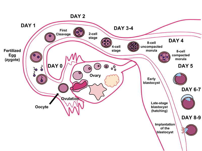
oocyte is produced in the ovary during female gametogenesis and the first meiotic division is completed.
The second meiotic division usually stops short of completion unless fertilization occurs( secondary)
Why can “tighty-whiteys” cause infertility?
temperature of the testes is at issue : HEAT
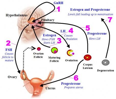
Effects of estrogen and progesterone on the menstrual cycle.

ESTROGEN
creates proliferative
endometrium
necessary for proper ovulation
decreases sex drive
PROGESTERONE
maintains secretory
endometrium
necessary for survival of embryo
restores sex
drive
precursor of corticosteroids (cortisol)
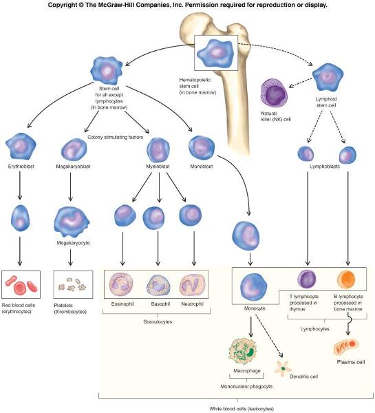
Function of T cells and B cells
T cells are involved in cell-mediated immunity
B cells are primarily responsible for humoral immunity (antibodies).
function of T cells and B cells is to recognize specific “non-self” antigens, during a process known as antigen presentation.
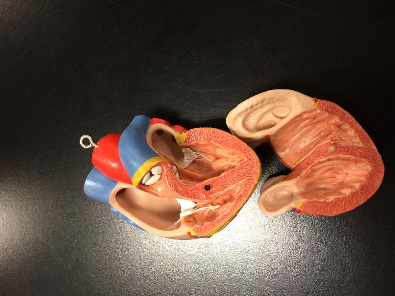
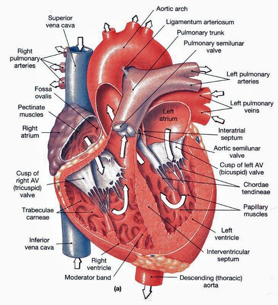
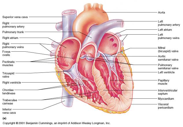
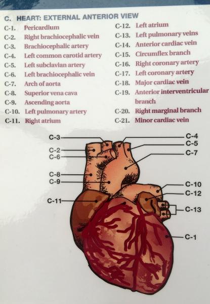
Flow of blood through the heart and all heart parts
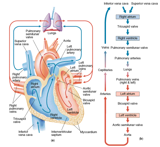
1. superior/inferior vena cava
2. right atrium
3. tricuspid valve
4. right ventricle
5. pulmonary valve
6. pulmonary artery
7. lungs
8. pulmonary veins
9. left atrium
10. mitral valve
11. left ventricle
12. aortic valve
13. aorta
14. body
What do the ductus deferens and the esophagus have in common?
peristalsis
Where is the egg fertilized?

FALLOPIAN TUBES
What hormones are in “the pill” and why?
estrogen and progestin
Functions of the liver
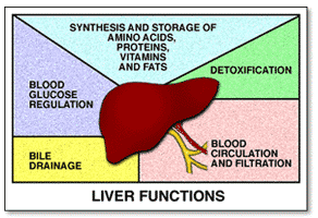
Bile production and excretion
Excretion of bilirubin, cholesterol, hormones, and drugs
Metabolism of fats, proteins, and carbohydrates
Enzyme activation
Storage of glycogen, vitamins, and minerals
Synthesis of plasma proteins, such as albumin, and
clotting factors
Blood detoxification and purification
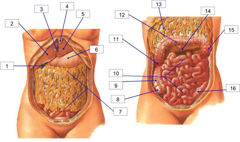
Identify all organs of the digestive system
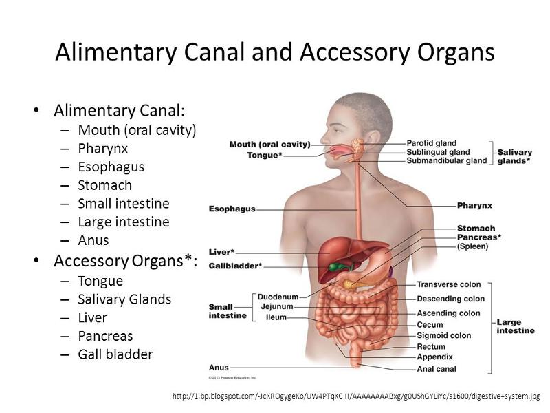
teeth, tongue, pharynx, stomach, gallbladder, liver, small intestine, colon, and pancreas.

Identify all organs of the reproductive system
Female: ovaries, uterus, and vagina.
Male: testes and penis.
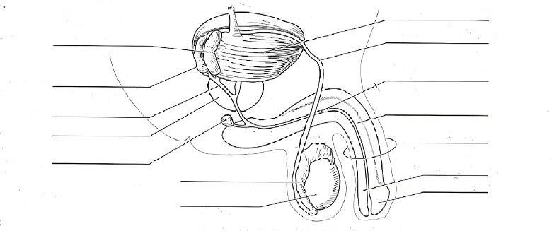
Identify all organs of the reproductive system MALE
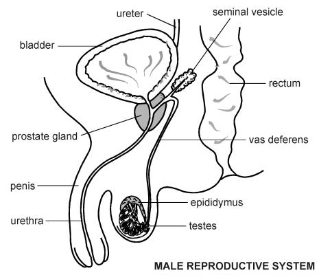
The male internal accessory organs
include:
Epididymides
Ductus
deferentia
Seminal vesicles
Prostate gland
Bulbourethral glands
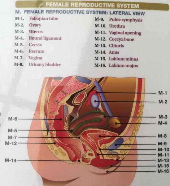
Identify all organs of the reproductive system FEMALE
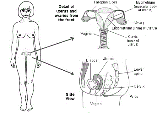
- Fallopian tube
- Ovary
- Uterus
- Cervix
- Vagina
- Clitoris
- Labia minora
- Labia majora.
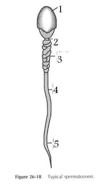
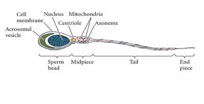
Structure of sperm
Sperm have three parts: a head chromatin; a midpiece filled with mitochondria to provide energy; and a flageullum or tail to move the sperm
Summarize the correct sequence from the formation of a drop of
urine to the elimination from the body(Essay)
Blood leaves the heart via the aorta and enters the renal artery where it flows into the interlobar arteries. From there it branches off to the arcuate artery, blood flows to the cortical radiate arteries then into the afferent arteriole. From the afferent arteriole, it goes into the glomerulus for filtering.
all waste goes to the proximal convoluted tubule, while blood that will stay in the body goes to the efferent arteriole. More blood filtering takes place in the peritubular capillaries. Blood that will stay in the body exits through the cortical radiate veins, then goes into the arcuate vein, then into the interlobar vein, then the renal vein, then back to the heart via the inferior vena cava.
At the same time this is happening, the waste that was sent to the proximal convoluted tubule travels down the descending loop of henle, then up the ascending loop of henle to the distal convoluted tubule where it is dumped into the collecting duct. From the collecting duct >>>, it goes into the ureters>>>>, travels to the bladder>>>>>, and then leaves the body via the urethra.
The dartos and cremaster muscles are important to the integrity of the male reproductive system. Which of the following is true about the role they play?
They regulate the temperature of the testes.
The ability of sperm cells to move along the ductus deferens is due to ________.
peristaltic contractions
female sex hormones
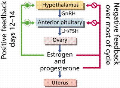
Hypothalamus →GnRH→Pituitary →FSH →Follicle →Estrogens
The ability of a male to ejaculate is due to the action of ________.
the bulbospongiosus muscles
Which of the gland are responsible for 60% of the synthesis of semen?
the seminal vesicles
Which of the following hormones controls the release of anterior pituitary gonadotropins?
GnRH
Uterine wall - has three layers
- Perimetrium - is a thin covering on the outside of the uterus. It is actually part of the peritoneum. OUTSIDE
- Myometrium - consists of three layers of smooth muscle. longitudinal, circular, and spiral. CONTRATIONS IN PREGNANCY
- Endometrium - is the inner mucosal lining. it consists of two layers:sheds at period
- Stratum functionale - contains secretory glands. This is the portion that is shed during mensus.
- Stratum basale - is a highly vascularized layer which serves to regenerate the stratum functionale.
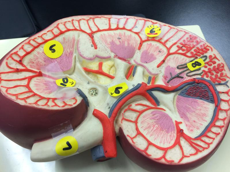
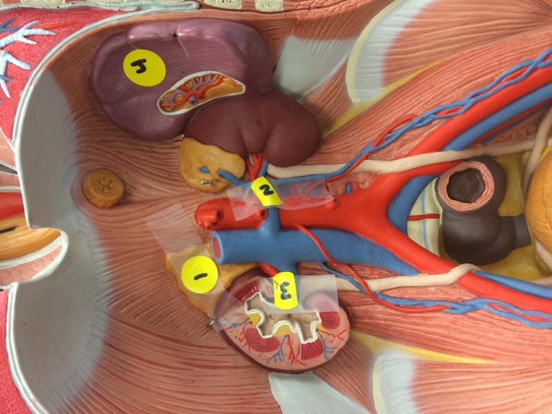
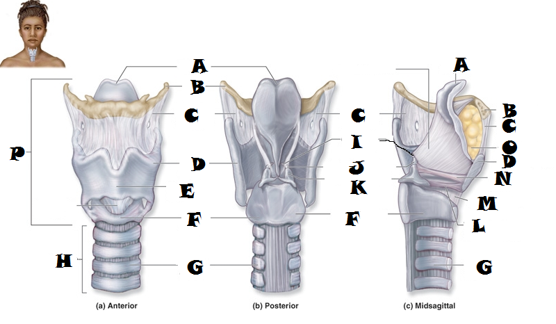
KIDNEY
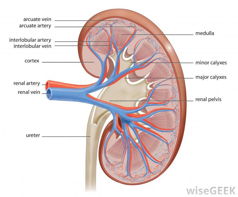
renal pyramid
renal corpuscle
RENAL ARTERY
ureter
nephron
major calyx
minor calyx
SEGMENTAL ARTERY
INTERLOBAR ARTERY
CORNICAL RADIATE
ARQUATE
POSTERIOR (BACK SIDE)
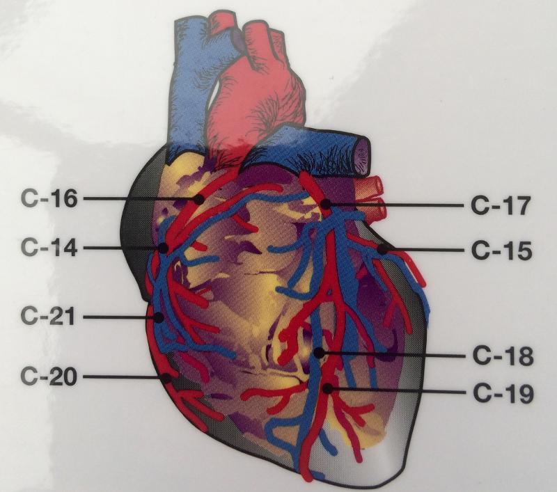
CORONARY SINUS- BIG FAT BACK
GREAT CARDIAC VEIN
INFERIOR VENA CAVA
L/R ORICAL
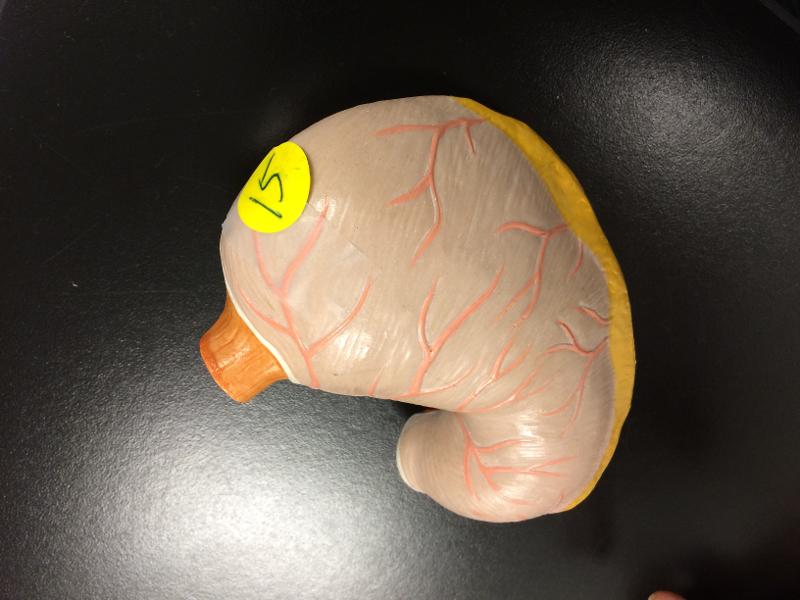
Stomach
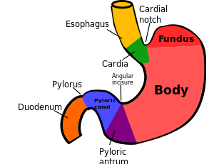
Top is fundus
layers of gastrointestinal tract, the stomach walls consist of an outer mucosa, and inner submucosa, muscularis externa, and serosa.
CARDIAC
PYLORIC REGION- PYLORIC CANAL
DUODEUOM
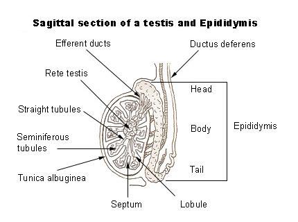
EPIPIMATES
Tightly coiled tubes
Connected to ducts within the testis
Promote maturation of sperm cells
Development of male reproductive structures depends on which of the following events?
secretion of male hormones prenatally and lasting into the first few months after birth
The primary function of the uterus is to ________.
receive, retain, and nourish a fertilized ovum
Why is the blood-testis barrier important?
cells produce surface antigens that are recognized as foreign by the immune system
The structures that receive the ovulated oocyte, providing a site for fertilization, are called the ________.
fallopian tubes
If gametes were diploid like somatic cells, how many chromosomes would the zygote contain?
twice the diploid number, and with every succeeding generation, the chromosome number would continue to double and normal development could not occur
Human egg and sperm are similar in that ________.
they have the same number of chromosomes
The constancy of the chromosome number from one cell generation to the next is maintained through ________.
meiosis
Spermiogenesis involves the ________.
formation of a functional sperm by the stripping away of superfluous cytoplasm
Which is not a part of the proliferative phase of the female menstrual cycle?
corpus luteum
What is the difference between metabolic alkalosis vs. acidosis?
Metabolic alkalosis develops when your body loses too much acid or gains too much base. This can be attributed to:
- excess vomiting, which causes a loss of electrolytes
- Gastric drainage, vomiting...loss of acids
Metabolic acidosis starts in the kidneys instead of the lungs. It occurs when they can’t eliminate enough acid or when they get rid of too much base. There are three major forms of metabolic acidosis:
- Diabetic acidosis occurs in people with diabetes
- Hyperchloremic acidosis results from a loss of sodium bicarbonate. This base helps to keep the blood neutral. Both diarrhea and vomiting can cause this type of acidosis.
- Lactic acidosis occurs when there’s too much lactic acid in your body.
- Kidney failure, excessive acidic ketones ( diabetes mellitus), prolonged diarrhea, prolonged vomiting...loss of bases
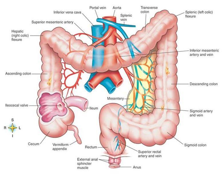
taenia coli Any of the three bands in which the longitudinal muscular fibers of the large intestine
sigmoid
epiploic appendices / omental appendices are small pouches of the peritoneum filled with fat and situated along the colon, but are absent in the rectum.
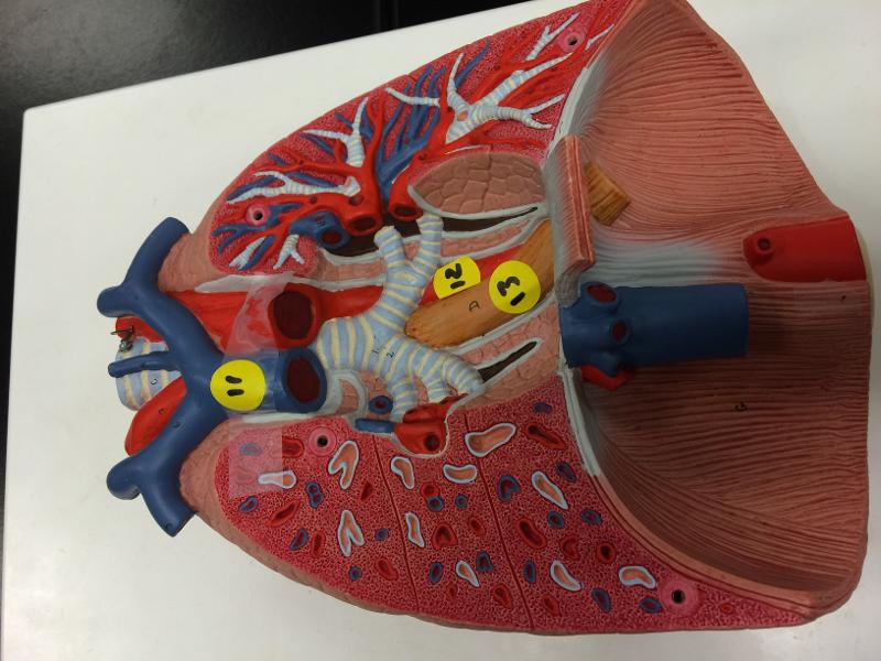
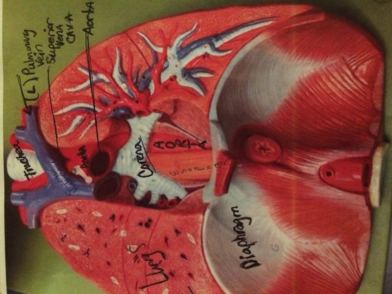
TRACHEA - L /R BRONCHI
AORTA
INFERIOR VENA CAVA
THYROID
ESOPHAGAS
SPLEEN
COMMON ILIAC- DIVISION
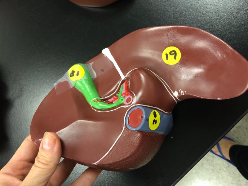
labels on liver
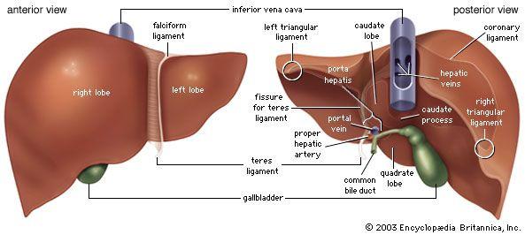
gallbladder
right lobe
inferior vena cava
hepatic portal

FRONTAL HEART

Endocardium - heart valves
Myocardium - Conduction system
- SA node >> AV node >>> bundle of Hise >> bundle branches >> Purkinje fibers
Pericardial cavity - Pericardial sinus
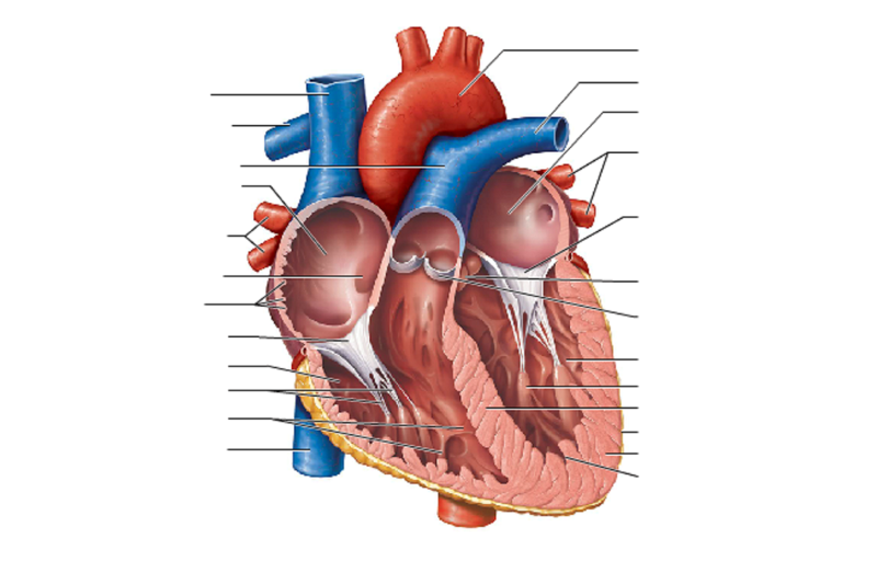
FRONTAL VIEW OF HEART
Vena Cava
Carries deoxygenated blood
from the body to the heart
Semilunar Valve
Flaps that prevent
backflow of blood
Left Atrium
Receives oxygenated
blood from the lungs
Left
Ventricle
Region of the heart that pumps
blood to the body
Pulmonary Artery
Carries blood to
the lungs
Right Ventricle
Region of the heart
that pumps blood to the lungs
Pulmonary Vein
Carries blood from
the lungs
Right Atrium
Segment of the heart
that receives deoxygenated blood
Aorta/ AORTIC ARCH
The main artery carrying blood to all parts of
the body
chordae tendineae : heart strings, are cord-like tendons that connect the papillary muscles to the tricuspid valve and the mitral valve
PAPIL
respiratory
ESOPHAGAS
L/R BRONCHI
TRACHEA
PULMONARY TRUNK
AORTIC ARCH
INFERIOR VENA CAVA
AORTA
DIAPHRAGM
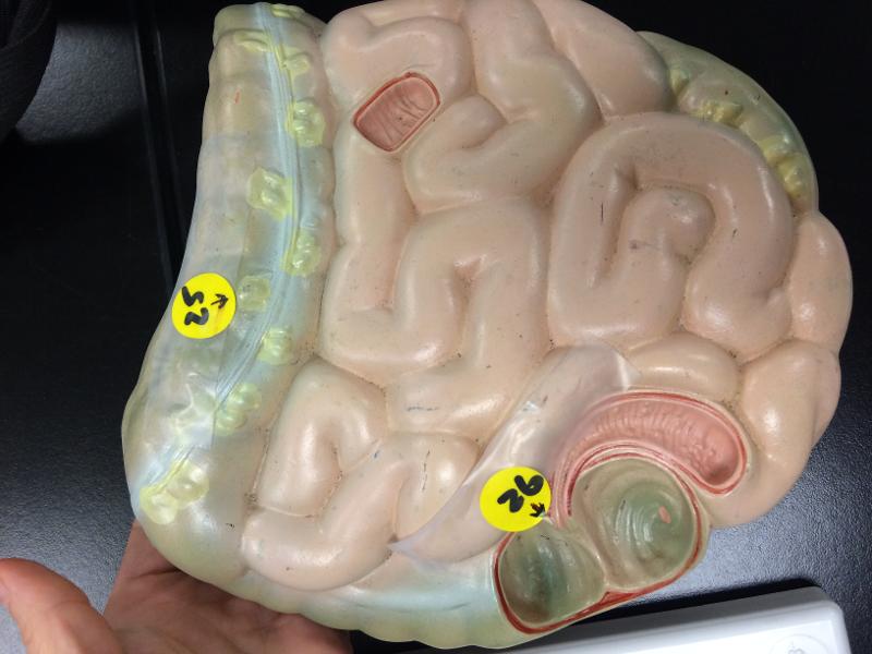
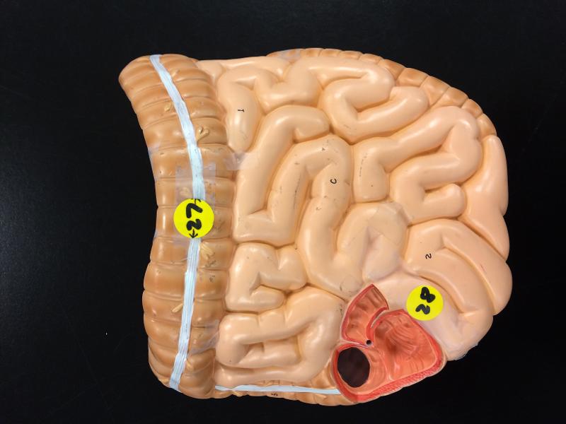
lower digestive/ small ; large intestine( front only )
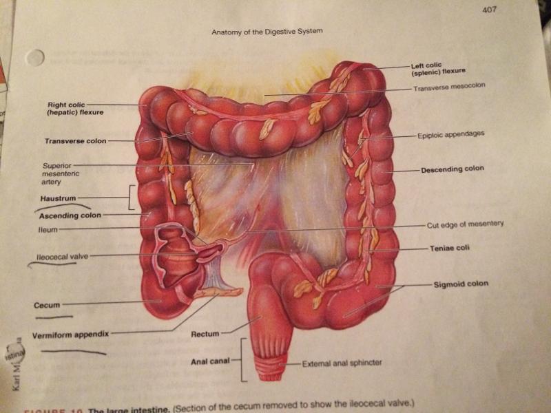
- appendix
- ascending colon
- transverse colon
- The descending colon
- The sigmoid colon – the v-shaped region of the large intestine Cecum – the first part of the large intestine Taeniae coli – three bands of smooth muscle Haustra – bulges caused by contraction of taeniae coli Epiploic appendages – small fat accumulations on the viscera
Ovarian hormones and cycle (essay)
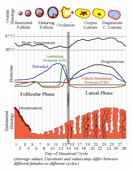
follice develops day 14 - ovulation (estrogen peaks)
day 12-14 estrogen drops - more frisky (surge)
day 20-21 progesterone goes up - mucus thickens - peaks
estrogen peaks again
pms estrogen and progesterone drop : repeat 28 days
FINAL product of oogenesis is called ______. (HINT: this is the ovulated secondary oocyte)
ovum
What is one of the hormones responsible for maintenance of corpus luteum after ferilization?
human chorionic gonadotropin (hCG)
Cortex of an ovary contains (4 things)
primordial follicles
developing follicles
corpus luteum (-->corpus albicans)
stromal cells
ovaries are supported by ______
1. Mesovarium (attaches to ovary to POSTERIOR surface of
broad ligaments)
2. A pair of supporting ligaments (ovarian ligaments
& suspension ligaments)
During the secretory phase of the menstrual cycle ________.
progesterone levels are at their highest
3 Major fx of ovary?
(1) synthesis & secretion of sex
hormones
(2) release a secondary oocyte every 28
days
(3) Secrete inhibin (which is involved in the negative
feedback ctrl of pituitary FSH production)
This membrane will eventually surround the oocyte
Zona pellucida
FINAL product of oogenesis is called ______. (HINT: this is the ovulated secondary oocyte)
ovum
United Secondary oocytes after the completion of meiosis II is called ____.
zygote
Select the correct statement about the uterine cycle.
A) The menstrual phase of the cycle is from day 1 to day 8.
B) During the secretory phase, estrogen
levels are at their highest.
C) During the
proliferative phase, levels of progesterone rise as the follicle
begins to produce more hormone.
D) If
fertilization occurs, the corpus luteum is maintained by a hormone
secreted by the developing embryo.
If fertilization occurs, the corpus luteum is maintained by a hormone secreted by the developing embryo.
Which of the choices below is not a part of the brain-testicular axis?
thalamus
Which of the following statements is true concerning the mammary glands of both males and females?
A) Both sexes are equally prone to
breast cancer.
B) All lumps identified in breast tissue are
malignant.
C) The only time hormones target breast tissue is
during pregnancy and lactation.
D) The mammary glands are
modified sweat glands that are actually part of the integumentary system.
The mammary glands are modified sweat glands that are actually part of the integumentary system.
The basic difference between spermatogenesis and oogenesis is that ________.
A) during
spermatogenesis two more polar bodies are produced
B) the
mature ovum is n, while the sperm is 2n
C) in oogenesis, one
mature ovum is produced, and in spermatogenesis four mature sperm are
produced from the parent cell
D) spermatogenesis involves
mitosis and meiosis, but oogenesis involves meiosis only
in oogenesis, one mature ovum is produced, and in spermatogenesis four mature sperm are produced from the parent cell
Occasionally three polar bodies are found clinging to the mature ovum. One came from an unequal division of the ovum, but from where did the other two arise?
The first polar body has also divided to produce two polar bodies.
Why doesn’t semen enter the urinary bladder during ejaculation?
The smooth muscle sphincter at the base of the urinary bladder closes.
Spermatogenesis ________.
involves a kind of cell division limited to the gametes
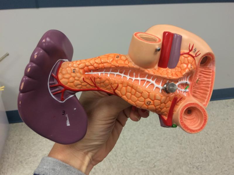
Combo pancreas / spleen
spleen - hepatic portal
pancreas leading to small intestine @ dueodium
mouth / upper head glands
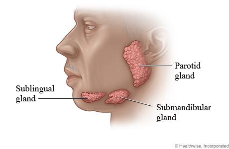
sublingual
submandibular
parotoid
ANTERIOR CAVITY
BLADDER
CURVED SIGMIOD
COMMON ILIAC ARTERY
INFERIOR VENA CAVA
AORTA
ADRENAL GLANDS
KIDNEY
SPLEEN
DIAPHRAGM
ESOPHAGUS
THYROID
Gland at neck
Thyroid
ARTERYS SUPERIOR LIMB
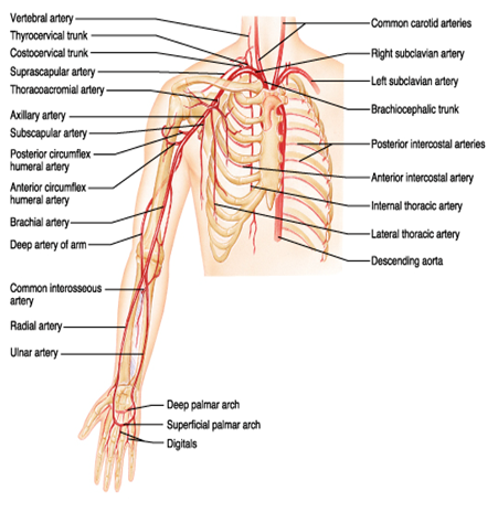
SUPERIOR LIMB- ARM
COMMON CAROTID-> SUBCLAVICAN-> AXILLARY->BRACHIAL-> DEEP ARTERY OF ARM-> RADIAL/ULNAR-> DEEP PALMAR-> SUPERFICIAL PALMAR->DIGITALS
VEINS SUPERIOR LIMB
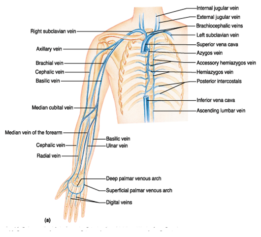
1. Palmar digital vein
2. Palmar metacarpal vein
3. Palmar venous arch vein
4. Radial and ulnar vein
5. Brachial vein
6. Axillary vein
7. Subclavian vein
8. Brachiocephalic vein
9. Superior vena cava
10. Right atrium
11. Tricuspid valve
12. Right ventricle
13. Pulmonary semilunar valve
14. Pulmonary trunk
15. Pulmonary artery
16. Lungs
17. Pulmonary veins
18. Left atrium
19. Mitral valve / bicuspid valve / Left AV Valve
20. Left ventricle
21. Aortic semilunar valve
22. Ascending aorta
23. Passes right and left coronary arteries
24. Aortic arch
25. Brachiocephalic trunk
26. Passes opening of the right common carotid artery
27. Right subclavian
28. Axillary artery
29. Brachial artery
30. Ulnar and radial artery
31. Palmar arch artery
32. Palmar metacarpal artery
33. Palmar digital vein
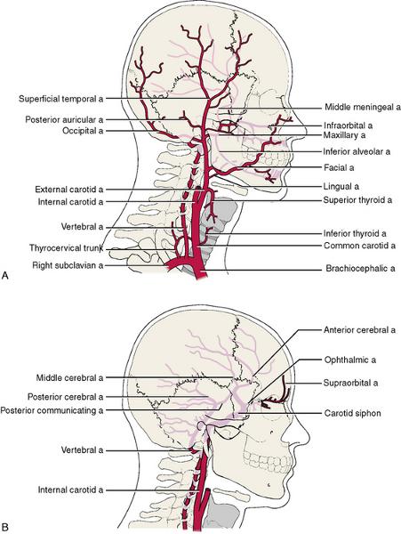
HEAD/NECK ARTERY

HEAD/NECK VEIN
HEPATIC PORTAL VEIN
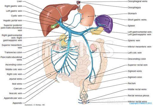
CELIAC TRUNK->LEFT GASTRIC-> COMMON HEPATIC->SPLEENIC R.GASTRIC->L. GASTROEPIPLOIC-> R. GASTROEPIPLOIC->GASTRODUODENAL

POSTERIOR MALE REPRODUCTIVE

NEPHRON
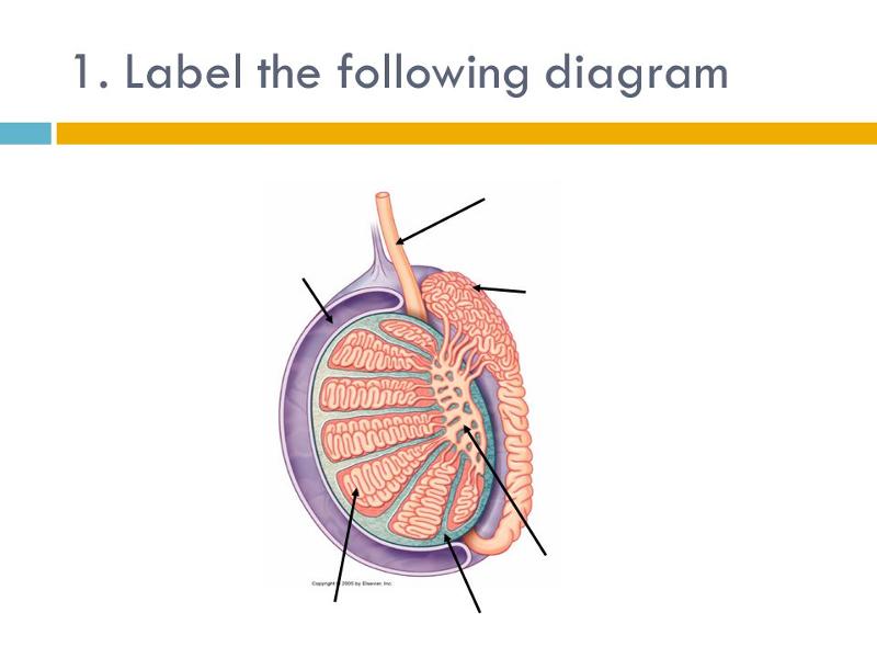
SAGITAL CUT TESTES
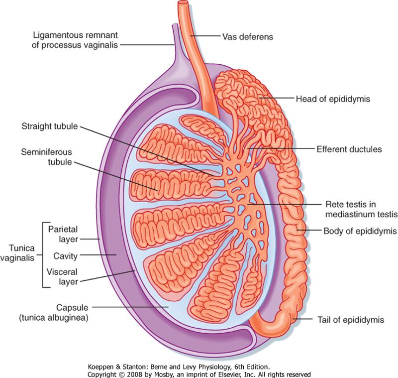
principal androgen produced by the testes is testosterone. Production of testosterone by the testes is stimulated by luteinizing hormone (LH), produced by anterior pituitary secretion of LH is stimulated by (GnRH), released from the hypothalamus, inhibited by testosterone, inhibits the secretion of GnRH. These hormones constitute the (hypothalamic-pituitary-testes axis).
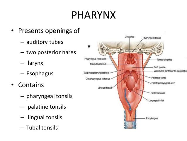
PHARYNX
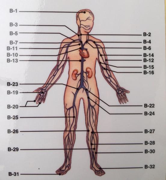
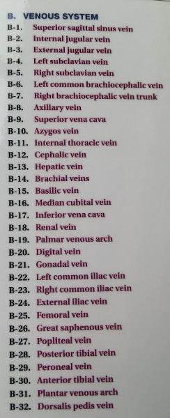
INFERIOR LIMB VEIN
External iliac
internal iliac
femoral
great saphenous
small saphenous
popliteal
perioneal (Fibula)
Anterior tibial
posterior tibial
WHAT IS THE MAIN BLOOD SUPPLY TO THE LOWER EXTREMITIES ?(ARTERY)
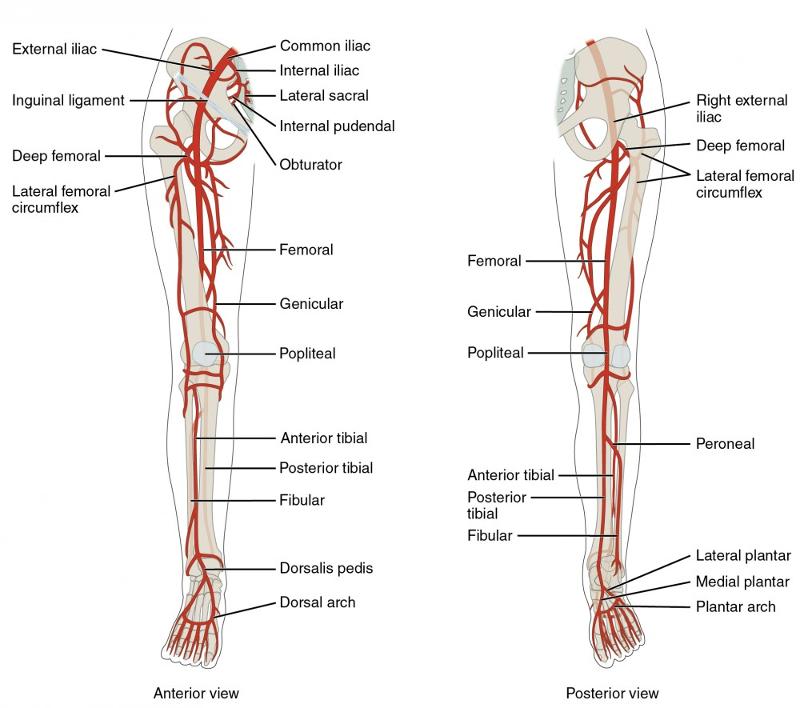
COMMON ILIAC -> FEMORAL-> DEEP ARTERY OF THE THIGH->POPLITIAL LOWER LEG->ANTERIOR TIBILIAR / POSTERIAL TIBULAR/ FIBULAR ->ARCULATE (DORSAL ASPECT OF FOOT ) _. PANTAR ARTERY->DIGITAL ARTERY
ELECTROLYTES
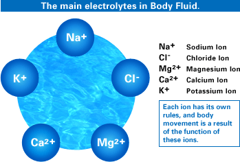
NA+
CI-
MG2+
CA2+
K+
CAUSES UTERINE CONTRACTIONS
prostaglandins- HELP SPERM MOVE
FUNCTIONS OF THE GALLBALDDER
BILE STORAGE
HYPERVENTILATION
CO2 DOWN PH UP- TOO LITTLE / DECREASE IN CO2 - RISE IN PH
WHAT DOES ADH DO TO THE COLLECTING TUBULE AND DISTAL ?
RE-ABSORPTION OF H2O: WHY ? DEHYDRATED AND NEED MORE RE-ABSORPTION OF FLUIDS
DIABETES INSIPIDUS
WHAT IS MOST IMPORTANT GAS FOR PH IN BLOOD ?
CO2
WHERE DOES GLUCOSE GET REABSORBED?
KIDNEY- PROXIMAL CONVOLUTED TUBULE - 70 % RE-ABSORPTION
what are the functions of the juxtaglomerular apparatus ?
FILTRATION RATE, BLOOD PRESSURE AND PH
DIABETES MILITIS: PRODUCING KEYTONE BODIES , WHAT CONDITION DOES IT LEAD TO?
METABOLIC ACIDOSIS
PROLONGED VOMITING OR TAKING TOO MANY ANTACIDS : WHAT DOES IT CAUSE ?
METABOLIC ALKALOSIS
OBSTRUCTION OR BLOCKING OF THE AIRWAY CAUSES?
RESPIRATORY ACIDOSIS
IF BLOOD PRESSURE SUDDENLY DROPS, WHAT HAPPENS TO FILTRATION?
DECREASES
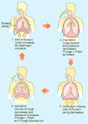
FORCE THAT IS RESPONSIBLE FOR NORMAL RESPIRATION IN THE LUNGS?
EXPAND THE LUNGS : NATURAL ELASTIC RECOIL
INCREASE PRESSURE / LOWER VOLUME = CO2 OUT
IF YOU HAVE ALOT OF CARBS IN YOUR MEAL? ENZYME NEEDED
AMYLASE
WHAT DOES CHYME DO?
MAKE BICARBONATE FROM PANCREAS BE RELEASED FOR SMALL INTESTINE
WHAT SEX HORMONE IN MALES PRODUCES SPERM ?
FSH- FOLLICLE STIMULATING HORMONE
SHORTER URETHRA IN A FEMALE COMPARED TO A MALE LEADS TO MORE SUSCEPTIBILITY TO?
UTI
WHAT IS THE MITRATION (URINATION) REFLEX CENTER CONTROLLED BY ?
SACRAL SEGMENTS OF THE SPINAL CORD
PROTEIN AND FAT IN THE SMALL INTESTINE LEADS TO THE SECRETION OF WHAT HORMONE?
CCK
WHICH HORMONE STIMULATES THE PANCREATIC JUICE TO RELEASE BICARBONATE TO MAKE CHYME LESS ACIDIC?
SECRETIN
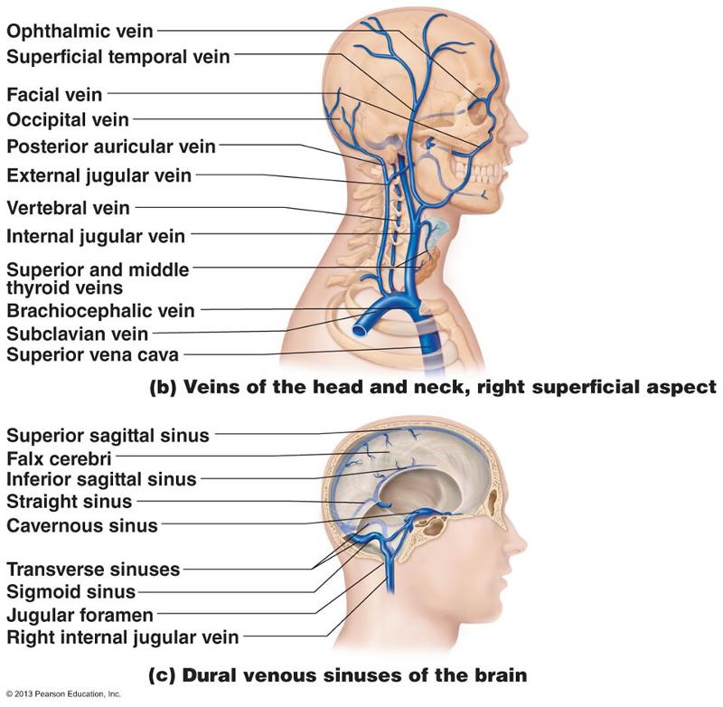
FACE AND SCALP IS DRAINED BY THE EXTERIOR JUGULAR THE DRAINS INTO WHAT VEIN ?
SUBCLAVIAN
SUBCLAVIAN AND INTERNAL JUGULAR MERGE TO FORM?
BRACIOCEPHALLIC
COMMON ILIAC MERGES TO FORM THE? ( 95 NORTH)
SUPERIOR VENA CAVA
WHEN WE REMOVE THE PREPUSE( FORESKIN)?
circumcise
UNDECENDED TESTES CAUSE INFERTILITY WHY?
heat of body will destroy it
MECHANISM EQUAL IN/ EQUAL OUT TO HEART ?
FRANK - STERLING
WHAT PROVIDES BLOOD SUPPLY TO THE LOWER EXTREMITIES ?
COMMON ILIAC
WHICH PROVIDE BLOOD SUPPLY TO THE THIGH?
FEMORAL
WHAT PROVIDES BLOOD SUPPLY TO THE KNEE?
POPLITEAL
WHICH PROVIDES BLOOD SUPPLY TO THE LOWER LEG? ( MORE THAN ONE)?
ANTERIOR TIBULAR/ POSTERIOR TIBULAR, FIBULAR
WHAT SUPPLY'S BLOOD TO THE FOOT ?
DORSALAS PEDIS
B CELLS DO WHAT
DIFFERENTIATES INTO PLASMA CELLS / CREATES ANTIBODIES
WHAT IS THE "SHRINKAGE"? REGULATES THE TESTES
dartos and cremaster muscle male shrinkage
ABILITY OF MALE TO EJACULATE IS DUE TO WHAT MUSCLES ?

SYMPATHETIC - CORPUS SPONGIOSUM
SITE OF FERTILIZATION IN A FEMALE?
IMPLANTATION OCCURS?
FALLOPIAN TUBES
UTERUS
MENSTRUATION ONLY WHEN BLOOD LEVELS OCCURS WHEN TWO HORMONES ARE DIMINISHED ?
ESTROGEN AND PROGESTERONE
WHAT CAUSES OVULATION ?
FIRST ESTROGEN SURGE DAY 12-14
WHICH IS THE HORMONE THAT PROMOTES THE UTERUS AND PREPS IT FOR PREGNANCY? PROMOTES THE UTERINE WALL
PROGESTERONE
2 POINT ESSAY - MUST KNOW
MENSTRAL : ESTROGEN ( UP ) DAY 12-14 >>> LH/ FSH ( UP) SURGE- EGG
COMES OUT >> LH/ FSH / ESTROGEN (DOWN) PROGESTRONE (UP) PEAKS AT
DAY 21 >>> 2ND SURGE DAY 22-25 ESTROGEN (UP) >>> ESTROGEN (DOWN)
PROGESTRON (DOWN) >>>> PMS UNTIL MENOPAUSE
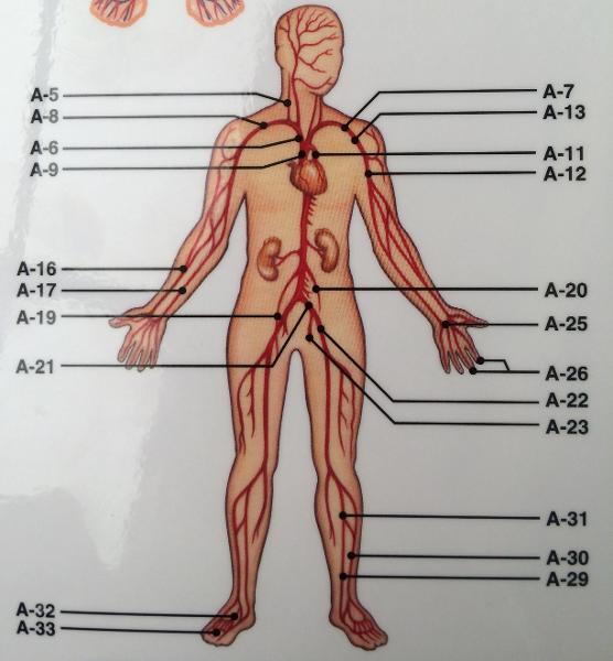
Blood >>> heart via >>>> aorta >>> renal artery >>>
interlobar arteries >>> arcuate artery >>>> pyramids >>>
arcuate artery >>> cortical radiate arteries >>>> afferent arteriole
>>> ADH- DCT DISTAL CONVELATED TUBE>>> proximal convoluted tubule
>>> efferent arteriole >>> peritubular capillaries >>> Blood >>>
in the body exits >>>cortical radiate veins >>>> arcuate vein>>> interlobar vein
>>> renal vein >> heart >>> inferior vena cava >>> waste >>
proximal convoluted tubule >>> descending loop of henle >>>
ascending loop of henle >>> distal convoluted tubule >>>
collecting duct>>>>ureters >>> bladder >>>> leaves the body via the urethra.
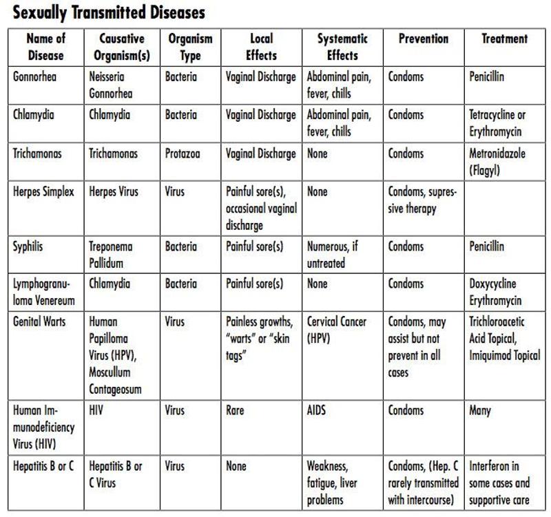
urogenital infection caused by the bacterium chlamydia trachomatis
chlamydia
STI that initially causes inflamation
gonorrhea
STI caused by a bacterium called treponema pallidum
syphilis
a vaginal infection caused by bacterial microorganisms
vaginitis
form of vaginitis caused by the one-celled protozoan trichomonas vaginalis
Trichomoniasis
How did it get so late so soon? Its night before its
afternoon. December is here before its June. My goodness how the
time has flewn. How did it get so late so soon?
Dr. Seuss
The more that you read, the more things you will know. The
more that you learn, the more places you'll go.
Dr. Seuss
ABNORMALLY FAST HEART BEAT= 100+ BEATS
ABNORMALLY LOW HEART BEAT = 60 - BEATS INADEQUATE BLOOD CIRCULATION
tachycardia
bradycardia
The right and left internal jugular veins and the right and left subclavian merge to form the
Brachiocephalic veins
Which of the following arteries provide blood to the lower extremities?
Common iliac arteries
A) Common hepatic artery
B) Renal artery
C) Inferior
mesenteric artery
D) Internal iliac artery
E) Aorta
Supplies the duodenum and stomach
Answer: A
Supplies the kidney.
Artery that does not anastomose.
Answer: B
Supplies the distal areas of the large intestine.
Answer: C
Supplies pelvic structures.
Answer: D
Largest artery of the body.
Answer: E
Veins that drain the lateral surface of the upper arm are the:
Cephalic veins
Which vessel drains the scalp?
External jugular vein
Which tunic of an artery is most responsible for maintaining blood pressure and continuous blood circulation?
tunica media
Which of the following is not a branch of the aorta?
right cartoid artery
Which of the following supplies blood to parts of the intestinal tract?
superior mesenteric artery
Which of the following are involved directly in pulmonary circulation?
right ventricle, pulmonary artery, and left atrium
The mechanism that ensures the volume of blood discharged from the heart is equal to the volume entering its chambers is ______ law of the heart.
Frank-Starling's
antenna- anterior communicating /posterior communicating /anterior cerebral
eyes- internal carotid
arms- posterior cerebral artery
body-basilar artery
leg- vertebral artery
circle of willis (brain)
WHAT HELPS RETURN THE BLOOD TO THE HEART ?
CONTRACTION OF SKELETAL MUSCLES AND THE DIAPHRAGM
GASTRAPHOLIAS : EXERCISE: SQUEEZES THE INFERIOR VENA CAVA
WHAT IS THE MAIN BLOOD SUPPLY TO THE BRAIN ?
INTERNAL CARTOID AND VERTERBRAL
Arch of aorta
Abdominal aorta
Coronary artery
Subclavian A/V
Axillary A/V
Brachial A/V
Radial A/V
Ulnar A/V
Vertebral artery
Hepatic A/V
Renal A/V
Lumbar A/V
To upper body
To abdominal cavity; legs
Supplies oxygenated blood to the heart muscle
To / From shoulder
To / From underarm area
To/From upper arm
To/From lateral lower arm used to take pulse @ wrist
To/From medial lower arm
(serving brain) To brain (posterior)
To/From liver
To/From kidneys
To/From posterior abdominal wall
Femoral A/V
Anterior/Posterior Tibial A/V
Dorsalis pedis A/V
Plantar A/V
Superior vena cava
Inferior vena cava
Internal jugular V
External jugular V
Cephalic V
Median cubital V
Great Saphenous V
External Iliac A/V
To/From upper leg- the external iliac artery entering the thigh
To/From lower leg
To/From top of foot
To/From bottom of foot
From upper body
From lower body
from brain
from superficial tissues of the head and neck
from superficial lateral arm
from superficial middle of arm (usually take blood from this vein)
from superficial medial leg (longest vein)
To/From superficial leg
List all of the veins and their tributaries of the trunk (abdomen, thorax and neck region)
- SVC
- L. and R. Brachiocephalic
- Internal Jugular
- External Jugular
- Subclavian
- IVC
- Hepatic veins
- R. Suprarenal
- R. gonadal
- R. Renal
- L. Suprarenal
- L. Renal
- L. Gonadal
- Common iliac
- Internal iliac
- External iliac
- Femoral
- R.Ascend

Major arteries and veins of torso

Descending aorta: descending thoracic aorta and descending abdominal aorta
Renal arteries
Inferior vena cava
Renal veins