Where is Cerebrospinal fluid (CSF) circulate?
lateral ventricles: line each hemisphere
3rd, 4th ventricle: in brain sterm
subrachnoid space
What cells make CSF
Neuroglia cells called ependymal cells
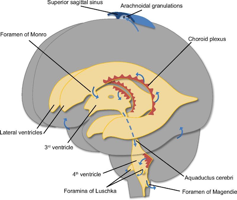
what is choroid plexus ?
Ependymal cells associated BV that produce CSF
What is the funtion of CSF?
protects brain from movement of the skull + vertebral column -> brain floats
buoyancy -> weight brain is reduced
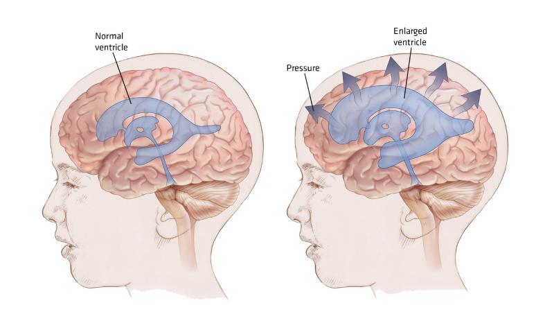
What is hydrocephalus?
Result from blockage of CSF through CNS (cerebral aqueduct)
When hydrocephalus occur ?
Elderly people
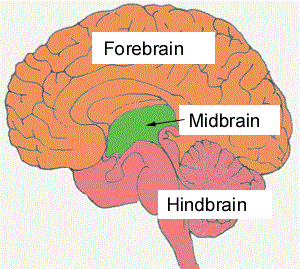
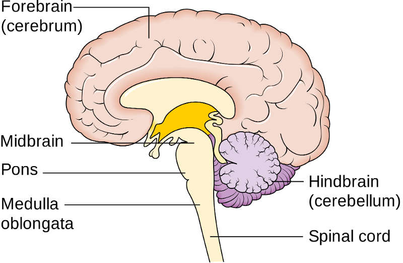
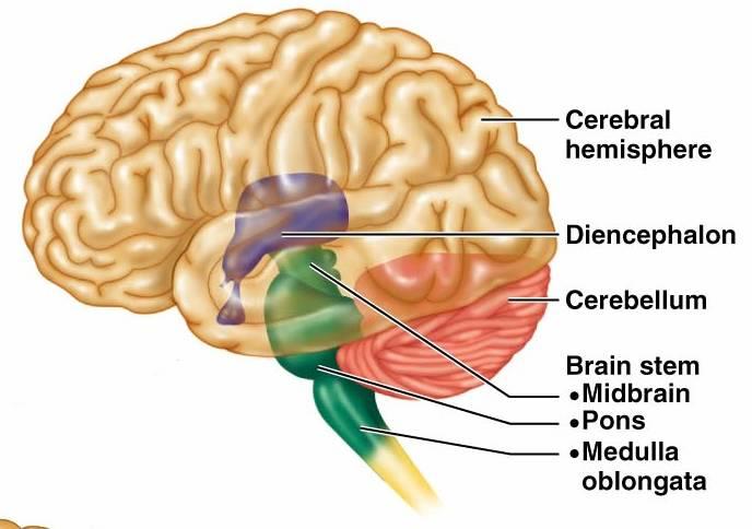
What are regions of the brain?
- Brain Stem
- Cerebellum
- DIencephalon
- Cerebrum
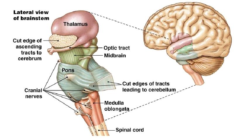
What is Brain Stem?
Connect the spinal cord and cerebellum
What is structure of brain sterm
Anterior part: contain descending tracts involved with motor control
Posterior: contain ascending tracts from the spinal cord, cerebellum and craial nerve
what are funtions of Brain stem
Control of hear rate, blood pressure, breathing
Part of the brain stem
Midbrain
Smallest region of brain stem
Composed of tracts of nerve fibers
what is corpora quadrigemina
the largest midbrain nuclei
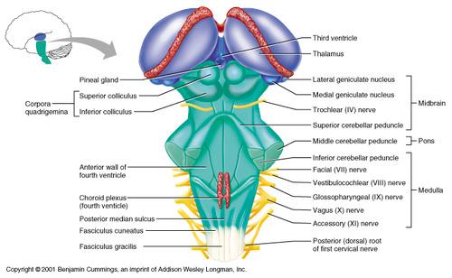
What are 2 superior colliculus?
Visual reflexes

What are 2 inferior colliculus?
Auditory reflexes
Midbrain contain pain perception
Receptors respond to opiates (thuoc phien)
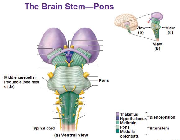
Part of the brain stem
Pons(bridge)
- Cerebral cortex communicate with pons
- Some nuclei relay info between cerebrum and pons
- lower part of pons control balance, breathing, swallowing,
part of brain stem
Medulla Oblongata
- Most inferior portion of brain stem, continuous spinal cord.
- Ra[idly fatal
What are funtions of Medualla Oblongata
- Heart rate control
- Blood pressure regulation
- Breathing
- Vomiting (non)
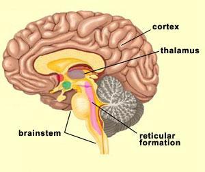
What is Reticular Formation?
- Begins at upper portion of medulla
- Some in brain stem
- Damage can result in coma opposite
half of the body
Funtion of Reticular Formation
- regulation of sleep
- arousal (hung phan)
- maintaining consciousness.
Part of the brain stem
Cerebellum
(little brain) smaller version of cerebrum
What part cerebellum attach to brain stem
Cerebellar peduncle
What is Cerebellar peduncle
- Superior cerebellar peduncle: connecting with the midbrain
- Middle cerebellar peduncle: connecting with the pons
- Inferior cerebellar peduncle: connecting with the medulla oblongata
Gray matter of cerebellum
consists of outer cortex and nuclei deep within the cerebellum
White matter of cerebellum
consists of tracts collectively called arbor vitae
Parts of Cerebellum
Flocculonodular lobe, vermis, lateral he,ispheres
Flocculonodular lobe:
• a small inferior part
• Help control balance and coordination
• Alcohol depresses cerebellum so, drinking driver test walking with imbalance
Lateral hemisphere
two large hemispheres
Funtion of lateral hemisphere
concert with the frontal lobes of
the cerebral cortex in
planning, and learning complex movements.
Part of brain stem
Diencephalon
Location: between brain stem and cerebrum
Main components: the thalamus, epithalamus, and hypothalamus
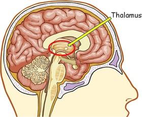
what is Thalamus
- The largest part if the diencephalon
- shape liek yo yo
- collection of sensory input
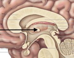
What is Interthamaic adhesion of thalamus ?
a small stalk in the center connecting two
lateral portions
What does Sensory area do?
Ascending axons carrying sensory information project to the thalamus
How does Sensory area work?
they synapse with thalamic neurons -> thalamic neurons send their axons to the cerebral cortex -> most awareness of sensory input occurs.
(audiotory, visual,, pain, touch pass through thalamus)
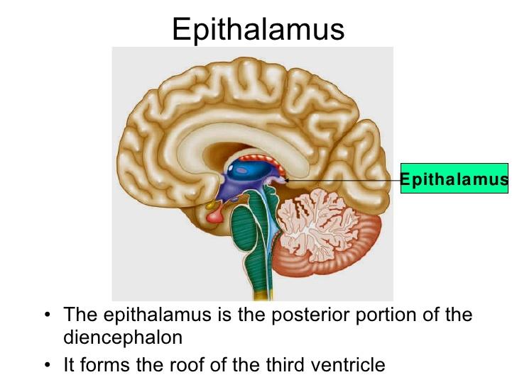
What is Epithlamus ?
Small area, superior and posterior to the thalamus
What does Epithalamus consit ?
habenular nuclei and pineal body
What is habenular nuclei
influenced by the sense of smell
involved in emotional
visceral responses to odors. (mui hoi)
What is pineal body
Influence the sleep-wake cycle
What is Hypothalamus
The most inferior part of the diencephalon •
A collection of nuclei: connect to many other parts of the brain and spinal cord
Involved with autonomic, endocrine, emotional (limbic system)
Autonomic nervous system (ANS)
helpingcontrol heart rate, blood vessel diameter, urine release from the urinary bladder, and the movement of food through the digestive tract.
Limbic system
HT is a part of the limbic system and affects mood, motivation, and emotions.
Feeling relaxed, sexual pleasure, rage, and fear are related to HT
What is Mammillary body?
involved in emotional response to order, olfactory
reflexes and memory
Cerebrum
Logitudinal fissure: divide into left and right hemispheres
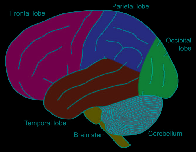
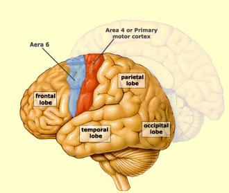
Frontal lobe
voluntary motor function, motivation, aggression, the sense of smell, and mood.
Parietal lobe
the reception and evaluation of most sensory information, such as touch, pain, temperature, balance, and taste.
• The frontal and parietal lobes are
separated by the central sulcus.
Occipital lobe
reception and integration of visual input.
Temporal lobe
Receive and evaluate input for smell and hearing.
Cortex
the gray matter on the outer surface of the cerebrum
Nuclei
clusters of gray matter deep inside the brain
Cerebral medulla
white matter of the brain between the cortex and nuclei
Consist of tracts that connect areas of the cerebral cortex to each other or to other parts of the CNS
Parts of Cerebral medulla
Association fibers, Commissural fibers, Projection fibers
Association fibers
connect areas of the cerebral cortex within the same hemisphere
Commissural fibers
connect one cerebral hemisphere to the other.
Projection fibers
between the cerebrum and other parts of the brain and spinal cord
Sensory area of the Cerebral Cortex
Sensory pathways project to specific regions of the cerebral cortex,
called primary
sensory areas.
primary sensory areas.
Visual cortex: processing visual images in the occipital lobe
auditory cortex: processing auditory stimuli in the temporal lobe
Taste area: perceive in the parietal lobe
Olfactory cortex: conscious and unconscious responses to odor on the inferior surface of the temporal lobe
Motor area of Cerebral Cortex
primary motor cortex and premotor area
primary motor cortex
controlling motor functions of the feet are in the most superior.
controlling the face are in the inferior region
premotor area
the staging area in which motor functions are organized before they are initiated in the motor cortex
5) Basal Nuclei
A group of functionally related nuclei located in the inferior cerebrum, diencephalon, and midbrain
Control of motor funtion
subthalamic nucleus
is located in the diencephalon.
substantial nigra
is located in the midbrain
6) LImbic system
Parts of the cerebrum and diencephalon are grouped together
Funtion of Limbic system
memory, reproduction, and nutrition