List the two layers of skin and briefly describe subcutaneous tissue.
Epidermis - composed of epithelial cells, is the outermost protective shield of the body.
Dermis - the only vascularized layer making up the bulk of the skin, is tough, leathery layer composed mostly of dense connective tissue.
Subcutaneous tissue > Hypodermis (superficial fascia)
- not a part of the skin but shares protective functions.
- superficial to the tough connective tissue wrapping, fascia, of the skeletal muscle
- Stores fat, anchors the skin mostly to muscle, loose enough to ensure "glancing blows"
The epidermis is composed of which of the following tissues?
Keratinized stratified squamous epithelial tissue
Which layer of the skin- dermis or epidermis- is better nourished?
The Dermis
- The dermis is connective tissue, which is vascular, so its cells would be better nourished than those of the epidermis, which is avascular epithelium.
Which Cell makes up most of the Epidermis and it's chief role is to produce keratin, the fibrous protein that helps give the epidermis it protective properties?
- a) Keratinocyte
- b) Melanocytes
- c) Dendritic Cells
- e) Tactile Cells
A
Which Cell of the Epidermis is a spider-shaped epithelial cell that synthesizes the pigment melanin?
- a) Keratinocyte
- b) Melanocytes
- c) Dendritic Cells
- d) Tactile Cells
B
Which star shaped Cell of the Epidermis, also known as a Langerhans cell, arise from bone marrow and migrate to the epidermis where they ingest foreign substance and are key activator of our immune system.
- a) Keratinocyte
- b) Melanocytes
- c) Dendritic Cells
- d) Tactile Cells
C
Which spiky hemisphere shaped Cell of the Epidermis, also known as a Merkel, occasionally are present at the epidermaldermal junction and functuions as a sensory receptor for touch.
- a) Keratinocyte
- b) Melanocytes
- c) Dendritic Cells
- d) Tactile Cells
D
While walking barefoot in a barn, Jeremy stepped on a rusty nail that penetrated the epidermis on the sole of his foot. Name the layers the nail pierced from the superficial skin surface to the junction with the dermis.
- a) stratum corneum, stratum lucidum, stratum spinosum, stratum granulosum stratum basale
- b) stratum lucidum, stratum corneum, stratum granulosum, stratum basale and stratum spinosum
- c) stratum corneum, stratum lucidum, stratum granulosum, stratum spinosum, and stratum basale
- d) stratum lucidum, stratum corneum, stratum granulosum, stratum spinosum, and stratum basale
C
Most Superficial layer; 20-30 layers of dead cells, essentially flat membranous sacs filled with keratin. Glycolipids in extracellular space.
- a) Stratum corneum
- b) Stratum Lucidum
- c) Stratum granulosum
- d) Stratum spinosum
- e) Stratum basale
A
Typically one to five layers of flattened cells, organelles deteriorating; cytoplasm full of lamellar granules (release lipids) and keratohyaline granules
- a) Stratum corneum
- b) Stratum Lucidum
- c) Stratum granulosum
- d) Stratum spinosum
- e) Stratum basale
C
Several layers of keratinocytes unified by desmosomes. Cells contain thick bundles of intermediate filaments made of pre-keratin.
- a) Stratum corneum
- b) Stratum Lucidum
- c) Stratum granulosum
- d) Stratum spinosum
- e) Stratum basale
D
Deepest epidermal layer; one row of actively mitotic stem cells; some newly formed cells become part of the more superficial layers. See occasional melanocytes and dendritic cells.
- a) Stratum corneum
- b) Stratum Lucidum
- c) Stratum granulosum
- d) Stratum spinosum
- e) Stratum basale
E
Layer of epidermal tissue that is only found in thick skin and subject to abrasion. ie: palms, fingertips, and the soles of the feet
- a) Stratum corneum
- b) Stratum Lucidum
- c) Stratum granulosum
- d) Stratum spinosum
- e) Stratum basale
B
The stratum basale is also called the stratum germinativum, a name that refers to its major function. What is that function?
the stratum basale undergoes almost continuous mitosis to replace cells lost by abrasion.
Why are the desmosomes connecting the keratinocytes so important?
The skin is subject to a lot of abrasion and physical trauma. The desmosomes, which are connecting junctions, help to hold the cells together during such stress.
Which of the following structures are primarily responsible for fingerprints?
- a) dermal ridges and epidermal ridges
- b) sweat pores and dermal ridges
- c) friction ridges and sweat pores
- d) reticular layer and sweat pores
- e)papillary layer and epidermal ridges
D
Name the tissue type/s composing the dermis.
- Areolar connective tissure (Parillary layer)
- Dense irregular connective tissue (Reticular Layer)
Which layer of the dermis is responsible for producing fingerprint patterns?
- a) Stratum corneum
- b) Papilary layer
- c) Stratum Lucidum
- d) Reticular Layer
B
Which tissue of the hypodermis make a good shock absorber?
- a) dense regular tissue
- b) areolar tissue
- c) adipose tissue
- d) dense irregular tissue
C
You have just gotten a paper cut. It is very painful, but it doesn't bleed. Which avasular layer of the skin has it only penetrated.
- a) dermis
- b) reticular layer
- c) hypodermis
- d) epidermis
D
What 3 factors contribute to skin color?
- a) melanin, carotene, and hemoglobin
- b) melanin, carotene, and folic acid
- c) carotene, hemoglobin, and Vitamin D
- d) carotene, hemoglobin and Vitamin C
A
What is cyanosis and what does it indicate?
- a) an abnormal yellow skin tone / liver disorders
- b) poorly oxygenated hemoglobin / heart failure and sever respiratory disorders
- c) richly oxygenated hemoglobin / heart failure and minor respiratory disorders
- d) reddened of the skin / fever, hypertension, inflammation, or allergy
B
Match the following condition with its description: Psoriasis
- a) Skin rashes resulting from allergic reactions
- b) Skin rashes resulting from allergic reactions
- c) Autoimmune condition, resulting in an over proliferation of the epidermis, characterized by reddened epidermal papules covered with dry, silvery scales
- d) A rare condition where hair follicles are attacked by the immune system, causing the hair to fall out in patches
C
Skin eruption produced by dilated small blood vessels of the face, especially on the nose and cheeks, is descriptive of which skin disorder?
- a) Melanocyte activity
- b) Port wine stain
- c) cyanosis
- d) raynaud disease
- e) rosacea
E
Which of the following cells would one not expect to find in the dermis?
- a) neutrophils
- b) mast cells
- c) macrophages
- d) fibroblasts
- e) keratinocytes
E
Which alteration in skin color may indicate a liver disorder?
- a) redness, or erythema
- b) pallor, or blanching
- c) jaundice
- d) bronzing
- e) black and blue marks
C
All of the following are a part of hair EXCEPT...
- a) medulla
- b) glassy membrane
- c) cortex
- d) cuticle
B

Labeled "A" indicates which portion of the skin.
- a) stratum spinosum
- b) stratum basale
- c) stratum corneum
- d) stratum granulosum
C
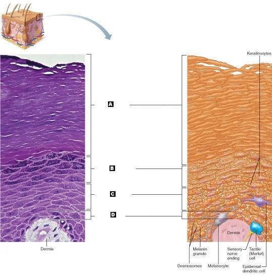
Labeled "B" indicates which portion of the skin.
- a) stratum spinosum
- b) stratum basale
- c) stratum corneum
- d) stratum granulosum
D

Labeled "C" indicates which portion of the skin.
- a) stratum spinosum
- b) stratum basale
- c) stratum corneum
- d) stratum granulosum
A
Labeled "D" indicates which portion of the skin.
- a) stratum spinosum
- b) stratum basale
- c) stratum corneum
- d) stratum granulosum
B
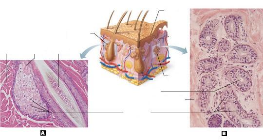
Labeled "A" indicates which cutaneous gland
a) eccrine (sweat) gland
b) sebaceous gland
B

Labeled "B" indicates which cutaneous gland
a) eccrine (sweat) gland
b) sebaceous gland
A
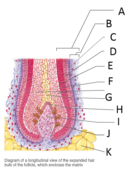
Labeled "A" indicates which hair or hair follicle structure.
- a) folicle wall
- b) peripheral connective tissue (fibrous) sheath
- c) glassy membrane
- d) epithelial root sheath (External)
- e) epithelial root sheath (internal)
- f) cuticle
- g) cortex
- h) medulla
- i) hair matrix
- j) hair papilla
- k) subcutaneous adipose tissue
A

Labeled "B" indicates which hair or hair follicle structure.
- a) folicle wall
- b) peripheral connective tissue (fibrous) sheath
- c) glassy membrane
- d) epithelial root sheath (External)
- e) epithelial root sheath (internal)
- f) cuticle
- g) cortex
- h) medulla
- i) hair matrix
- j) hair papilla
- k) subcutaneous adipose tissue
B

Labeled "C" indicates which hair or hair follicle structure.
- a) folicle wall
- b) peripheral connective tissue (fibrous) sheath
- c) glassy membrane
- d) epithelial root sheath (External)
- e) epithelial root sheath (internal)
- f) cuticle
- g) cortex
- h) medulla
- i) hair matrix
- j) hair papilla
- k) subcutaneous adipose tissue
C

Labeled "D" indicates which hair or hair follicle structure.
- a) folicle wall
- b) peripheral connective tissue (fibrous) sheath
- c) glassy membrane
- d) epithelial root sheath (External)
- e) epithelial root sheath (internal)
- f) cuticle
- g) cortex
- h) medulla
- i) hair matrix
- j) hair papilla
- k) subcutaneous adipose tissue
D

Labeled "E" indicates which hair or hair follicle structure.
- a) folicle wall
- b) peripheral connective tissue (fibrous) sheath
- c) glassy membrane
- d) epithelial root sheath (External)
- e) epithelial root sheath (internal)
- f) cuticle
- g) cortex
- h) medulla
- i) hair matrix
- j) hair papilla
- k) subcutaneous adipose tissue
E

Labeled "F" indicates which hair or hair follicle structure.
- a) folicle wall
- b) peripheral connective tissue (fibrous) sheath
- c) glassy membrane
- d) epithelial root sheath (External)
- e) epithelial root sheath (internal)
- f) cuticle
- g) cortex
- h) medulla
- i) hair matrix
- j) hair papilla
- k) subcutaneous adipose tissue
F

Labeled "G" indicates which hair or hair follicle structure.
- a) folicle wall
- b) peripheral connective tissue (fibrous) sheath
- c) glassy membrane
- d) epithelial root sheath (External)
- e) epithelial root sheath (internal)
- f) cuticle
- g) cortex
- h) medulla
- i) hair matrix
- j) hair papilla
- k) subcutaneous adipose tissue
G

Labeled "H" indicates which hair or hair follicle structure.
- a) folicle wall
- b) peripheral connective tissue (fibrous) sheath
- c) glassy membrane
- d) epithelial root sheath (External)
- e) epithelial root sheath (internal)
- f) cuticle
- g) cortex
- h) medulla
- i) hair matrix
- j) hair papilla
- k) subcutaneous adipose tissue
H

Labeled "I" indicates which hair or hair follicle structure.
- a) folicle wall
- b) peripheral connective tissue (fibrous) sheath
- c) glassy membrane
- d) epithelial root sheath (External)
- e) epithelial root sheath (internal)
- f) cuticle
- g) cortex
- h) medulla
- i) hair matrix
- j) hair papilla
- k) subcutaneous adipose tissue
I

Labeled "J" indicates which hair or hair follicle structure.
- a) folicle wall
- b) peripheral connective tissue (fibrous) sheath
- c) glassy membrane
- d) epithelial root sheath (External)
- e) epithelial root sheath (internal)
- f) cuticle
- g) cortex
- h) medulla
- i) hair matrix
- j) hair papilla
- k) subcutaneous adipose tissue
J

Labeled "K" indicates which hair or hair follicle structure.
- a) folicle wall
- b) peripheral connective tissue (fibrous) sheath
- c) glassy membrane
- d) epithelial root sheath (External)
- e) epithelial root sheath (internal)
- f) cuticle
- g) cortex
- h) medulla
- i) hair matrix
- j) hair papilla
- k) subcutaneous adipose tissue
K
Which of the following human integumentary system glands is believed to be analogous to the sexual scent glands of an animal?
- a) apocrine sudoriferous gland
- b) cerunimous gland
- c) eccrine sudoriferous gland
- d) mammary gland
- e) sebaceous gland
A
Match the following glands with their secretion: Sudoriferous glands
- a) milk
- b) sebum
- c) sweat
- d) cerumen
C
Which of the following is untrue with regards to finger nails?
- outward concavity of the nail (spoon nail) may signal an iron deficiency
- horizontal lines (Beau's lines) across the nails may hint of malnutrituion
- thickened yellow nails may signal a fungal infection
- nails ar primarily used to protect the capillary beds in the tip of the fingers and toes
- yellow-tinged nail may indicate a respiratory or thyroid gland disorder
D
Match the following hair follicle-associated structure with its description: Hair bulb
- actively dividing cellular area of the bulb that produces the hair
- bundle of smooth muscle tissue running from the superficial dermis to the hair follicle; responsible for producing goose bumps
- the expanded, deep region of a hair follicle
- superficial wall of the hair follicle that is derived from the dermis
- a knot of sensory nerve ending wrapped around the base of a hair follicle
C
Match the following type of burns with its description: First-degree burn
- Entire thickness of the skin is consumed, resulting in the burned area appearing white, red, or blackened
- Injury to the epidermis and the superficial region of the dermis resulting in redness, swelling, pain, and blisters
- Damage to only the epidermis that results in redness, swelling, and pain
C
Match the following: Tactile cells
- sensory detection
- protect from UV radiation
- activate the immune system
- make fibrous proteins for skin protection
A
Match the following: Melanocytes
- sensory detection
- protect from UV radiation
- activate the immune system
- make fibrous proteins for skin protection
B
Match the following: Dendritic cells
- sensory detection
- protect from UV radiation
- activate the immune system
- make fibrous proteins for skin protection
C
Match the following: Keratinocytes
- sensory detection
- protect from UV radiation
- activate the immune system
- make fibrous proteins for skin protection
D
Match the following skin functions with the mechanism that accomplishes them: Metabolic functions
- Provides a chemical barrier and a mechanical barrier to ward off bacterial invasion and to provide “waterproofing”
- Potential to hold about 5% of the body's blood volume in numerous blood vessels
- Synthesis of vitamin D; destruction of cancer-causing chemicals; activation of some steroid hormones
- Activation of numerous receptors that are part of the nervous system
- Dilation of blood vessels and secretion of sweat that evaporates from body surface
C