
Identify the letter that indicates the left common carotid
artery.
A) A
B) B
C) C
D) D
E) E
A

Identify the letter that indicates the left auricle.
A)
A
B) B
C) C
D) D
E)
D

Identify the letter that indicates the ligamentum arteriosum.
A)
A
B) B
C) C
D) D
E) E
C
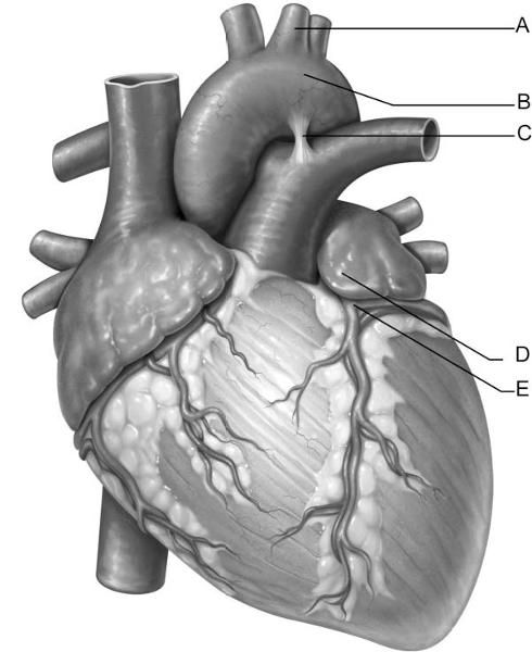
Identify the letter that indicates the left coronary artery.
A)
A
B) B
C) C
D) D
E) E
E

Identify the letter that indicates the aortic arch.
A) A
B)
B
C) C
D) D
E) E
B

Identify the letter that indicates anchor points for chordae
tendineae, comprised of cells from
the myocardium.
A)
A
B) B
C) C
D) D
E) E
B

Identify the letter that indicates the tissue layer of the heart
known as the epicardium.
A) A
B) B
C) C
D)
D
E) E
E

Identify the letter that indicates the left atrioventricular
valve.
A) A
B) B
C) C
D) D
E) E
A

Identify the letter that indicates the location of the bundle
branches.
A) A
B) B
C) C
D) D
E) E
C
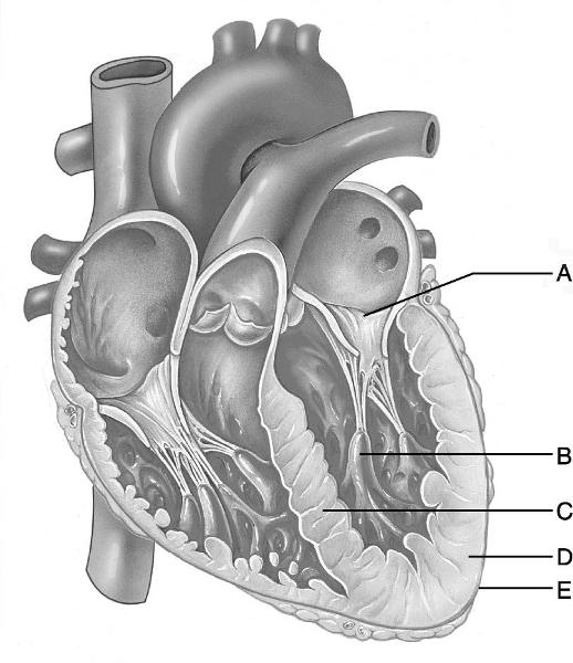
Identify the letter that indicates the thicker myocardial layer of
the left ventricle.
A) A
B) B
C) C
D) D
E) E
D
The region between the right and left pleural cavities is the
A)
pulmonary cavity.
B) peritoneal cavity.
C) pericardial
cavity.
D) vertebral cavity.
E) mediastinum.
E
The accumulation of pericardial fluid due to inflammation or the
accumulation of blood in
the pericardial cavity can lead
to
A) pericarditis.
B) pleuritis.
C) cardiac
tamponade.
D) fasciae adherens.
E) mitral valve prolapse.
C
The heart chamber that receives oxygenated blood from the pulmonary
veins.
A) right atrium
B) right ventricle
C) left
atrium
D) left ventricle
E) right auricle
C
The heart chamber that receives blood from the superior vena cava,
inferior vena cava, and
coronary sinus.
A) right
atrium
B) right ventricle
C) left atrium
D) left
ventricle
E) left auricle
A
The internal C-shaped crest of the right atrium which indicates the
openings for the Superior
vena cava and Inferior vena cava
is
A) ligamentum arteriosum.
B) crista terminalis.
C)
trabeculae carneae.
D) pectinate muscles.
E) fossa ovalis.
B
Threadlike structures of the endocardium that prevent prolapse of the
atrioventricular valves.
A) ligamentum arteriosum
B)
pectinate muscles
C) trabeculae carneae
D) chordae
tendineae
E) fossa ovalis
D
The valve responsible for preventing backflow of blood from the lungs
into the heart.
A) tricuspid valve
B) bicuspid valve
C)
aortic semilunar valve
D) pulmonary semilunar valve
E)
pectinate muscles
D
Heart valve with two cusps.
A) mitral valve
B) pulmonary
semilunar valve
C) aortic semilunar valve
D) fossa
ovalis
E) atrioventricular bundle
A
Contraction of these structures tightens the chordae tendineae,
preventing valve prolapse.
A) trabeculae carneae
B)
pectinate muscles
C) crista terminalis
D) papillary
muscles
E) atrioventricular bundle
D
Cells of the conducting system located between the AV node and bundle
branches.
A) trabeculae carneae
B) pectinate muscles
C)
crista terminalis
D) papillary muscles
E) atrioventricular bundle
E
Large cardiac cells of the conducting system embedded in the
ventricular walls between the
endocardium and myocardium.
A)
atrioventricular bundle
B) atrioventricular branches
C)
subendocardial conducting network (Purkinje fibers)
D) sinoatrial
node
E) atrioventricular node
C
Parasympathetic impulses to the SA node are transmitted on this
cranial nerve.
A) Glossopharyngeal nerve
B) Accessory
nerve—spinal part
C) Vagus nerve
D) Trigeminal nerve
E)
Hypoglossal nerve
C
Coronary artery that supplies the left atrium.
A) circumflex
artery
B) left anterior descending artery
C) marginal
artery
D) posterior descending artery
E) pulmonary artery
A
Death of heart musculature due to lack of oxygen.
A) cardiac
tamponade
B) valve insufficiency
C) heart block
D)
myocardial infarction
E) ventricular fibrillation
D
The pericardial cavity lies between
A) the fibrous pericardium
and the parietal pericardium.
B) the parietal pericardium and the
visceral pericardium.
C) the serous pericardium and the
epicardium.
D) the fibrous pericardium and the diaphragm.
B
How did the sinoatrial (SA) node most likely get its name?
A) It
is on the side of the atrium.
B) It lies at the opening of the
coronary sinus.
C) It is damaged by sinus infections (head
colds).
D) It develops from the sinus venosus and lies in an atrium.
D
The inner endothelial layer that lines the heart is the
A)
epicardium.
B) pericardium.
C) myocardium.
D) endocardium.
D
Which of the following vessels does not carry oxygen-poor blood to
the heart?
A) the superior vena cava
B) the inferior vena
cava
C) the pulmonary vein
D) the coronary sinus
C
The superior corner of the right atrium of the heart is located at
the
A) costal cartilage of the sixth rib, a finger's width
lateral to the sternum.
B) costal cartilage of the third rib
where it attaches to the sternum.
C) fifth intercostal space
along a line extending inferiorly from the midpoint of the
clavicle.
D) midpoint of the jugular notch.
B
The auricles are
A) earlike flaps on the surface of the
ventricles.
B) earlike flaps on the surface of the atria.
C)
projections of the endothelium into the ventricles.
D)
modifications of the pectinate muscles on the inner surface of the atria.
B
What structures anchor the chordae tendineae?
A) trabeculae
carneae
B) papillary muscles
C) pectinate muscles
D)
semilunar valves
B
Which structure develops from the embryological chamber called the
bulbus cordis?
A) the left atrium
B) the right
ventricle
C) the left ventricle
D) the sinoatrial node
B
Which of the following structures is not found in the left
ventricle?
A) the pectinate muscles
B) the mitral
valve
C) the trabeculae carneae
D) the papillary muscles
A
The right ventricle pumps blood into which vessel?
A) the
aorta
B) the pulmonary vein
C) the pulmonary trunk
D)
the superior vena cava
C
A drop of blood returning to the heart from the head region would
enter the heart through
which vessel?
A) a pulmonary
vein
B) the inferior vena cava
C) the superior vena
cava
D) the coronary sinus
C
The cusps of the valves of the heart are covered by
A)
epicardium.
B) myocardium.
C) endocardium.
D) modified pericardium.
C
Semilunar valves are located
A) between the atria and the
ventricles.
B) between the ventricles and the great
arteries.
C) between the great veins and the atria.
D) only
between the left ventricle and the aorta.
B
A condition in which the ventricles are unable to pump blood
efficiently because of rapid,
random contraction of cardiac
muscle fibers is called
A) atrial fibrillation.
B)
ventricular fibrillation.
C) pulmonary arterial
hypertension.
D) congestive heart failure.
B
At which corner point of the heart does one listen for the sound of
the closing aortic
semilunar valve?
A) superior
right
B) inferior right
C) superior left
D) inferior left
A
What is the effect of the parasympathetic fibers carried by the vagus
nerve?
A) They speed up the heartbeat.
B) They increase the
force of cardiac contractions.
C) They slow the
heartbeat.
D) The heartbeat is not influenced by the vagus nerve.
C
The crista terminalis can be used to locate all of the following
structures except the
A) opening of the coronary sinus.
B)
opening of the inferior vena cava.
C) opening of the pulmonary
veins.
D) opening of the superior vena cava.
C
Which vessel returns most of the venous blood from the heart to the
right atrium?
A) the great cardiac vein
B) the coronary
sinus
C) the anterior cardiac vein
D) the posterior
interventricular vein
B
During ventricular systole, blood is
A) flowing from the
systemic and pulmonary circuits into both the atria and
ventricles.
B) forced from the atria into the ventricles.
C)
forced from the ventricles into the aorta and pulmonary trunk.
D)
not flowing into or out of the heart.
C
Blood within the pulmonary veins returns to the
A) right
atrium.
B) left atrium.
C) right ventricle.
D) left ventricle.
B
Blood is carried to capillaries in the myocardium by way of
the
A) coronary sinus.
B) fossa ovalis.
C) coronary
arteries.
D) coronary veins.
C
Which of the following veins does not deliver blood directly to the
right atrium?
A) superior vena cava
B) inferior vena
cava
C) coronary sinus
D) the great cardiac veins
D
The desmosome-like structures that attach adjacent cardiac muscle
cells are called
A) fasciae adherens.
B) gap
junctions.
C) intercalated disks.
D) T tubules.
A
Oxygen-poor blood returns to the heart and enters the
A) right
atrium.
B) left atrium.
C) left ventricle.
D) right ventricle.
A
Which structure develops from the embryological chamber called the
sinus venosus?
A) sinoatrial (SA) node
B) the right
ventricle
C) the left atrium
D) the pulmonary trunk
A
The small cardiac vein is present on the
A) right
ventricle.
B) left ventricle.
C) right atrium.
D) left atrium.
A
The epicardium is the same as the
A) visceral layer of serous
pericardium.
B) pericardium.
C) endocardium.
D) fibrous pericardium.
A
A specific coronary vessel that lies in the anterior interventricular
sulcus is the
A) anterior interventricular artery/Left anterior
descending artery (LAD).
B) middle cardiac vein.
C) coronary
sinus.
D) circumflex artery.
A
The artery that nourishes the walls of the left atrium is the
A)
anterior interventricular.
B) circumflex.
C) posterior
interventricular.
D) right coronary.
B
The heart chamber with the thickest wall is the
A) right
atrium.
B) left atrium.
C) right ventricle.
D) left ventricle.
D
A specific coronary vessel that lies in the coronary sulcus is
the
A) posterior interventricular artery.
B) right coronary
artery.
C) small cardiac vein.
D) right marginal artery.
B
The heart chamber that pumps oxygenated blood around the systemic
circuit is the
A) right atrium.
B) left atrium.
C)
right ventricle.
D) left ventricle.
D
If the beating heart makes a "lub-dup" sound, the
"dup" sound is caused by
A) the apex of the heart
hitting the anterior chest wall.
B) a stenotic atrioventricular
valve.
C) the large force of the contracting ventricles.
D)
vibrations that result from the semilunar valves slamming shut.
D
The base of the aorta derives from which of these
"original" heart chambers in the embryo?
A) sinus
venosus
B) atrium
C) ventricle
D) bulbus cordis
D
Destruction of which structure will result in electrical signals
traveling to only one ventricle?
A) atrioventricular
bundle
B) bundle branch
C) internodal pathway
D)
sinoatrial node
B
Clinically, the posterior interventricular artery is referred to as
the
A) left artery ascending.
B) left artery
descending.
C) posterior descending artery.
D) posterior
ascending artery.
C
Of the three layers of the heart wall, the layer that contains the
cardiac muscle is the
A) epicardium.
B) visceral layer of
serous pericardium.
C) myocardium.
D) endocardium.
C
Cells of the subendocardial conducting network
A) are larger and
have fewer myofilaments than other cardiac cells.
B) are
nonconducting cells that electrically insulate the bundle branches of
the interventricular
septum.
C) are pacemaker cells located
in the SA node that initiate each heartbeat.
D) are sensory cells
that monitor the stretch of the myocardium to prevent overexpansion by
high
blood pressure.
A
There is a foramen ovale in the skull and another one in the heart.
The foramen ovale in the
heart gives rise to the
A) openings
between the atria and ventricles.
B) openings between the
ventricles.
C) fossa ovalis.
D) aortic semilunar valve.
C
Insertion of a stent to treat coronary artery disease (CAD)
A)
involves grafting a portion of the saphenous vein that contains the
stent into the occluded
artery.
B) is accomplished by
laparoscopic incision at the jugular notch.
C) occurs through a
catheter inserted in the femoral artery.
D) requires open heart surgery.
C
Pericarditis can lead to all of the following except
A) excess
fluid in the pericardial cavity.
B) pericardial friction
rub.
C) adhesions.
D) a myocardial infarction.
D
The "heartstrings" are
A) cusps of the
atrioventricular valves.
B) chordae tendineae.
C) trabeculae
carneae.
D) papillary muscles.
B
The semilunar valves are closed when
A) the ventricles are
contracting.
B) the ventricles are relaxing.
C) the
atrioventricular valves are closed.
D) atria are contracting.
B
The atrioventricular node is located in the
A) right atrium,
just inferior to the opening of the superior vena cava.
B)
inferior part of the interatrial septum.
C) interventricular
septum, near the heart base.
D) walls of the ventricles, with the
other Purkinje fibers.
B
Of the following heart chambers, which is most affected by
hypertrophic cardiomyopathy?
A) right atrium
B) right
ventricle
C) left ventricle
D) left atrium
C
The left ventricular wall of the heart is thicker than the right
ventricular wall so that it can
A) accommodate a greater volume
of blood.
B) expand the thoracic cage during diastole.
C)
pump blood with greater pressure.
D) pump blood through a smaller valve.
C
During left ventricular systole, blood exits the heart to enter
the
A) aorta.
B) pulmonary trunk.
C) pulmonary
vein.
D) venae cavae.
A
To listen for the aortic semilunar valve on the chest wall, one would
place the stethoscope in
the
A) second intercostal space to
the right of the sternum.
B) second intercostal space to the left
of the sternum.
C) fifth intercostal space inferior to the left
nipple.
D) fifth right intercostal space.
A
Which of the following statements about fetal heart development is
false?
A) The four heart chambers first develop during the third
trimester.
B) The heart begins as a pair of tubes in the midline
of the thorax.
C) The heart develops from mesodermal
mesenchyme.
D) The two atria are connected by a foramen ovale
until birth.
A
The tricuspid valve is closed
A) while the ventricle is in
diastole.
B) by movement of blood from atrium to
ventricle.
C) while the atrium is contracting.
D) when the
ventricle is in systole.
D
Which of the following is not an age-related change in the
heart?
A) thinning of the valve cusps
B) decline in cardiac
reserve
C) fibrosis of cardiac muscle
D) atherosclerosis
A
In the pericardial sac, the ________ lies directly deep to the
fibrous pericardium.
A) pericardial cavity
B) visceral layer
of serous pericardium
C) parietal layer of the serous
pericardium
D) epicardium
C
This blood vessel is located in the anterior interventricular
sulcus.
A) anterior cardiac vein
B) great cardiac
vein
C) middle cardiac vein
D) small cardiac vein
B
The cardiac centers that control heart rate are located in
the
A) cerebral cortex of the temporal lobe.
B) medulla
oblongata of the brain stem.
C) pituitary gland of the
diencephalon.
D) thoracic segments of the spinal cord.
B
Sounds of the aortic valve are heard in the second intercostal space at the right sternal margin.
A) True
B) False
A
The correct sequence of heart wall layers from superficial to deep is
epicardium, endocardium,
and myocardium.
A) True
B) False
B
Heart block interferes with the ability of the ventricles to receive
the pacing impulses of the
conducting system.
A) True
B) False
A
Papillary muscles are horizontal ridges in the walls of the atrium.
A) True
B) False
B
Formation of the interatrial and interventricular septa occurs during
the second month of
embryonic development.
A) True
B) False
A
Prolapse of the atrioventricular valves is prevented by the chordae tendineae.
A) True
B) False
A
Oxygen-rich blood returning from the lungs enters the left atrium
through two right and two
left pulmonary veins.
A) True
B) False
A
Contraction of the heart proceeds first on the right side of the heart and second on the left.
A) True
B) False
B
The middle cardiac vein lies alongside the anterior interventricular artery.
A) True
B) False
B
The electrical event that begins each heartbeat occurs at the sinoatrial (SA) node.
A) True
B) False
A
Contraction of the ventricles begins at the apex and proceeds superiorly.
A) True
B) False
A
Parasympathetic fibers innervate the SA node, AV node, and cardiac musculature.
A) True
B) False
B
The right and left coronary arteries arise from the descending aorta.
A) True
B) False
B
The fibrous skeleton of the heart forces the transmission of
electrical signals from the atria to
the ventricles via the
atrioventricular bundle.
A) True
B) False
A