Which network of blood vessels carries blood to and from the gas exchange surfaces in the lungs?
Pulmonary Circuit
Which vessels have very thin walls and are often called exchange vessels because they allow for the exchange of nutrients, gases, and wastes with surrounding tissues?
Capillaries
Which layer of the heart wall is the visceral pericardium?
Epicardium
Which chamber of the heart receives blood from the superior and inferior venae cavae?
Right Atrium
Which chamber contains trabeculae carneae?
Right and Left Ventricles
The left ventricle pumps blood into the __________.
Ascending Aorta
Which valve is found between the right atrium and the right ventricle?
Tricuspid Valve
The right and left coronary arteries originate at the base of the __________.
Ascending Aorta
A large vein that opens into the right atrium and brings in venous blood from the heart tissue is the __________.
Coronary Sinus
Which of the following is called the cardiac pacemaker?
The sinoatrial node is called the cardiac pacemaker because it establishes heart rate.
What structures in the conduction system conduct impulses very rapidly to the ventricular myocardium?
Purkinje Fibers
The QRS complex on the ECG represents __________.
Ventricular Depolarization
The plateau in the action potential is caused by the entry of ________ ions.
Calcium
What is the term for contraction of a heart chamber?
Systole
What occurs during the first phase of ventricular systole?
The AV valves close, and ventricular pressure rises.
What produces the "lubb" of the first heart sound?
closing of the atrioventricular valves
What is measured in milliliters per beat?
stroke volume
What effect does epinephrine have on the SA node?
increases heart rate
What could increase the strength of the contraction of a ventricle?
increased stretch on the ventricle
Which of the following statements about autonomic tone is FALSE?
Resting heart rate will be 70–80 beats per minute.
Parasympathetic rates dominate in a resting healthy adult.
ACh and NE are released at the nodes.
Sympathetic effects dominate in a resting healthy adult.
Sympathetic effects dominate in a resting healthy adult.
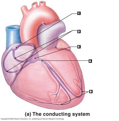
Label the following structures of the conducting system of the heart.
A. AV Bundle
B. Purkinje Fibers
C. SA Node
D. Internodal Pathways
E. AV Node
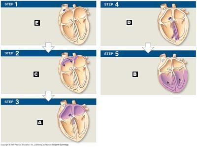
Label the following activities of the impulse conduction through the heart.
E. SA node activity and atrial activation begin.
C. Electrical impulses spread across both atria.
A. Atrial contraction begins.
D. The electrical impulse moves down the interventricular septum.
B. Atrial contraction is over and ventricular contraction begins.
Put the following steps to blood flow through the heart in the correct order:
A. venae cavae
B. right ventricle
C. pulmonary arteries
D. pulmonary veins
E. pulmonary trunk
F. right atrium
A. venae cavae
F. right atrium
B. right ventricle
E. pulmonary trunk
C. pulmonary arteries
D. pulmonary veins
Beginning at the natural pacemaker region, arrange the components of the heart's conducting system in the order that an action potential would pass, by matching (1) through (6):
a. AV bundle (bundle of His)
b. internodal pathways
c. Purkinje fibers
d. atrioventricular (AV) node
e. sinoatrial (SA) node
f. left and right bundle branches
E. SA node
B. Internodal pathways
D. AV node
A. AV bundle
F. left and right bundle branches
C. Purkinje Fibers
Put the steps of the cardiac cycle in order, beginning with the resting period between heart beats.
a. Ventricular diastole early
b. Ventricular diastole late
c. Atrial systole
d. Ventricular systole first phase
e. Ventricular systole second phase
f. Atrial diastole
C. Atrial Systole
F. Atrial Diastole
D. Ventricular Systole 1st phase
E. Ventricular Systole 2nd phase
A. Ventricular Diastole Early
B. Ventricular Diastole Late
P wave
Depolarization of the atria
P-R Interval
Transmission of the impulse to contract from the SA node to the AV node and through the ventricles
QRS complex
Ventricular depolarization
Q-T interval
Time required for the ventricles to undergo a single cycle of depolarization and repolarization
T wave
Repolarization of ventricles
Vessels that carry blood away from the heart are called __________.
Arteries
When the heart beats, the __________ contract first.
atria
The left atrium collects blood from the __________ and empties into the left ventricle.
pulmonary circuit
When blood is ejected from the heart, it is pushed from the ____ to the _____.
atria; ventricle
apex; base
ventricle; atria
base; apex
apex; base
The heart has _____ chambers and ______ valves.
4;4
The epicardium of the heart is the same as the __________.
visceral pericardium
On the outside of the heart, the boundaries between the right and left ventricles are marked by the __________.
anterior interventricular sulcus and the posterior interventricular sulcus
The expandable outer portion of each atrium is called __________.
an auricle
The right atrium receives blood from the __________.
superior vena cava
coronary sinus
inferior vena cava
All of the listed responses are correct.
All of the listed responses are correct.
The AV valves prevent the backflow of blood into the _____, and the semilunar valves prevent backflow of blood into the _____.
atria; ventricles
The right and left AV valves are also called the ______ and _______, respectively.
tricuspid; bicuspid
The free edges of the AV valves are attached to fibers called __________.
chordae tendineae
The left atrium receives blood from the __________.
pulmonary veins
The innermost layer of the heart is called the __________.
endocardium
Deoxygenated blood leaves the right ventricle through a semilunar valve and enters the __________.
pulmonary trunk
Which of the following statements concerning the heart is FALSE?
The heart sits at an angle to the longitudinal axis of the body.
The heart lies slightly to the left of midline.
The heart is rotated toward the right.
The heart is surrounded by the pericardial cavity.
The heart is rotated toward the right.
Compared to the right ventricle, the left ventricle __________.
develops less pressure during contraction
holds less blood
has a thicker wall
increases in diameter during contraction
has a thicker wall
The valve known as the mitral valve is located __________.
between the left atrium and left ventricle
Marginal branches are extensions of the __________.
right coronary artery
In contrast to skeletal muscle, cardiac muscle cells have a _______ phase as part of their action potential.
Plateau
The SA node acts as the pacemaker of the heart because these cells are __________.
the cells that depolarize and reach threshold first
The refractory period of cardiac muscles is __________ than that of skeletal muscles.
longer
If undisturbed, the cells of the AV node will depolarize about __________ times per second.
40–60
Without external interactions, the cells of the SA node depolarize __________ times per minute.
80-100
The Purkinje fibers __________.
conduct impulses very slowly
do not conduct impulses
conduct impulses rapidly
spontaneously depolarize 100 times per second
conduct impulses rapidly
The bundle branches ___________.
extend to the apex of the heart
transmit impulses to the left and right ventricle.
conduct impulses to the Purkinje fibers
All of the listed responses are correct.
All of the listed responses are correct
The QRS complex represents __________.
Ventricular Depolarization
The P wave represents __________.
Atrial Depolarization
The T wave represents _________.
Ventricular Repolarization
Which of the following correctly describes conducting cells of the SA node?
They are smaller than contractile cells.
They cannot maintain a stable resting potential.
They generate actions potentials at a rate of 80–100 per minute.
All of the listed responses are correct.
All of the listed responses are correct.
The contractile cells of the myocardium reach threshold because of an influx of ____.
cations
Which of the following statements concerning contractile cells and the heartbeat is INCORRECT?
The opening of fast channels in the membranes of contractile cells results in a rapid depolarization.
During the absolute refractory period, the membrane is incapable of responding to a new stimulus.
The plateau phase of the contractile cell's action potential is the result of calcium ions moving out of the cell.
Contractile cells form the bulk of the atrial and ventricular walls.
The plateau phase of the contractile cell's action potential is the result of calcium ions moving out of the cell.
********
The conduction delay that occurs at the AV node allows __________.
the atria to contract before the ventricles begin to contract
An elderly man is brought into the hospital on a hot, sunny day complaining of light-headedness. You administer an ECG and notice that the P–R interval is 350 msec. His heart sounds are normal. His blood tests indicate that the LDH, SGOT, CK, and CK-MB levels are all normal. What is wrong with this patient?
The man has a problem with the conduction system of the atria or possibly with the AV node.
During ventricular filling, the AV valves are ____, and the semilunar valves are _______.
open;closed
Contraction of the chambers of the heart is called _____, and relaxation of the chambers of the heart is called _____.
systole;diastole
When atrial contraction begins, the ventricles are __________.
relaxed and filling
The dicrotic notch marks the point when the __________.
aortic valve closes
The first heart sound, "lubb," marks the point when __________.
the atrioventricular valves close
There is (are) __________ heart sound(s).
4
Which of the following statements concerning atrial systole is FALSE?
The volume of blood in the ventricle at the end of atrial systole is end-diastolic volume, or EDV.
During atrial systole, blood moves from the atrium to the ventricle through the AV valve.
During atrial systole, the pressure in the atrium is higher than the pressure in the ventricle.
At the start of atrial systole, there is very little blood in the ventricles.
At the start of atrial systole, there is very little blood in the ventricles.
Which of the following statements concerning ventricular diastole is FALSE?
The elasticity of the connective tissues of the heart and fibrous skeleton causes ventricles to expand during ventricular diastole.
At the beginning of ventricular diastole, both the AV valves and semilunar valves are closed.
As the ventricles begin to fill with blood, the pressure in the chamber rises.
When the AV valves initially open, both the atria and the ventricles are in diastole.
As the ventricles begin to fill with blood, the pressure in the chamber rises.
The stroke volume of the heart is approximately __________.
80mL
Stroke volume is defined as __________.
the amount of blood ejected from each ventricle during ventricular systole
Cardiac output is defined as __________.
HR × (EDV − ESV)
HR × EDV
HR × SV
both HR × SV and HR × (EDV − ESV)
None of the listed responses is correct.
both HR × SV and HR × (EDV − ESV)
Which statement(s) is (are) true with regard to preload?
If EDV is greater and ESV is lower, then the stroke volume increases.
Preload is increased with a rapid heartbeat.
During exercise, increased venous return increases EDV.
If EDV is greater and ESV is lower, then the stroke volume increases; AND during exercise, increased venous return increases EDV.
All of these statements are true.
If EDV is greater and ESV is lower, then the stroke volume increases; AND during exercise, increased venous return increases EDV.
Starling's law of the heart refers to the relationship between __________.
EDV and SV
Which of the following would be considered a positive inotropic agent?
beta-blocking medications
acetylcholine
calcium-blocking medications
digitalis
digitalis
In a normal resting adult, the effects of the __________ division of the autonomic nervous system dominate.
parasympathetic
The cardiac output (CO) is equal to __________.
SV x HR
Which of the following is NOT a factor that controls stroke volume?
sympathetic stimulation of the myocardium
cardiac output
contractility of the heart
end-systolic volume
end-diastolic volume
cardiac output
What is the term for an abnormally slow heart rate?
bradycardia
What does the Frank–Starling principle state?
There is a direct relationship between the EDV and the SV.
True/False. Bradycardia is the term used to describe a faster than normal heart beat.
False. Tachycardia is the term used to describe a faster than normal heart rate.
True/False. Starling’s law of the heart states that increasing the EDV leads to an increase in the stroke volume.
True
True/False. The AV valves are part of the heart’s fibrous skeleton.
True
True/False. The coronary arteries are branches of the superior vena cava.
False. The coronary arteries are the first branches of the aorta.
True/False. The pericardial sac is lined by the visceral pericardium.
False. The visceral pericardium covers or is attached directly to the heart.
Since the absolute refractory period in cardiac muscle is nearly as long as the contraction phase, _____ contractions cannot occur in normal cardiac muscle cells.
tetanic
The end-diastolic volume (EDV) is affected by the filling time and the _____. (use two words)
Venous return
Calcium entry into the cardiac muscle cell creates a _____ in the action potential that lasts about 175 msec.
Plateau
When _____ is released from autonomic neurons at the heart, the repolarization period is shortened and nodal cells reach threshold more quickly.
norepinephrine; NE
The blood vessels in the cardiovascular system are subdivided into three circuits known as the __________.
coronary, pulmonary, and systemic circuits
Which statement is true?
If the pacemaker of the heart stops, the AV node will take over.
The AV node can generate electrical impulses just as quickly as the SA node.
The autonomic nervous system has no effect on the heart.
If the pacemaker of the heart fails, then the heart will stop beating.
If the pacemaker of the heart stops, the AV node will take over.
The left atrium receives blood from the pulmonary circuit and empties it into the __________.
conus arteriosus
right atrium
left ventricle
right ventricle
left ventricle
The "double pump" function of the heart includes the right side, which serves as the __________ circuit pump, while the left side serves as the __________ pump.
pulmonary; hepatic portal
pulmonary; systemic
hepatic portal; cardiac
systemic; pulmonary
pulmonary; systemic
The coronary arteries emerge at the base of the __________.
pulmonary trunk
aorta
inferior vena cava
circumflex branch
aorta
Which blood vessels are known as exchange vessels?
veins
capillaries
arteries
All of the listed responses are correct.
capillaries
Blood from the coronary circuit is collected on the posterior aspect of the heart in a blood vessel known as the __________.
interventricular vein
great cardiac vein
circumflex branch
coronary sinus
coronary sinus
During the action potential in a contractile cardiac muscle cell, the opening of slow calcium channels results in the______ phase.
repolarization
depolarization
plateau
hyperpolarization
plateau
When deoxygenated blood leaves the right ventricle through a semilunar valve, it is forced into the __________.
aortic arch
lung capillaries
pulmonary arteries
pulmonary veins
pulmonary arteries
The passageways between cardiac muscle cells that allow ions to pass freely are called __________.
trabeculae carneae
anastomoses
gap junctions
desmosomes
gap junctions
Which of the following are characteristics of cardiac muscle cells?
nonstriated, multinucleated, and involuntary
striated, multinucleated, and voluntary muscle
striated, single central nucleus, and involuntary
striated, single central nucleus, and voluntary
striated, single central nucleus, and involuntary
Cardiac muscle tissue __________.
will not contract unless stimulated by autonomic nerves
will not contract unless stimulated by somatic motor neurons
is innervated mostly by neurons associated with the sympathetic division of the ANS
has its own intrinsic conduction system that can set the pace of the beating heart
has its own intrinsic conduction system that can set the pace of the beating heart
The right coronary artery generally gives rise to __________.
the posterior interventricular artery
the circumflex branch
the marginal branches
the marginal branches and the posterior interventricular artery
All the listed responses are correct.
the marginal branches and the posterior interventricular artery
The left coronary artery supplies blood to __________.
the circumflex branch
the anterior interventricular artery
the posterior descending artery
the anterior interventricular artery and the circumflex branch
All the listed responses are correct.
the anterior interventricular artery and the circumflex branch
What is the correct sequential path of a normal action potential in the heart?
SA node, AV node, bundle branches, AV bundle, Purkinje fibers
AV node, SA node, AV bundle, bundle of His
SA node, AV node, bundle of His, bundle branches, Purkinje fibers
SA node, AV bundle, AV node, Purkinje fibers
SA node, AV node, bundle of His, bundle branches, Purkinje fibers
Blood flows from the left atrium through the _______ to the left ventricle.
aortic valve
tricuspid valve
pulmonary valve
mitral valve
mitral valve
The P wave on the ECG indicates __________.
the electrical events spreading out over both atria
the spread of the electrical events down the interventricular septum
the contraction of the atria
the spread of electrical events over both ventricles
the electrical events spreading out over both atria
After the AV node is depolarized and the impulse spreads through the atria, there is a slight delay before the impulse spreads to the ventricles. The reason for this delay is to allow __________.
the ventricles to contract
a greater venous return
the atria to contract
the atria to fill with blood
the atria to contract
Valvular heart disease can be a result of __________.
congenital malformation
rheumatic fever
carditis
All of the listed responses are correct.
All of the listed responses are correct.
The QRS complex of the ECG appears as the __________.
ventricles repolarize
atria depolarize
ventricles depolarize
atria repolarize
ventricles depolarize
When a chamber of the heart fills with blood and prepares for the start of the next cardiac cycle, the chamber is in __________.
diastole
systole
ventricular ejection
isovolumetric contraction
diastole
During the isovolumetric contraction phase, the pressure in the _____ has to rise above aortic pressure for ventricular ejection to occur.
right atria
left atria
right ventricle
left ventricle
left ventricle
How would you define cardiac output?
the amount of blood pumped out of both ventricles in one minute
the amount of blood pumped out of the left ventricle in one minute
the amount of blood pumped out of the atria and ventricles in one minute
the end-diastolic volume plus the end-systolic volume
the amount of blood pumped out of the left ventricle in one minute
The amount of blood pumped out of each ventricle during a single beat is the __________.
end-systolic volume
cardiac output
end-diastolic volume
stroke volume
stroke volume
Under normal circumstances, the factors responsible for making delicate adjustments to the heart rate as circulatory demands change are __________.
cardiac output and stroke volume
nerve activity and muscular activity
autonomic activity and circulatory hormones
All of the listed responses are correct.
autonomic activity and circulatory hormones
The cardiac centers in the medulla oblongata monitor baroreceptors and chemoreceptors innervated by the __________.
glossopharyngeal N IX and vagus N X
accessory N XI and hypoglossal N XII
facial N VII and vesitbulocochlear N VIII
trochlear N IV and trigeminal N V
glossopharyngeal N IX and vagus N X
The difference between the end-diastolic volume (EDV) and the end-systolic volume (ESV) is the __________.
preload and afterload
cardiac output
stroke volume
cardiac reserve
stroke volume
Parasympathetic stimulation from the vagus nerve results in __________.
a decrease in heart rate
an increased heart rate and cardiac output
no effect on the heart rate
more forceful ventricular contractions
a decrease in heart rate
Which of the following statements is part of Starling's law of the heart?
A slightly larger and better-contracting heart will increase stroke volume.
An increase in venous return will increase blood flow into the heart.
The greater the ventricular stretch, the more forceful the contraction.
All of the listed responses are correct.
All of the listed responses are correct.
Physicians are interested in cardiac output because it provides a useful indication of __________.
valvular malfunctions
atrial efficiency in respect to time
ventricular efficiency over time
the amount of blood ejected by each ventricle
ventricular efficiency over time
Which heart chamber has the thickest muscular walls?
left ventricle
right ventricle
right atrium
left atrium
left ventricle
Which of the following medications serves as a positive treatment by interfering with the removal of calcium ions from the sarcoplasm of cardiac muscle cells?
digitalis
beta blockers
acetylcholine
verapamil
digitalis
What is the function of the chordae tendineae?
to anchor the aortic valve flaps and prevent backflow into the ventricles
to anchor the AV valve flaps and prevent backflow of blood into the atria
to anchor the bicuspid valve flaps and prevent backflow of blood into the ventricle
to anchor the semilunar valve flaps and prevent backward flow of blood into the ventricles
to anchor the AV valve flaps and prevent backflow of blood into the atria
Which of the following would NOT show up on an electrocardiogram?
conduction deficits
SA node damage
cardiac arrhythmias
murmurs
murmurs
During ventricular systole , what occurs when the pressure in the left ventricle rises above that in the left atrium?
The aortic valve closes.
The left AV valve opens.
All the valves close.
The left AV valve closes.
The left AV valve closes.
During ventricular systole, the blood volume in the atria is __________, and the volume in the ventricle is __________.
decreasing; decreasing
increasing; decreasing
increasing; increasing
decreasing; increasing
increasing; decreasing
Cardiac muscle cells are nourished via blood supply provided from the __________.
pulmonary circuit
coronary circuit
systemic circuit
All of the listed responses are correct
coronary circuit
Pulmonary arteries carry blood to the __________.
lungs
aorta
left atria
right atria
lungs
Which of the following statements is (are) true?
If you increase heart rate, then you will increase SV.
During ventricular systole, the papillary muscles contract to keep the AV valves shut and prevents them from swinging up into the atria.
If you increase ESV, then you will increase SV.
None of these statements is true.
During ventricular systole, the papillary muscles contract to keep the AV valves shut and prevents them from swinging up into the atria.
The heart sound associated with S2 occurs as the ventricles ____ and the semilunar valves ______.
relax; open
relax; close
contract; close
contract; open
relax; close
During isovolumetric systole, pressure is highest in the __________.
left atrium
pulmonary veins
aorta
left ventricle
left ventricle
Blood pressure in the large systemic arteries is greatest during __________.
isovolumetric systole
atrial systole
isovolumetric diastole
ventricular ejection
ventricular ejection
Decreased parasympathetic (vagus) stimulation to the heart results in a situation known as __________.
stenosis
bradycardia
carditis
tachycardia
tachycardia
Serious arrhythmias that reduce the pumping efficiency of the heart may indicate __________.
injury to the SA and AV nodes
variations in the ionic composition of the extracellular fluids
damage to the myocardium
All of the listed responses are correct.
All of the listed responses are correct.
During exercise, the most important control mechanism to increase cardiac output is __________.
increased end-systolic volume
increased acetylcholine release from the vagus nerve
increased body temperature
increased sympathetic activity to the ventricles
increased sympathetic activity to the ventricles
In the diastole phase of the ventricles, __________.
presystolic pressures are high
the ventricles are "resting"
the semilunar valves are open
the AV valves are closed
the ventricles are "resting"
Which of the following does NOT control the movement of blood through the heart?
the conducting system
size of the atria and ventricles
contraction of the myocardium
opening and closing of the valves
size of the atria and ventricles
Valvular malfunction in the heart __________.
interferes with ventricular contraction
interferes with movement of blood through the heart
increases the cardiac output
causes an increase in the amount of blood pumped out of each ventricle
interferes with movement of blood through the heart
If the bicuspid valve is defective and valvular regurgitation occurs, the end result is __________.
an insufficient amount of blood available to be moved into the aorta and systemic circulation
an overstretching of the left ventricle, causing a decrease in the force of contraction
gushing of blood into the left ventricle, causing excessive pressure
All of the listed responses are correct.
an insufficient amount of blood available to be moved into the aorta and systemic circulation
Which of the following is NOT a characteristic of cardiac muscle cells?
multiple nuclei
calcium entry is a trigger for contraction
mitochondria account for 25 percent of cell volume
short T tubules
multiple nuclei