The Skeletal System
• Skeletal system includes:
– bones of the skeleton
– cartilages, ligaments, and connective tissues
Functions of the Skeletal System
1. Support
2. Storage of minerals (calcium)
3. Storage of lipids (yellow marrow)
4. Blood cell production (red marrow)
5. Protection
6. Leverage (force of motion)
Classification of Bones
• Bone are identified by:
– shape
– internal tissues
– bone markings
Bone Shapes
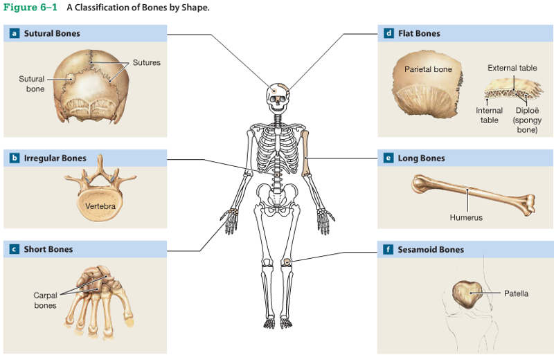
1. Long bones
2. Flat bones
3. Sutural bones
4. Irregular bones
5. Short bones
6. Sesamoid bones
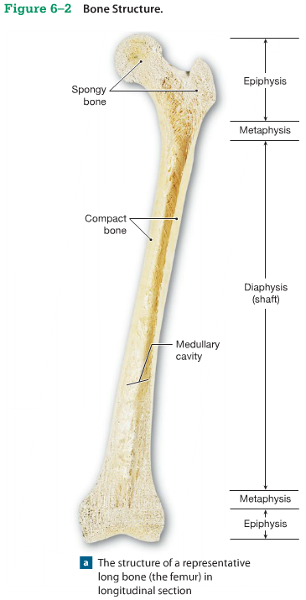
Long Bones
(Bone Shapes)
• Are long and thin
• Are found in arms, legs, hands, feet, fingers, and toes
• Diaphysis:
– the shaft
- A heavy wall of compact bone, or dense bone
- A central space called marrow cavity
• Epiphysis:
– wide part at each end
– articulation with other bones
• Mostly spongy (cancellous)bone
• Covered with compact bone (cortex)
• Metaphysis:
– where diaphysis and epiphysis meet
Flat Bones
(Bone Shapes)
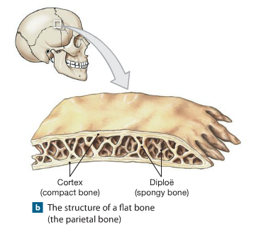
• Are thin with parallel surfaces
• Are found in the skull, sternum, ribs,and scapula
• Resembles a sandwich of spongy bone
• Between 2 layers of compact bone
Sutural Bones
(Bone Shapes)
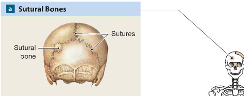
• Are small,irregular bones
• Are found between the flat bones of the skull
Irregular Bones
(Bone Shapes)
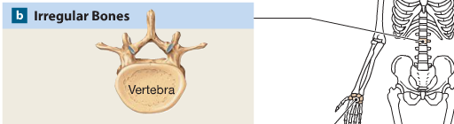
• Have complex shapes
• Examples:
– spinal vertebrae
– pelvic bones
Short Bones
(Bone Shapes)
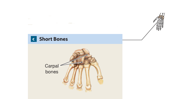
• Are small and thick
• Examples:
– ankle
– wrist bones
Sesamoid Bones
(Bone Shapes)
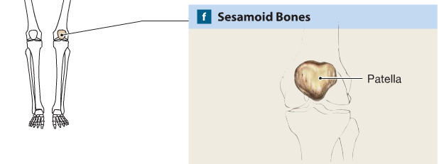
• Are small and flat
• Develop inside
tendons near joints of knees, hands, and feet
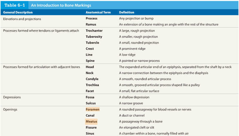
Bone Markings
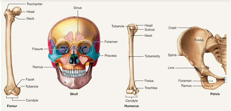
• Depressions or grooves:
‐ along bone surface
– Projections:
– where tendons and ligaments attach
– at articulations with other bones
• Tunnels:
– where blood and nerves enter bone
Bone Markings
Bone (Osseous) Tissue
• Dense, supportive connective tissue
• Contains specialized cells
• Produces solid matrix of calcium salt deposits
• Around collagen fibers
Characteristics of Bone Tissue
• Dense matrix, containing:
– deposits of calcium salts
– bone cells within lacunae organized around blood vessels
• Canaliculi:
– form pathways for blood vessels
– exchange nutrients and wastes
• Periosteum:
– covers outer surfaces of bones
– consist of outer fibrous and inner cellular layers
Matrix Minerals
• 2/3 of bone matrix is calcium phosphate, Ca3(PO4)2:
– reacts with calcium hydroxide, Ca(OH)2 to form – crystals of hydroxyapatite, Ca10(PO4)6(OH)2
– which incorporates other calcium salts and ions
Matrix Proteins
• 1/3 of bone matrix is protein fibers (collagen)
Bone Cells
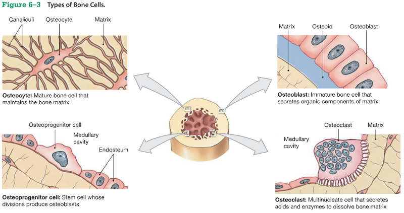
• Make up only 2% of bone mass:
- Osteocytes
- Osteoblasts
– osteoprogenitor cells
– osteoclasts
Osteocytes
(Bone Cells)
• Mature bone cells that maintain the bone matrix
• Live in lacunae
• Are between layers (lamellae) of matrix
• Connect by cytoplasmic extensions through canaliculi in lamellae
• Do not divide
Functions
• To maintain protein and mineral content of
matrix
• To help repair damaged bone
Osteoblasts
(Bone Cells)
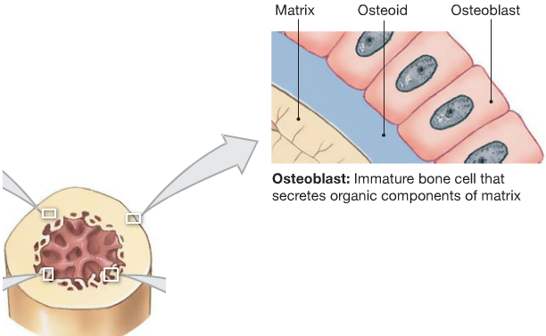
• Immature bone cells that secrete matrix compounds
(osteogenesis)
Osteoid
• Matrix produced by osteoblasts, but not yet
calcified to form bone
• Osteoblasts surrounded by bone become
osteocytes
Osteoprogenitor Cells
(Bone Cells)
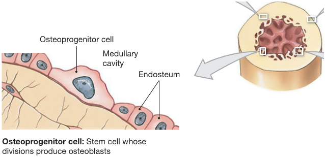
• Mesenchymal stem cells that divide to produce
osteoblasts
• Are located in inner,cellular layer of periosteum
(endosteum)
• Assist in fracture repair
Osteoclasts
(Bone Cells)
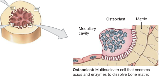
• Secrete acids and proteindigesting enzymes
• Giant, mutlinucleate cells
• Dissolve bone matrix and release stored minerals
(osteolysis)
• Are derived from stem cells that produce macrophages
Homeostasis
• Bone building (by osteocytes) and bone recycling (by osteoclasts) must balance:
– more breakdown than building, bones become weak
– exercise causes osteocytes to build bone
Compact Bone
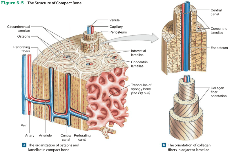
Osteon
• The basic unit of mature compact bone
• Osteocytes are arranged in concentric lamellae
• Around a central canal containing blood vessels
Perforating Canals
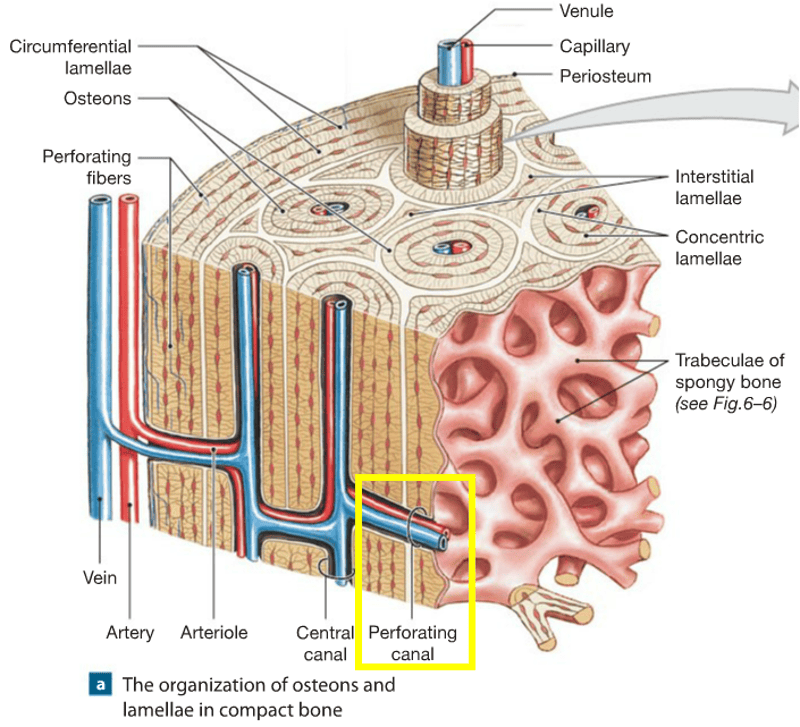
• Perpendicular to the central canal
• Carry blood vessels into bone and marrow
Circumferential Lamellae
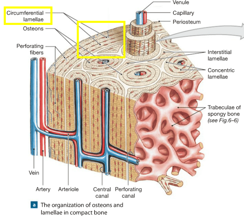
• Lamellae wrapped around the long bone
• Binds osteons together
Spongy Bone

• Does not have osteons
• The matrix forms an open network of trabeculae
• Trabeculae have no blood vessels
Red Marrow
• The space between trabeculae is filled with red bone marrow:
– which has blood vessels
– forms red blood cells
– and supplies nutrients to osteocytes
Yellow Marrow
• In some bones, spongy bone holds yellow bone marrow:
– is yellow because it stores fat
Weight–Bearing Bones
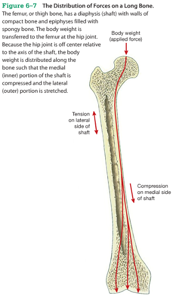
• The femur transfers weight from hip joint to knee joint:
– causing tension on the lateral side of the shaft
– and compression on the medial side
Periosteum and Endosteum
• Compact bone is covered with membrane:
– periosteum on the outside
– endosteum on the inside
Periosteum
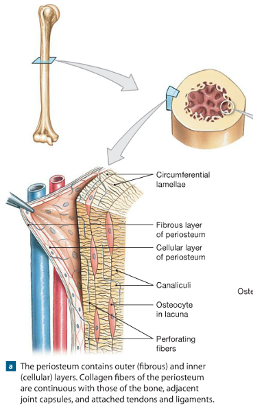
• Covers all bones:
– except parts enclosed in joint capsules
• It is made up of:
– an outer, fibrous layer
– and an inner,cellular layer
Functions of Periosteum
1. Isolate bone from surrounding tissues
2. Provide a route for circulatory and nervous
supply
3. Participate in bone growth and repair
Perforating Fibers
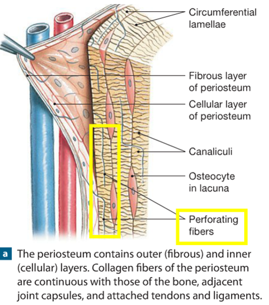
• Collagen fibers of the periosteum:
– connect with collagen fibers in bone
– and with fibers of joint capsules,attached tendons,and ligaments
Endosteum
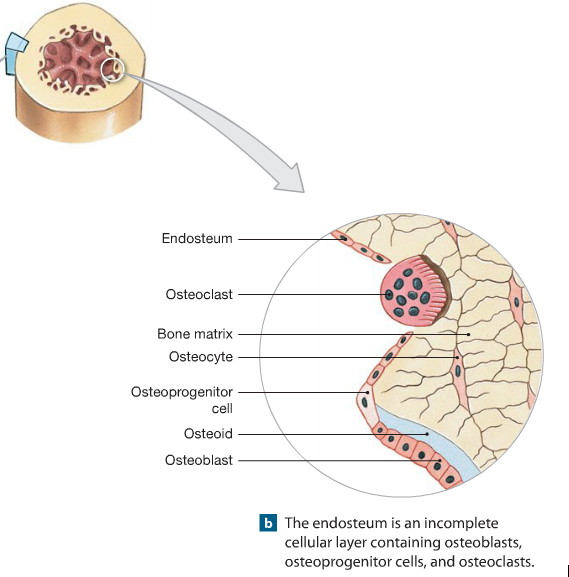
• An incomplete cellular layer:
– lines the marrow cavity
– covers trabeculae of spongy bone
– lines central canals
• Contains osteoblasts,osteoprogenitor cells, and osteoclasts
• Is active in bone growth and repair
Bone Development
• Human bones grow until about age 25
• Osteogenesis:
– bone formation
• Ossification:
– the process of replacing other tissues
with bone
Calcification
• The process of depositing calcium salts
• Occurs during bone ossification and in other
tissues
Ossification
• The 2 main forms of ossification are:
– intramembranous ossification
– endochondral ossification
Intramembranous Ossification
• Also called dermal ossification:
– because it occurs in the dermis
– produces dermal bones such as mandible and
clavicle
• There are 3 main steps in intramembranous
ossification
Intramembranous Ossification: Step 1
• Mesenchymal cells aggregate:
– differentiate into osteoblasts
– begin ossification at the ossification center
– develop projections called spicules
Intramembranous Ossification: Step 2
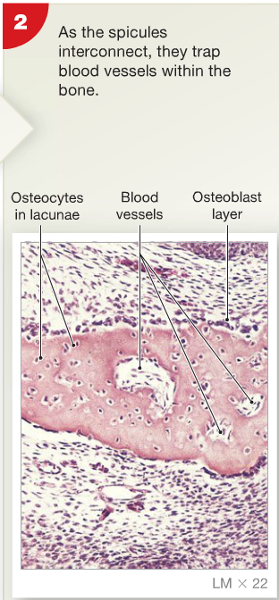
• Blood vessels grow
into the area:
– to supply the osteoblasts
• Spicules connect:
– trapping blood vessels inside bone
IntramembranousOssification: Step 3
• Spongy bonedevelops and is remodeled into:
– osteons of compact bone
– periosteum
– or marrow cavities
Endochondral Ossification
• Ossifies bones that originate as hyaline
cartilage
• Most bones originate as hyaline cartilage
• Growth and ossification of long bones occurs in 6 steps
• Appositional growth:
– compact bone thickens and strengthens long bone with layers of circumferential lamellae
Endochondral Ossification: Step 1
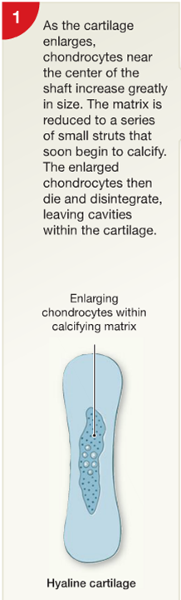
• Chondrocytes in the center of hyaline cartilage:
– enlarge
– form struts and calcify
– die, leaving cavities in cartilage
Endochondral Ossification: Step 2
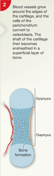
• Blood vessels grow around the edges of the cartilage
• Cells in the perichondrium change to osteoblasts:
– producing a layer of superficial bone around the shaft which will continue to
grow and become compact bone (appositional growth)
Endochondral Ossification: Step 3
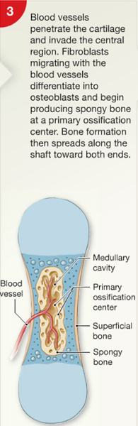
• Blood vessels enter the cartilage:
– bringing fibroblasts that become osteoblasts
– spongy bone develops at the primary ossification center
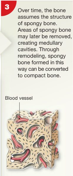
Endochondral Ossification:
Step 4
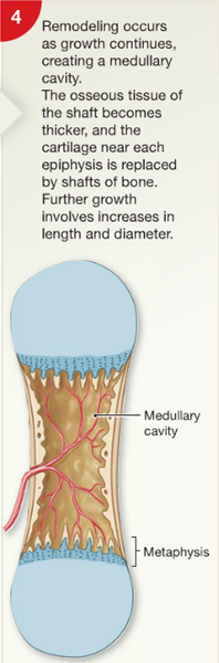
• Remodeling creates a marrow cavity:
– bone replaces cartilage at the metaphyses
Endochondral Ossification: Step 5
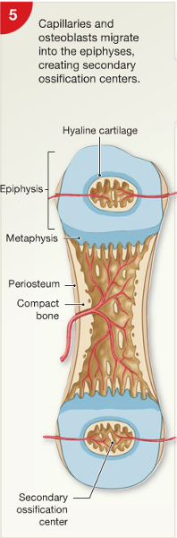
• Capillaries and osteoblasts
enter the epiphyses:
– creating secondary ossification centers
Endochondral Ossification: Step 6
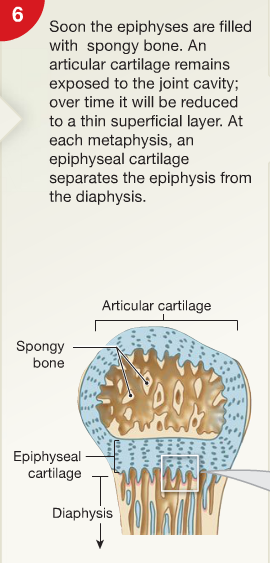
• Epiphyses fill with spongy bone:
– cartilage within the joint cavity is articulation cartilage
– cartilage at the metaphysis is epiphyseal
cartilage
Endochondral Ossification: Step 7
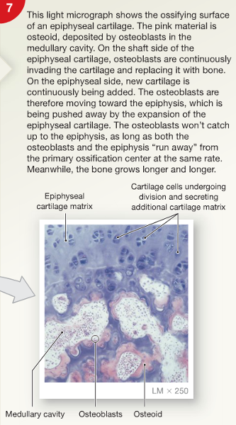
• As long as the epiphyseal cartilage continues to grow at its epiphyseal surface, the bone will continue to increase in length.
Bone Growth in Length
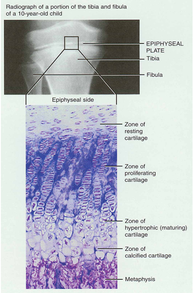
• Epiphyseal plate or cartilage growth plate
- cartilage cells are produced by mitosis on epiphyseal side of plate
- cartilage cells are destroyed and replaced by bone on diaphyseal side of plate
• Between ages 18 to 25, epiphyseal plates close.
- cartilage cells stop dividing and bone replaces the cartilage (epiphyseal line)
• Growth in length stops at age 25
Zones of Growth in Epiphyseal Plate
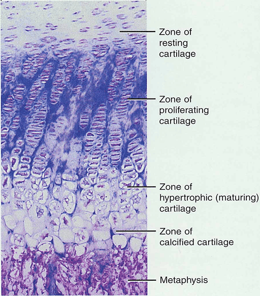
• Zone of resting cartilage
- anchors growth plate to bone
• Zone of proliferating cartilage
- rapid cell division (stacked coins)
• Zone of hypertrophic cartilage
- cells enlarged & remain in columns
• Zone of calcified cartilage
- thin zone, cells mostly dead since matrix calcified
- osteoclasts removing matrix
- osteoblasts & capillaries move in to create bone over calcified cartilage
Bone Growth in Width
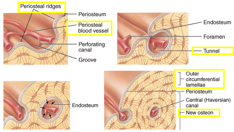
• Only by appositional growth at the bone’s surface
• Periosteal cells differentiate into osteoblasts and form bony ridges and then a tunnel around periosteal blood vessel.
• Concentric lamellae fill in the tunnel to form an osteon.
Epiphyseal Lines
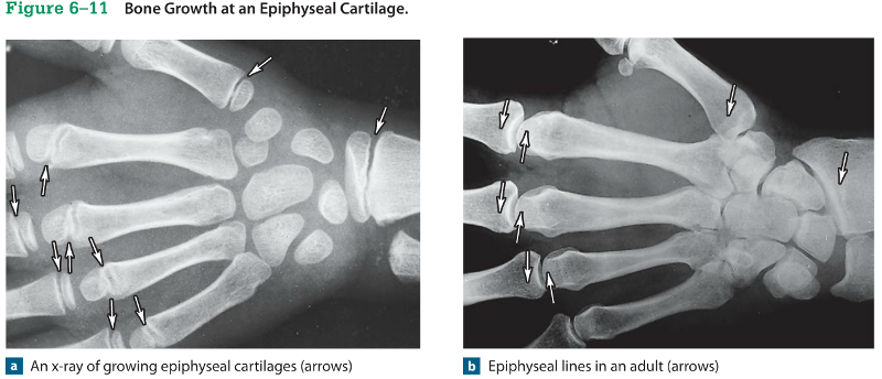
• When long bone stops growing, after puberty:
– epiphyseal cartilage disappears
– is visible on X‐rays as an epiphyseal line
Mature Bones
• As long bone matures:
– osteoclasts enlarge marrow cavity
– osteons form around blood vessels in compact
bone
Blood Supply of Mature Bones
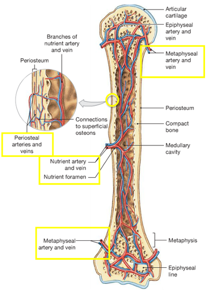
• 3 major sets of blood vessels develop
1) Nutrient artery and vein:
– a single pair of large blood vessels
– enter the diaphysis through the nutrient foramen
– femur has more than 1 pair
2) Metaphyseal vessels:
– supply the epiphyseal cartilage
– where bone growth occurs
3) Periosteal vessels provide:
– blood to superficial osteons
– secondary ossification centers
Lymph and Nerves
• The periosteum also contains:
– networks of lymphatic vessels
– sensory nerves
Remodeling
• The adult skeleton:
– maintains itself
– replaces mineral reserves
• Remodeling:
– recycles and renews bone matrix
– involves osteocytes, osteoblasts, and osteoclasts
• Bone continually remodels, recycles, and replaces
• Turnover rate varies
• If deposition is greater than removal, bones get stronger
• If removal is faster than replacement, bones get weaker
Effects of Exercise on Bone
• Mineral recycling allows bones to adapt to stress
• Heavily stressed bones become thicker and stronger
Bone Degeneration
• Bone degenerates quickly
• Up to 1/3 of bone mass can be lost in a few weeks of inactivity
• What you don’t use, you lose
• Stresses applied to bones during physical activity are essential to maintain bone strength and mass
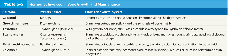
Effects of Hormones and Nutrition on Bone
• Normal bone growth and maintenance
requires nutritional and hormonal factors
Minerals
Effects of Hormones and Nutrition on Bone
• A dietary source of calcium and phosphate salts:
– plus small amounts of magnesium, fluoride, iron,
and manganese
Vitamins
Effects of Hormones and Nutrition on Bone
• Vitamin C is required for collagen synthesis,
and stimulates osteoblast differentiation
• Vitamin A stimulates osteoblast activity
• Vitamins K and B12 help synthesize bone proteins
Calcitriol
Effects of Hormones and Nutrition on Bone
• The hormone calcitriol:
– is made in the kidneys
– helps absorb calcium and phosphorus from digestive tract
– synthesis requires vitamin D3 (cholecalciferol)
Other Hormones
Effects of Hormones and Nutrition on Bone
• Growth hormone and thyroxine stimulate
bone growth
• Estrogens and androgens stimulate osteoblasts
• Calcitonin and parathyroid hormone regulate
calcium and phosphate levels
Hormones for Bone Growth and Maintenance
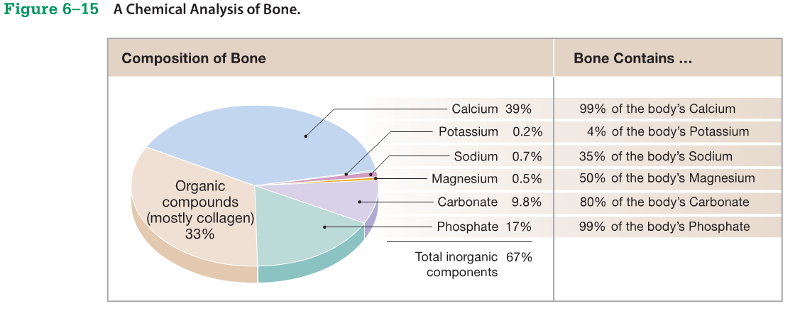
The Skeleton as Calcium Reserve
• Bones store calcium and other minerals
• Calcium is the most abundant mineral in the
body
Chemical Composition of Bone
Functions of Calcium
• Calcium ions are vital to:
– membranes
– neurons
– muscle cells, especially heart cells
Calcium Regulation
• Calcium ions in body fluids:
– must be closely regulated
• Homeostasis is maintained:
– by calcitonin and parathyroid hormone
– which control storage, absorption, and excretion
KEY CONCEPTS
• Calcium and phosphate ions in blood are lost in urine
• Ions must be replaced to maintain homeostasis
• If not obtained from diet, ions are removed
from the skeleton, weakening bones
• Exercise and nutrition keep bones strong
Calcitonin and Parathyroid Hormone Control
• Bones:
– where calcium is stored
• Digestive tract:
– where calcium is absorbed
• Kidneys:
– where calcium is excreted
Parathyroid Hormone (PTH)
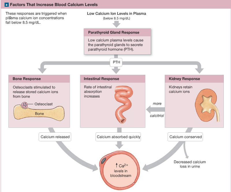
• Low calcium ion levels in the blood cause the parathyroid glands in neck to secrete Parathyroid Hormone (PTH)
• Increases calcium ion levels by:
– stimulating osteoclasts to release stored calcium ions from the bone
– increasing intestinal absorption of calcium
- kidneys retain calcium ions
– decreases calcium loss in urine
- calcium is absorbed quickly in intestines
Calcitonin
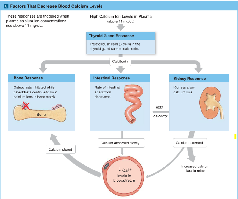
• High calcium ion levels in blood cause Calcitonin to be secreted by C cells (parafollicular cells) in thyroid
• Decreases blood calcium ion levels by:
– inhibiting osteoclast activity
- rate of intestinal absorption decreases
- Kidneys allow calcium loss
– increasing calcium excretion at kidneys and increased calcium loss in urine
- Calcium is absorbed slowly in intestines
- Calcium is stored in bone matrix
Fractures
• Fractures:
– cracks or breaks in bones
– caused by physical stress
• Fractures are repaired in 4 steps
Fracture Repair: Step 1

• Bleeding:
– produces a clot (fracture hematoma)
– establishes a fibrous network
• Bone cells in the area die
Fracture Repair: Step 2
• Cells of the endosteum and periosteum:
– Divide and migrate into fracture zone
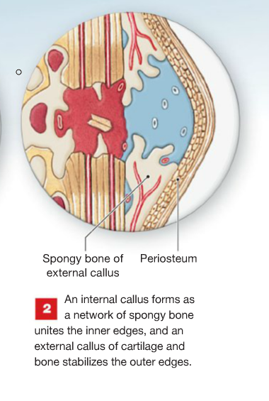
• Calluses stabilize the break:
– external callus of cartilage and bone surrounds break
– internal callus develops in marrow cavity
Fracture Repair: Step 3

• Osteoblasts:
– replace central cartilage of external callus
– with spongy bone
Fracture Repair: Step 4

• Osteoblasts and osteocytes remodel the fracture for up to a year:
– reducing bone calluses
Pott’s fracture
The Major Types of Fractures
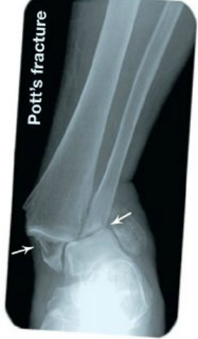
- occurs at the ankle and affects both bones of the leg
Comminuted fractures
The Major Types of Fractures
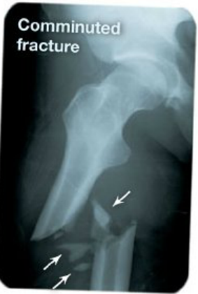
- shatter the affected area into a multitude of bony fragments.
Transverse fractures
The Major Types of Fractures
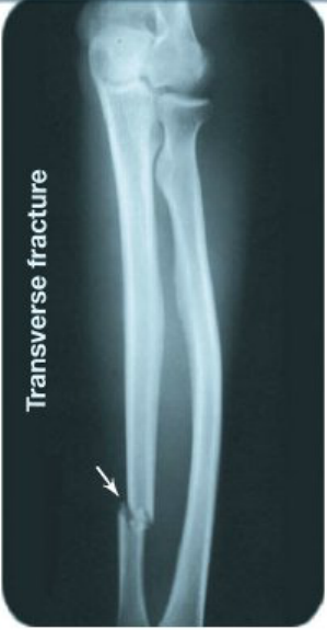
- break a bone shaft across its long axis
Spiral fractures
The Major Types of Fractures
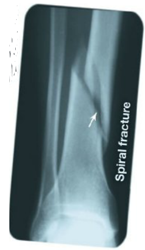
- twisting stresses that spread along the length of the bone
Displaced fractures
The Major Types of Fractures
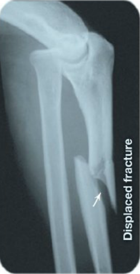
- produce new and abnormal bone arrangements, non-displaced fractures retain the normal alignment of the bones or fragments
Colles’ fracture
The Major Types of Fractures
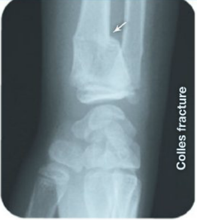
- break in the distal portion of the radius (usually from reaching to cushion a fall)
Greenstick fracture
The Major Types of Fractures
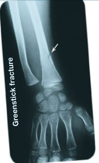
- one side of the shaft is broken, and the other side is bent.
Epiphyseal fractures
The Major Types of Fractures
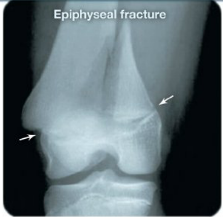
- where bone matrix is undergoing calcification and chondrocytes are dying. A clean transverse fracture along this line can generally heal well. Unless carefully treated, fractures between the epiphysis and the epiphyseal cartilage can permanently stop growth at this site.
Compression fractures
The Major Types of Fractures
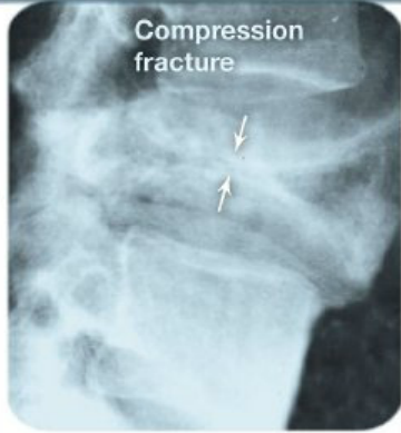
- occur in the vertebrae subjected to extreme streses
Age and Bones
• Bones become thinner and weaker with age
• Osteopenia begins between ages 30 and 40
• Women lose 8% of bone mass per decade, men 3%
Effects of Bone Loss
• The epiphyses, vertebrae, and jaws are most
affected:
– resulting in fragile limbs
– reduction in height
– tooth loss
Osteoporosis
• Severe bone loss
• Affects normal function
• Over age 45, occurs in:
– 29% of women
– 18% of men
Hormones and Bone Loss
• Estrogens and androgens help maintain bone mass
• Bone loss in women accelerates after menopause
Cancer and Bone Loss
• Cancerous tissues release osteoclastactivating
factor:
– that stimulates osteoclasts
– and produces severe osteoporosis
Name the five primary functions of the skeletal system. p170
The 5 primary functions of the skeletal system are: - support,
- storage of minerals and lipids,
- blood cell production,
- protection,
- and leverage.
Identify the six broad categories for classifying a bone according to shape. p173
The 6 broad categories for classifying bones according to shape are:
- flat
- irregular
- long
- sesamoid
- short
- sutrural
Define bone marking. p173
A bone marking, (surface feature) is an area on the surface of a bone structured for a specific function, such as:
- joint formation,
- muscle attachment,
- or the passage of nerves and blood vessels.
Mature bones cells are known as _____, bone-building cells are called __________, and ______ are bone-resorbing cells. p175
Mature bones cells are known as OSTEOCYTES, bone-building cells are called OSTEOBLASTS, and OSTEOCLASTS are bone-resorbing cells.
How would the compressive strength of a bone be affected if the ration of collagen to hydroxyapatite increased? p175
IF the ration of collagen to hydroxyapatite in a bone increased, the bone would become less strong (as well as more flexible.)
If the activity of osteoclasts exceeds the activity of osteoblasts in a bone, how will the mass of the bone be affected? p175
because osteoclasts break down or demineralize bone, the bone would have a reduced mineral content (less mass), as a result, it would be weaker.
Compare the structures and functions of compact bone and spongy bone. p179
Compact bone consists of osteons (Haversian systems) with little space between them. Compact bone lies over spongy bone and makes up most of the diaphysis. It functions to protect, support, and resist stress. Spongy bone consists of trabeculae with numerous red marrow-filled spaces. Spongy bone makes up most of the structure of short, flat, and irregular bones and is also found at the epiphyses of long bones. Spongy bone functions in storing marrow and providing some support.
A sample of bone has lamellae, which are not arranged in osteons, Is the sample most likely taken from the epiphysis or diaphysis? p179
The presence of lamellae that are not arranged in osteons is indicative of spongy bone, which is located in an epiphysis.
During intramembraneous ossification, which type of tissue is replaced by bone? p183
During intramembranous ossification, fibrous connective tissue is replaced by bone.
In endochondral ossification, what is the original source of osteoblasts? p183
In endochondral ossification, cells of the inner layer of the pericondrium differentiate into osteoblasts, and a cartilage model is gradually replaced by bone.
How could x-rays of the femur be used to determine whether a person has reached full height? p183
Long bones of the body, such as the femur, have an epiphyseal cartilage, a plate of cartilage that separates the epiphysis from the diaphysis so long as the bone is still growing lengthwise. An x-ray would indicate whether the epiphyseal cartilage is still present. If it is, growth is still occurring; if it is not, the bone has reached its adult length.
Describe bone remodeling. p184
Bone remodeling refers to the process whereby old bone is continuously being destroyed by osteoclasts while new bone is being constructed by osteoblasts.
Explain how heavy-metal ions could be incorporated into bone matrix. p184
The biochemistry of some heavy-metal ions, such as strontium, cobalt, uranium, and plutonium, is very similar to that of calcium. Osteoblasts cannot differentiate these abnormal heavy-metal ions from normal calcium ions, so the heavy metal ions become incorporated into the bone matrix. Over time, these dangerous ions can be released into circulation during normal bone remodeling.
Why would you expect the arm bones of a weight lifter to be thicker and heavier than those of a jogger? p186
The larger arm muscles of the weight lifter would apply more mechanical stress to the bones of the upper limbs. in response to that stress, the bones would grow thicker.
A child who enters puberty several years later than the average age is generally taller than average as an adult, Why? p186
Growth continues throughout childhood. At puberty, a growth spurt occurs and is followed by the closure of the epiphyseal cartilages. The later puberty begins, the taller the child will be when the growth spurt begins, so the taller the individual will be when growth is completed.
A 7yr old child has a pituitary gland tumor involving the cells that secrete growth hormone (GH), resulting in increased levels of GH. How will this condition affect the child's growth? p186
increased levels of growth hormone prior to puberty will result in excessive bone growth, making the individual taller.
Identify the hormones involved in stimulating and inhibiting the release of calcium ions from bone matrix. p188
Parathyroid hormone (PTH) influences osteoclast activity to cause a release of stored calcium ions from the bone. Under the influence of calcitonin, osteoclast activity is inhibited, while osteoblasts continue to lock calcium ions in the bone matrix. Therefore, PTH serves to increase blood calcium levels by causing its release from bone, and calcitonin decreases blood calcium levels by causing calcium to remain in bone.
Why does a child who has rickets have difficulty walking? p188
The bones of children who have rickets are poorly mineralized and as a resulte are quite flexible. under the weight of the bondy, the leg bones bend. The instability makes walking difficult and can lead to other problems of the legs and feet.
What effect would increased PTH secretion have on blood ion calcium levels? p188
Parathyroid hormone (PTH) stimulates osteoclasts to release calcium ions from bone and enhances calcitriol's effect on the intestinal absorption of calcium. Increase PTH secretion would result in an increase in the level of calcium ions in the blood.
How does calcitonin help lower the calcium ion concentration of blood? p188
Calcitonin lowers blood calcium levels by inhibiting osteoclast activity and increasing the rate of calcium excretion by the kidneys.
List the steps involved in fracture repair, beginning at the onset of the bone break. p192
Immediately following a fracture, extensive bleeding occurs at the site of injury. after several hours, a large blood clot called a fracture hematoma develops. Next, an internal callus forms as a network of spongy bone unties the inner edges, and an external callus of cartilage and bone stabilizes the outer edges. The cartilaginous external callus is eventually replaced by one, and the struts of spongy bone now unite the broken ends. With time, the swelling that initially marks the location of the fracture is remodeled, and little evidence that a break occurred remains.
At which point in fracture repair would you find an external callus? p192
An external callus forms early in the healing process, when cells from the endosteum and periosteum migrate to the area of the fracture. These cells form an enlarged collar (external Callus) that encircles the bone in the area of the fracture.
Define osteopenia. p193
Osteopenia is inadequate ossification and is common to the aging process. It results as a consequence of decreasing osteoblast activity accompanied with normal osteoclast activity.
Why is osteoporosis more common in older women that in older men? p193
In women, the sex hormones known as estrogens play an important role in moving calcium into bones. after menopause, the level of these hormones decreases dramatically; as a result, older women have difficulty replacing the calcium in bones that is being lost due to normal aging. In men, the level of sex hormones (androgens) does not decrease until much later in life.
Which of the following is NOT a function of the skeletal system?
protection
contraction
support
blood cell production
contraction
The femur and the humerus are examples of __________.
long bones
The carpals or wrist bones are examples of __________.
short bones
What is the term for the extended tubular shaft of a long bone?
diaphysis
Which of the following types of cells are the mature bone cells that maintain the bone matrix?
osteocytes
Which of the following statements about bone tissue is FALSE?
It is made primarily of cells.
It contains collagen.
It is made primarily of calcium phosphate.
It contains four different cell types.
It is made primarily of cells.
Which of the following types of bone cells is responsible for removing and recycling bone?
osteoclasts
Osteoclasts are responsible for removing and recycling bone.
Which of the following are NOT structural components of compact bone?
trabeculae
osteons
concentric lamellae
central canals
trabeculae
Which component of bone is responsible for blood cell formation?
Red bone marrow
Red bone marrow is responsible for blood cell formation.
What is the name of the membrane that covers the outer surface of the bones?
periosteum
Which of the following forms the flat bones of the skull?
intramembranous ossification
In which of the following does bone replace existing cartilage?
endochondral ossification
Which of the following allows a bone to increase in diameter or width?
appositional growth
Appositional growth allows a bone to increase in diameter.
What is the term for the process in which the organic and mineral components of bone are continuously recycled and renewed?
remodeling
Which of the following is an effect of stress on a bone?
The bone will become thicker.
Bones become thicker and stronger in response to stress.
Text: __________ is required for collagen synthesis, and a deficit results in a condition called scurvy.
Vitamin C
Which two hormones play opposing roles in regulating the calcium level in blood and body fluids?
calcitonin and parathyroid hormone
Which of the following is the term for a fracture in which the broken bone breaks through the skin?
open or compound
An open or compound fraction occurs when the broken bone projects through the skin.
Which of the following is the last step of fracture repair?
remodeling to return the bone to its normal shape
Remodeling is the last step of fracture repair.
What is the term for a reduction in bone mass that is sufficiently large that it compromises the normal function of the bone?
osteoporosis
Osteoporosis is a reduction in bone mass that is sufficiently large to compromise the normal function of bone. It results in weakening of the bones, making them likely to break.