
When the semilunar valves are open, which of the following are occurring?
1.coronary arteries fill
2.AV valve are closed
3.Ventricles are in systole
4.Ventricles are in diastole
5.Blood enters aorta
6.Blood enters pulmonary arteries
7.Atria contract
A)2, 3, 5, 6
B)1, 2, 3, 7
C)1, 3, 5, 6,
D)2, 4, 5, 7
A)2, 3, 5, 6
The portion of the intrinsic conduction system located in the superior interventricular septum is the
A)AV node
B)SA node
C)AV bundle
D)Subendocardial conducting network
C)AV bundle
An ECG provides information about
A)Cardiac output
B)Movement of the excitation wave across the heart
C)Coronary circulation
D)Valve impairment
B)Movement of the excitation wave across the heart
The sequence of contraction of the heart chamber is
A)Random
B)Left chambers followed by the right chambers
C)Both atria followed by both ventricles
D)Right atrium, right ventricle, left atrium, left ventricle
C)Both atria followed by both ventricles
The fact that the left ventricle wall is thicker than the right reveals that
A)Pumps greater volume of blood
B)Pumps blood against greater resistance
C)Expands the thoracic cage
D)Pumps blood through a smaller valve
B)Pumps blood against greater resistance
The chordae tendinease
A)Close the atrioventricular valves
B)Prevents the AV valve flaps from everting
C)Contracts the papillary muscle
D)Open the semilunar valve
B)Prevents the AV valve flaps from everting
In the heart, which of the following apply?
(1)Action potentials are conducted from cell to cell across the myocardium via gap junctions
(2) the SA node sets the pace for the heart as a whole,
(3) spontaneous depolarization of cardiac cells can occur in the absence of nerve stimulation,
(4) cardiac muscle can continue to contract for long periods in the absence of oxygen.
A)all of the above,
B)1, 3, 4,
C)1, 2, 3,
D)2, 3.
C)1, 2, 3,
The activity of the heart depends on intrinsic properties of cardiac muscle and on neural factors. Thus,
A)vagus nerve stimulation of the heart reduces heart rate,
B)sympathetic nerve stimulation of the heart decreases time available for ventricular filling,
C)sympathetic stimulation of the heart increases its force of contraction,
D)all of the above.
D)all of the above.
Freshly oxygenated blood is first received by the
A)right atrium,
B)left atrium,
C)right ventricle,
D)left ventricle.
B)left atrium,
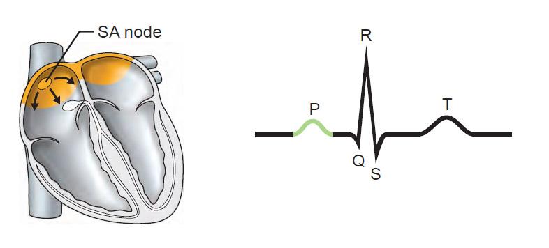
Atrial depolarization, initiated by the SA node, causes the P wave.
Describe the location and position of the heart in the body.
The heart is enclosed within the mediastinum. It lies anterior to the vertebral column and posterior to the sternum. It tips slightly to the left.
Describe the pericardium and distinguish between the fibrous and the serous pericardia relative to histological structure and function
The pericardium has two layers, a fibrous and a serous layer. The outer fibrous layer is a fibrous connective tissue that protects the heart and anchors it to surrounding structures. The inner serous layer (squamous epithelial cells) lines the fibrous layer as the parietal serous pericardium and at the base of the heart continues over the heart surface as the visceral serous pericardium. The visceral serous pericardium is the outermost layer of the heart wall, that is, the epicardium.
Trace one drop of blood from the time it enters the right atrium until it enters the left atrium. What is the circuit called?
•Pulmonary circuit
Right atrium
tricuspid valve
right ventricle
pulmonary semilunar valve
pulmonary trunk
pulmonary arteries
lungs
pulmonary veins
left atrium
(a)Describe how the heart contraction and relaxation influences coronary blood flow
(b) Name the major branches of coronary arteries, and note the heart regions served by each
a. When the ventricles begin to relax following contraction, blood flows back toward the ventricles, getting caught in the semilunar valves. During this time, the coronary arteries are actively delivering blood to the myocardium. During ventricular contraction, the coronary vessels are compressed and ineffective in blood delivery. (pp. 667, 670)
b. The major branches of the coronary arteries and the areas they serve are as follows: the left coronary artery runs toward the left side of the heart and divides into the anterior interventricular artery and the circumflex artery. The anterior interventricular artery supplies blood to the interventricular septum and anterior walls of both ventricles, and the circumflex artery serves the left atrium and the posterior walls of the left ventricle. The right coronary artery splits to the right side of the heart, where it divides into the marginal artery and the posterior interventricular artery. The marginal artery serves the myocardium of the lateral part of the right side of the heart and the posterior interventricular artery, which runs to the heart apex and supplies the posterior ventricular walls. (pp. 668, 670)
The refractory period of cardiac muscle is much longer than that of skeletal muscle. Why is a desirable functional property?
A longer refractory period of cardiac muscle is desirable because it prevents the heart from going into prolonged or tetanic contractions, which would stop its pumping action. (p. 673)
(a)What are the elements of the intrinsic conduction system in order beginning with pacemaker?
(b) What is the important function of this conductor system?
a. The elements of the intrinsic conduction system of the heart, beginning with the pacemaker, are: the SA node or pacemaker, AV node, AV bundle, right and left bundle branches, and the subendocardial conducting network. (pp. 675–676)
b. This system functions to initiate and distribute impulses throughout the heart so that the myocardium depolarizes and contracts in an orderly, sequential manner from atria to ventricles. (p. 674)
Draw a normal ECG pattern. Label and explain the significance of its waves.
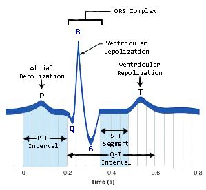
The P wave results from impulse conduction from the SA node through the atria during atrial depolarization. The QRS complex results from ventricular depolarization and precedes ventricular contraction. Its shape reveals the different size of the two ventricles and the time required for each to depolarize. The T wave is caused by ventricular repolarization. (pp. 677–678)
Define cardiac cycle, and follow the events of one cycle.
The cardiac cycle includes all events associated with the flow of blood through the heart during one complete heartbeat. One cycle includes a period of ventricular filling (mid-to-late diastole at the end of which atrial systole occurs), isovolumetric contraction (early ventricular systole), ventricular ejection (mid to late ventricular systole), and isovolumetric relaxation (early ventricular diastole). (pp. 679–680)
What is cardiac output, and how is it calculated?
Cardiac output is the amount of blood pumped out by the left ventricle in one minute. It can be calculated by the following equation: cardiac output = heart rate × stroke volume. (p. 681)
Discuss how Frank-Starling law of the heart helps to explain the influence of venous return on stroke volume
The Frank-Starling law explains that the critical factor controlling stroke volume is the degree of stretch of the cardiac muscle cells just before they contract. The important factor in the stretching of cardiac muscle is the amount of blood returning to the heart and distension of the ventricles. (p. 682)
Describe the common function of the foramen ovale and the ductus arteriosus in a fetus.
What problems result if these shunts remain patent (open) after birth?
a. In a fetus, the common function of the foramen ovale and the ductus arteriosus is to allow blood to bypass the pulmonary circulation, and move directly into the systemic circulation.
b. If these shunts remain patent after birth, the opening prevents adequate gas exchange, O2 loading and CO2 unloading, in the pulmonary circulation. This would occur because a large volume of blood returning to the heart would simply bypass the pulmonary circuit. (p. 686)
Critical Thinking
A gang member was stabbed in the chest during a street fight. He was cyanotic and unconscious from lack of blood delivery to the brain. The diagnosis was cardiac tamponade.
What is cardiac tamponade and how does it cause the observed symptoms.
Cardiac tamponade is the compression of the heart that occurs when blood or fluid builds up in the space between the myocardium (the muscle of the heart) and the pericardium (the outer covering sac of the heart) (p. 661)
Critical Thinking
You have been called upon to demonstrate the technique for listening to valve sounds.
(a) Explain where you would position your stethoscope to auscultate
(1) aortic valve of a patient with severe aortic valve insufficiency
(2) Stenotic mitral valve.
(b) During which periods would you hear these abnormal heart sounds most clearly? (During atrial diastole, ventricular systole, ventricular diastole, or atrial systole?
(c) What cues would you use to differentiate between an insufficient and a stenosed valve?
a. To auscultate the aortic valve, place the stethoscope over the second intercostal space at the right sternal margin. To auscultate the mitral valve, place the stethoscope over the heart apex, in the fifth intercostal space in line with the middle of the clavicle. (p. 679)
b. These abnormal sounds would be heard most clearly during ventricular diastole for the aortic valve and during atrial systole for the mitral valve. (p. 679)
c. An incompetent valve has a swishing sound after the valve has supposedly closed. A stenosed valve has a high-pitched sound when blood is being forced through its constricted opening during systole just before valve closure. (p. 679)
Critical Thinking
Florita Santos, a middle-aged woman, is admitted to the coronary care unit with a diagnosis of Left ventricular failure resulting from myocardial infarction. Her history indicated that she was aroused in the middle of the night by severe chest pain. Her skin is pale and cold, and moist sounds are heard over the lower regions of both lungs. Explain how failure of the left ventricle can cause these signs and symptoms.
Failure of the left ventricle (which pumps blood to the body) can result in chest pain due to dying or dead ischemic cardiac cells; pale, cold skin due to lack of circulation of blood from blocked ventricular contraction; and moist sounds in the lower lungs due to high pressure and pooling of blood in the pulmonary circulation because of nonfunction of the left ventricle. (pp. 668–669)
Critical Thinking
Heather, a newborn baby, needs surgery because she was born with an aorta that arises from the right ventricle and a pulmonary trunk that issues from the left ventricle, a condition called transposition of the great vessels. What are the physiological consequences of this defect?
Oxygen-deficient blood returning from the systemic circulation to the right heart will pass repeatedly around the systemic circuit, while oxygenated blood returned from the lungs is continually recycled through the pulmonary circuit. (pp. 686–687)
Critical Thinking
Gabriel, a heroin addict, feels tired, is weak and feverish, and has vague aches and pains. Terrified that he has AIDS, he goes to a doctor and is informed that he is suffering not from AIDS, but from a heart murmur accompanied by endocarditis. What is the most likely way that Gabriel contracted endocarditis?
Gabriel, being a user of an injectable drug, probably was infected by a bacteria-contaminated (“dirty”) needle used to administer heroin. (p. 690)