Exercise 15: Gross Anatomy of the Muscular System
location of muscle relative to a bone or body region; relative size of muscle
gluteus maximus
action of the muscle; relative size of muscle
adductor magnus
number of origins; location of muscle relative to a bone or body region
biceps femoris
location of muscle relative to a bone or body region; direction in which the muscle fibers run relative to some imaginary line
transversus abdominis
action of muscle; location of the origin and or insertion of the muscle; location of muscle relative to bone or body region
extensor carpi ulnaris
shape of muscle
trapezius
location of the muscle relative to a bone or body region; direction in which the muscle fibers run relative to some imaginary line
rectus femoris
location of the muscle reltive to a bone or body region; direction in which the muscle fibers run relative to some imaginary line
external oblique
term for the biceps brachii during elbow flexion
prime mover (agonist)
term that describes relation of brachialis to biceps brachii during elbow flexion
synergist
term for the triceps brachii during elbow flexion
antagonist
term for iliopsoas during hip extension
antagonist
term for the gluteus maximus during hip extension when walking up the stairs
prime mover (agonist)
terms for the rotator cuff muscles and deltoid when the elbow is flexed and the hand grabs a tabletop to lift the table
fixator
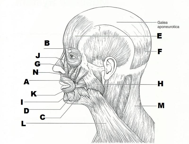
START FROM LEFT SIDE AND GO DOWN, THEN START ON THE UPPER RIGHT SIDE AND GO DOWN.
B. CORRUGATOR SUPERCILII
J. ORBICULARIS OCULI
G. LEVATOR LABII SUPERIORIS
N. ZYGOMATICUS MAJOR AND MINOR
A. BUCCINATOR
K. ORBICULARIS ORIS
I. MENTALIS
D. DEPRESSOR LABII INFERIORIS
C. DEPRESSOR ANGULI ORIS
L. PLATYSMA
RIGHT:
E. EPICRANIUS (FRONTAL BELLY)
F. EPICRANIUS (OCCIPITAL BELLY)
H. MASSETER
M. TRAPEZIUS
used in smiling
zygomaticus
used to suck in your cheeks
buccinator
Used in blinking and squinting (closes eye)
Orbicularis Oculi
used to pout (pulls the corners of the mouth downward)
Depressor Anguli Oris Muscle and Mentalis Muscle
raises your eyebrows for a questioning expression
epicranius (frontal belly)
used to form the vertical frown crease on your forehead
corrugator supercilii
your "kisser"
orbicularis oris
prime mover to raise the mandible
masseter
tenses skin of the neck during shaving
platysma
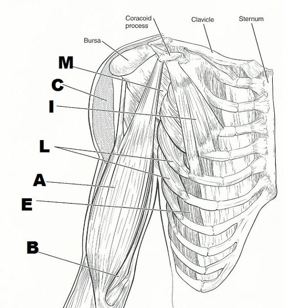
FROM TOP TO BOTTOM:
M. SUBSCAPULARIS
C. DELTOID (CUT)
I. PECTORALIS MINOR
L. SERRATUS ANTERIOR
A. BICEPS BRACHII
E. EXTERNAL OBLIQUE
B. BRACHIALIS
a major spine flexor
rectus abdominus
prime mover for arm extension
latissimus dorsi
prime mover for arm flexion
pectoralis major
assume major responsibility for forming the abdominal girdle
external oblique, internal oblique, transverse abdominus, rectus abdominus
prime mover of shoulder abduction
deltoid
important in shoulder adduction; antagonists of the shoulder abductor
latissimus dorsi, pectoralis major
moves the scapula forward and rotates scapula upwards
serratus anterior
small, inspiratory muscles between the ribs; elevate the ribs
external intercostals
extends the head
trapezius
pull the scapula medially
rhomboids
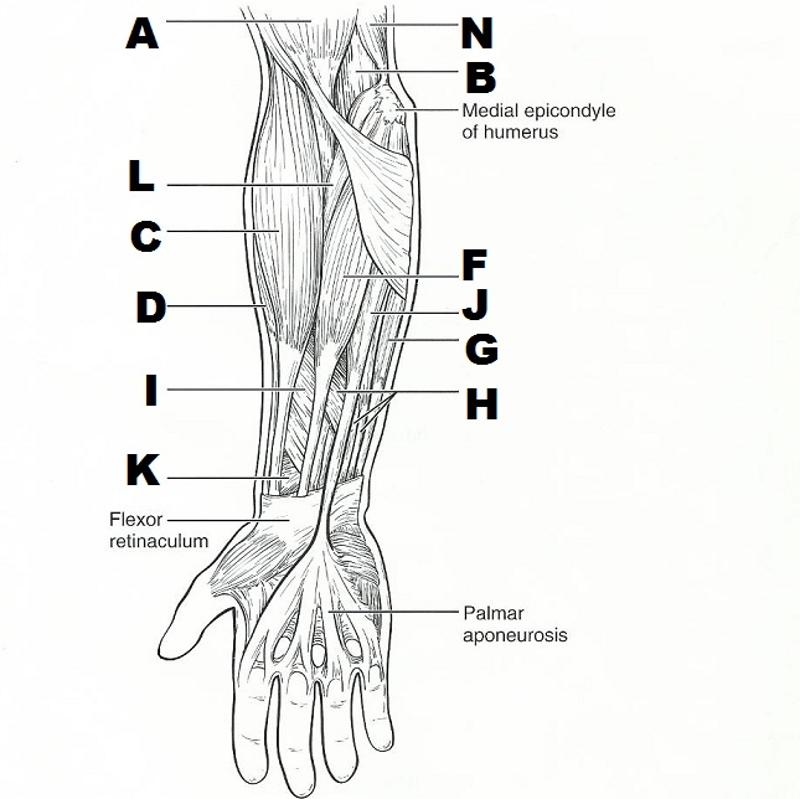
FROM LEFT TOP TO BOTTOM, THEN RIGHT TOP TO BOTTOM:
LEFT:
A. BICEPS BRACHII
L. PRONATOR TERES
C. BRACHIORADIALIS
D. EXTENSOR CARPI RADIALIS LONGUS
I. FLEXOR POLLICIS LONGUS
K. PRONATOR QUADRATUS
RIGHT:
N. TRICEPS BRACHII
B. BRACHIALIS
F. FLEXOR CARPI RADIALIS
J. PALMARIS LONGUS
G. FLEXOR CARPI ULNARIS
H. FLEXOR DIGITORUM SUPERFICIALIS
flexes the forearm and supinates the hand
biceps brachii
synerhist for supination of hand
supinator
forearm flexors; no role in supination
brachialis; brachioradialis
elbow extensor
triceps brachii
power wrist flexor and abductor
flexor carpi radialis
flexes wrist and middle phalanges
flexor digitorum superficialis
pronates the hand
pronator quadratus, pronator teres
flexes the thumb
flexor pollicis longus
extends and abducts the wrist
extensor carpi radialis longus
extends the wrist and digits
extensor digitorum
flat muscle that is a weak wrist flexor; tenses skin of the palm
palmaris longus
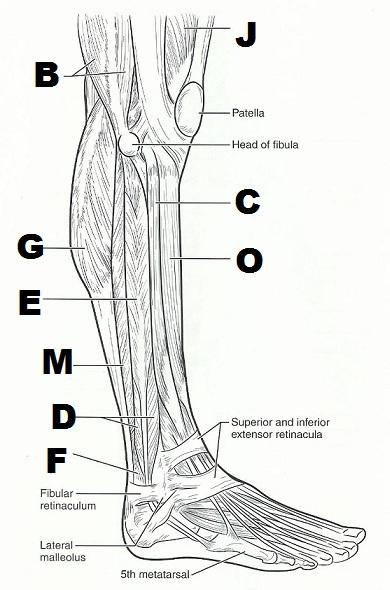
LEFT FROM TOP TO BOTTOM:
B. BICEPS FEMORIS
G. GASTROCNEMIUS
E. FIBULARIS LONGUS
M. SOLEUS
D. FIBULARIS BREVIS
F. FLEXOR HALLUCIS LONGUS
RIGHT FROM TOP TO BOTTOM:
J. RECTUS FEMORIS
C. EXTENSOR DIGITORUM LONGUS
O. TIBIALIS ANTERIOR
flexes the great toe and inverts the foot
flexor hallucis longus
lateral compartment muscles that plantar flex and evert the foot
fibularis brevis, fibularis longus
abduct the thigh to take the "at ease" stance
gluteus medius, tensor fasciae latae
used to extend the hip when climbing stairs
gluteus maximus
prime movers of plantar flexion of the foot
gastrocnemius, soleus
prime mover of inversion of the foot
tibialis posterior
prime mover of dorsiflexion of the foot
tibialis anterior
adduct the thigh, as when standing at attention
adductor group
extends the toes
extensor digitorum longus
entend thigh and flex knee
biceps femoris, semimembranosus, semitendinosus
extends knee and flexes thigh
rectus femoris
A. TEMPORALIS
B. MASSETER
C. PLATYSMA
D. TRAPEZIUS
E. DELTOID
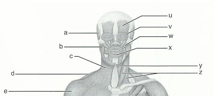
U. FRONTALIS
V. ORBICULARIS OCULI
W. ZYGOMATICUS MAJOR
Y. STERNOTHYROID
Z. STERNOCLEIDO-MASTOID
11
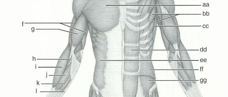
AA - PECTORALIS MAJOR
BB - SERRATUS ANTERIOR
CC - LATISSIMUS DORSI
DD - RECTUS ABDOMINIS
EE - EXTERNAL ABDOMINAL OBLIQUE
FF - INTERNAL ABDOMINAL OBLIQUE
GG - TARASVERSUS ABDOMINIS
F - BRACHOALIS
G - BICEPS BRACHII
H - PRONATOR TERES
I - BRACHIORADIALIS
J - FLEXOR CARPI RADIALIS
K - PALMARIS LONGUS
L - FLEXOR CARPI ULNARIS
11
M - ILIOPSAS
N - PECTINEUS
O - RECTUS FEMORIS
P - VASTUS LATERALIS
Q - VASTUS MEDIALIS
HH- TENSOR FASCIAE LATAE
II - SARTORIUS
JJ - ADDUCTOR LONGUS
KK - GRACILIS

11
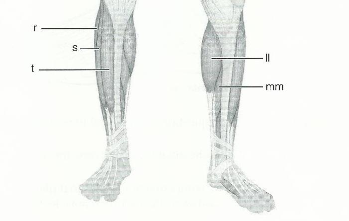
R - PERONEUS LONGUS
S - EXTENSOR DIGITORUM LONGUS
T - TABIALIS ANTERIOR
LL - GASTROCNEMIUS
MM - SOLEUS
11
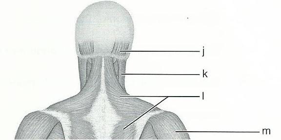
J - OCCIPITALIS
K - STERNOCLOIDOMASTOID
L - TRAPEZIUS
M - DELTOID
12
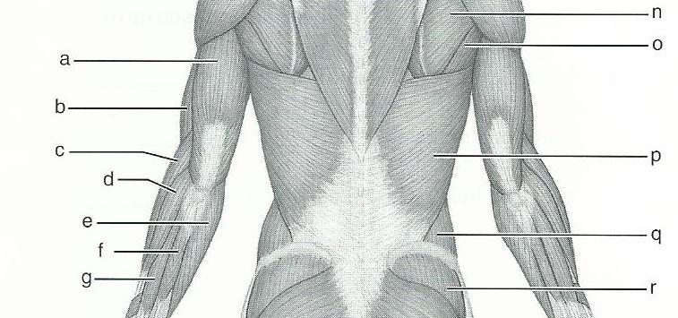
A. TRICEPS BRACHII
B. BRACHIALIS
C. BRACHIORADIALIS
D. EXTENSOR CARPI RADIALIS LONGUS
E. FLEXOR CARPI ULNARIS
F. EXTENSOR CARPI ULNARIS
G. EXTENSOR DIGITORUM
N - TERES MINOR
O - TERES MAJOR
P - LATISSIMUS DORSI
Q- EXTERNAL ABDOMINAL OBLIQUE
R - GLUTEUS MEDIUS
12
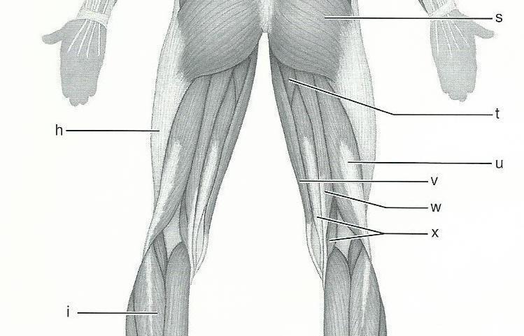
H - ILIOTIBIAL TRACT
I - GASTROCNEMIUS
S- GLUTEUS MAXIMUS
T - ADDUCTOR MAGNUS
U - BICEPS FEMORIS
V - GRACILIS
W - SEMITENDINOSUS
X - SEMIMEMBRANOSUS
12
Deltoid, Vastus Lateralis, Gluteus Medius, Gluteus Maximus
What are the 4 muscles commonly used for intramuscular injections?
The insertion tendon of the ____ group contains a large sesamoid bone, the patella.
Quadriceps
The triceps surae insert in common into the ____ tendon.
calcanal
The bulk of the tissue of a muscle tends to lie ______ to the part of the body it causes to move.
mediala proximal
The extrinsic muscles of the hand originate on the ______
forearm
Most flexor muscles are located on the _____ aspect of the body;
anterior
most extensors are located ______
posteria
An exception to this generalization is the extensor-flexor musculature of the ____
knee