Lower Extremity Muscles
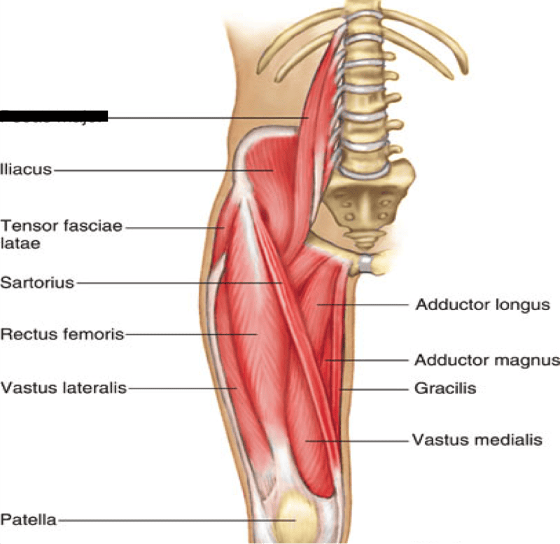
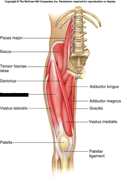
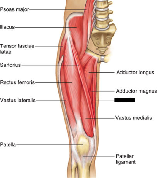
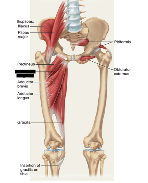
Iliacus
O: Iliac Fossa
I: Lesser trochanter of femur
A: Flexion of Hip
A: Assists with adduction & lateral rotation of hip
A: Anterior rotation of pelvis with femur stabilized
N: Femoral nerve (L2-L4)
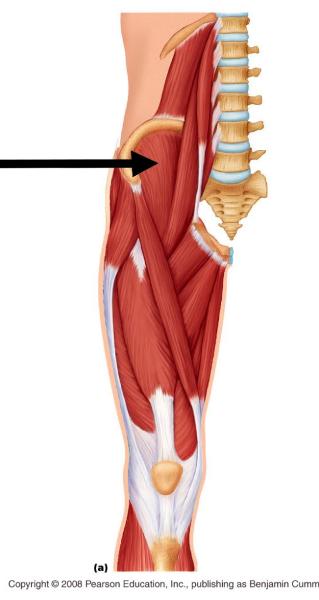
Psoas Major
O: L1-L4 - bodies and transverse process, IV discs at same levels
I: Lesser trochanter
A: Flexion of hip
A: Assists with adduction & lateral rotation of hip; some rotation of the lumbar spine
A: Flexion of lumbar spine with femur stabilized
N: Femoral Nerve (L2-L4)
Psoas Minor
A: Assists psoas major with flexion of lumbar spine
*Not a true muscle of the hip joint (coxofemoral joint)
*Not present in some people
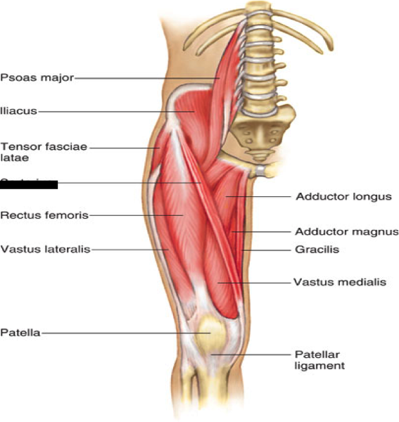
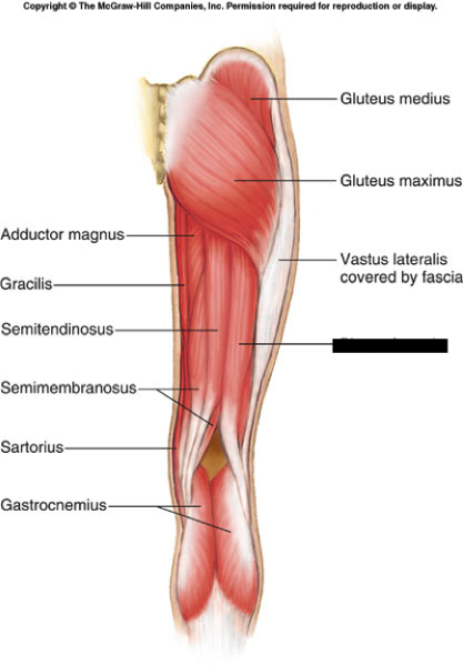
Sartorius "Tailor's Muscle"
O: Anterior superior iliac spine (ASIS)
I: Medial surface of tibia (pes anserine)
A: Flexes hip & knee: crosses both joints
A: Also acts to laterally rotate the knee at the tibiofemoral joint
N: Femoral Nerve (L2-L4)
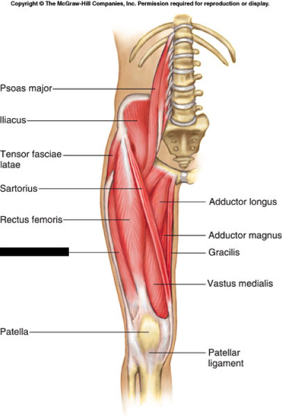
Rectus Femoris
O: Anterior inferior iliac spine
I: Tibial tuberosity
A: Assists iliopsoas with flexion of hip
A: Knee extension and stabilization
N: Femoral Nerve (L2-L4)
Vastus Lateralis
O: Lateral lip of linea aspera & greater trochanter
I: Tibial tuberosity
A: Knee extension
N: Femoral nerve (L2-L4)
*Largest muscle of the quads
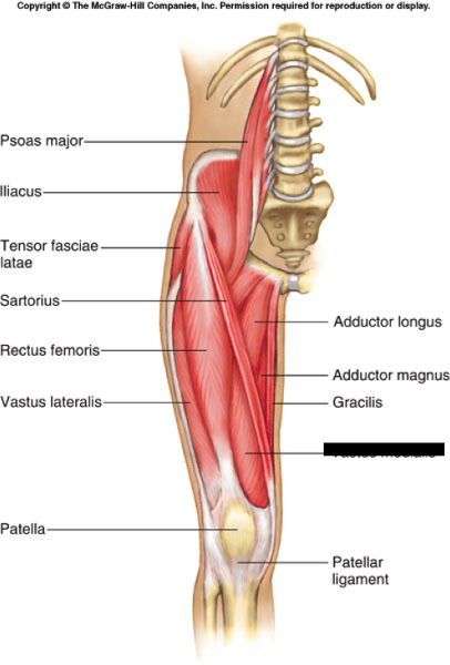
Vastus Medialis
O: Medial lip of linea aspera
I: Tibial tuberosity
A: Knee extension
N: Femoral Nerve (L2-L4)
*Vastus medialis obliquus
Vastus Intermedius
O: Proximal, anterolateral shaft of femur
I: Tibial Tuberosity
A: Knee extension
N: Femoral Nerve (L2-L4)
Pectineus
O: Superior Ramus of Pubis
I: Intertrochanteric line to line aspera
A: Adduction & Flexion of hip
N: Femoral Nerve
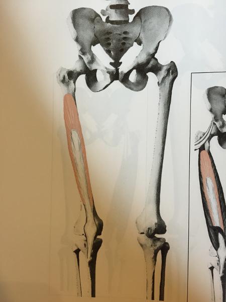
Gracilis
O: Inferior ramus & body of pubis
I: Medial surface of tibia (pes anserine)
A: Adduction & flexion of femur
A: Medial rotation of tibia
N: Obturator nerve (L2-L4)
Adductor Brevis
O: Inferior ramus & body of pubis
I: Upper 1/3 of linea aspera
A: Adduct & laterally of the hip
N: Obturator (L2-L4)
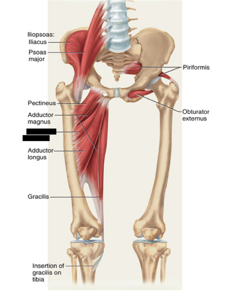
Adductor Longus
O: Inferior ramus & body of pubis
I: Middle 1/3 of linea aspera
A: Adducts & laterally rotates femur
N: Obturator Nerve (L2-L4)
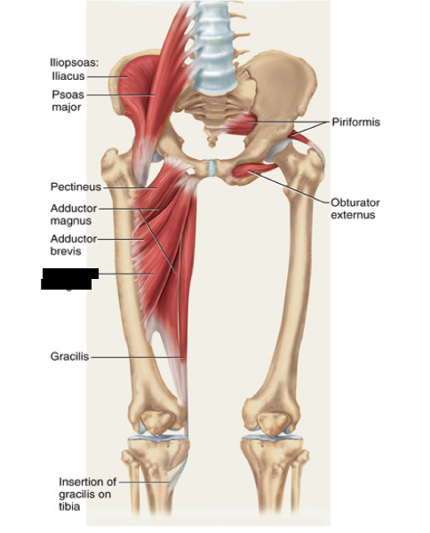
Adductor Magnus
O: Ischial tuberosity & Pubis
I: Linea aspera & adductor tubercle - anterior & posteior portions (BIG!)
A: Adduction of femur
Anterior = flex & lateral rotation
Posterior = extend & medial rotation
N: Obturator (L2-L4)
N: Sciatic nerve (L4-S3)
Tensor Fascia Latae (TFL)
O: Anterior iliac crest
I: Fascia of the thigh-Iliotibial (IT) Band
A: Abduct femur
A: Flex hip joint
A: Helps depress femoral head into acetabulum
N: Superior gluteal nerve (L4-S1)
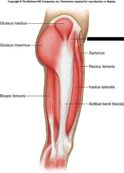
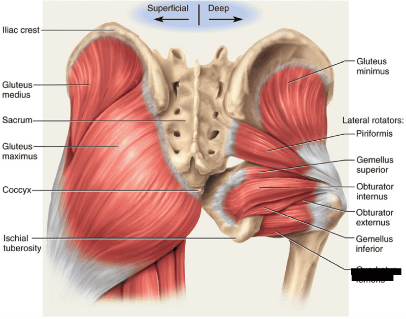
Gluteus Maximus
O: Iliac crest, Sacrum, Coccyx
I: Posterior portion of femur - blends with IT band
A: Hip extension (prime mover)
A: Also laterally rotates
A: Also abducts & adducts
N: Inferior Gluteal Nerve (L5-S1)
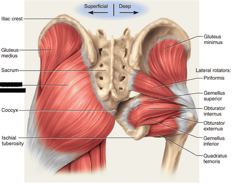
Gluteus Medius
O: Posterior aspect of ilium
I: Greater trochanter of femus
-portion of gluteus medium is deep to the gluteus maximus
A: Abduct & medially rotate femur
N: Superior gluteal nerve (L4-S1)
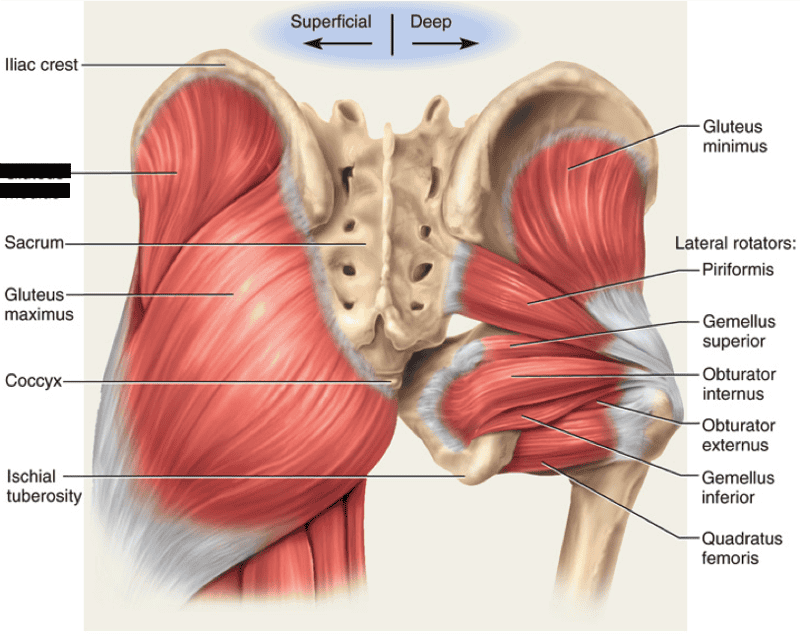
Gluteus Minimus
O: Posterior aspect of ilium
I: Greater trochanter of femus
-portion of gluteus medium is deep to the gluteus maximus
A: Abduct & medially rotate femur
N: Superior gluteal nerve (L4-S1)
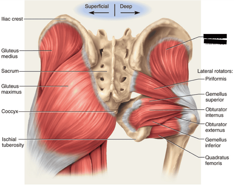
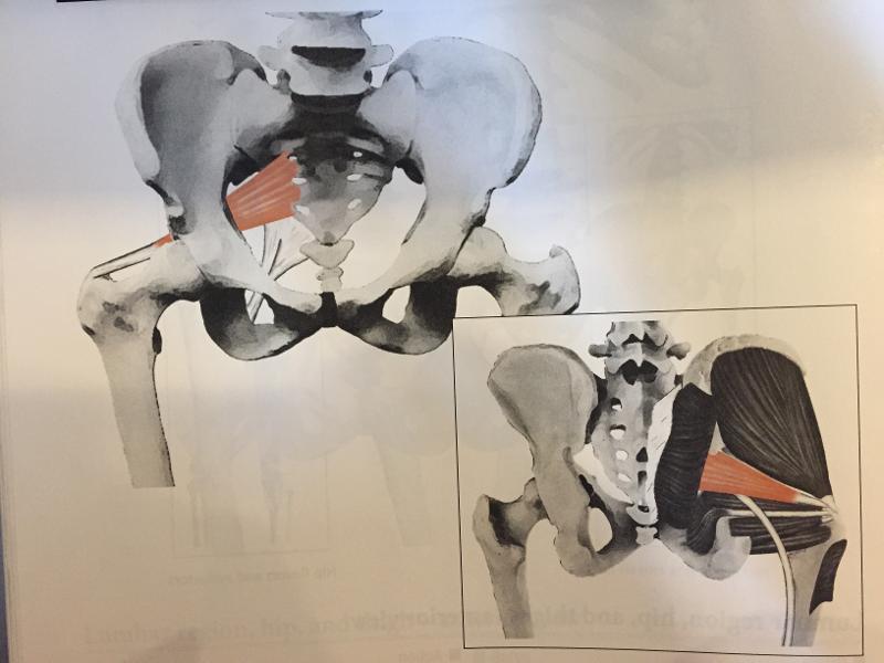
Piriformis
O: Upper portion of sacrum
I: Greater trochanter - crosses greater sciatic notch
A: Lateral rotation of femur
N: Anterior rami of first and second sacral nerves
Gemellus superior
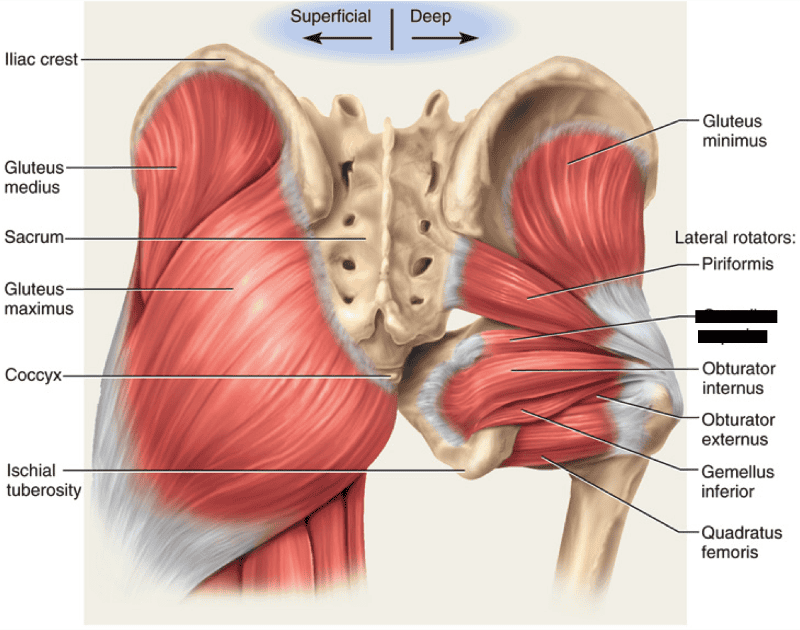
Obturator internus
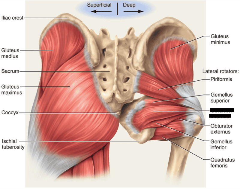
Obturator externus

Gemellus inferior
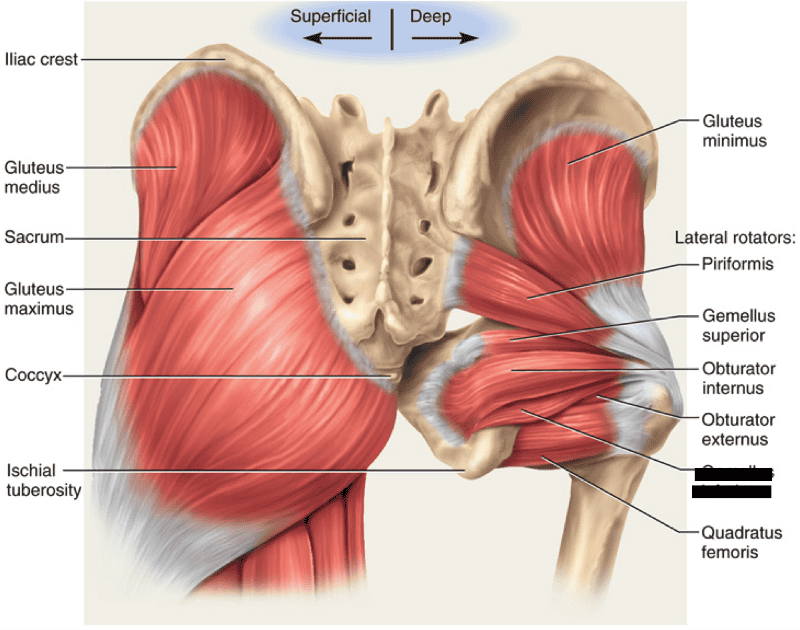
Quadratus femoris
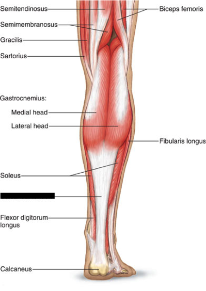
Semitendinosus
O: Ischial tuberosity
I: Medial surface of tibia (pes anserine)
A: Knee flexion
A: Medial rotation of the tibia
A: Hip extension
N: Sciatic Nerve (L5, S1, S2)
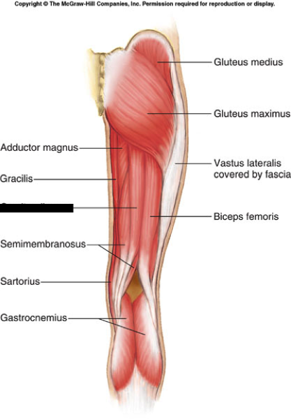
Semimembranosus
O: Ischial tuberosity
I: Posterior, medial condyle of tibia
A: Flex knee & medially rotate femur
N: Sciatic Nerve (L5, S1, S2)
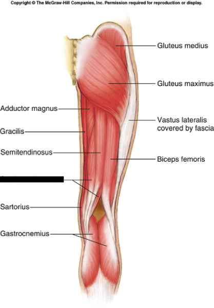
Biceps Femoris
O: Long head - ischial tuberosity
O: Short head - posterior femur (line aspera) - not a hip extender
I: Fibular head, lateral condyle of tibia
A: Flex knee & laterally rotate femur
N: LH - tibial part of sciatic nerve (S1-S3)
N: SH - common perennial part of sciatic nerve (L5, S1, S2)
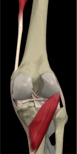
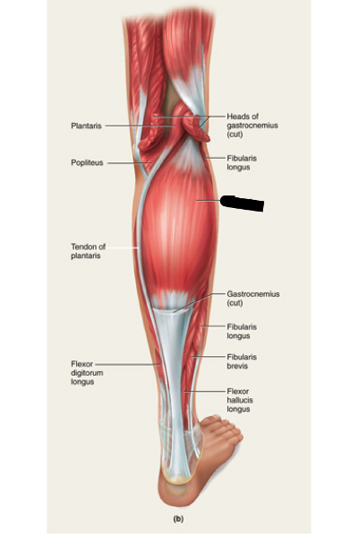
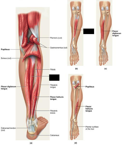
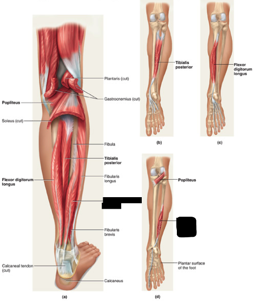
Popliteus
O: Lateral epicondyle of femur
I: Shaft of tibia
A: Unlocks knee to allow flexion to occur
Open kinetic chain (OKC) position: Medial rotation of the tibia
Closed kinetic chain (CKC) position: Lateral rotation of the femur
N: Tibial Nerve
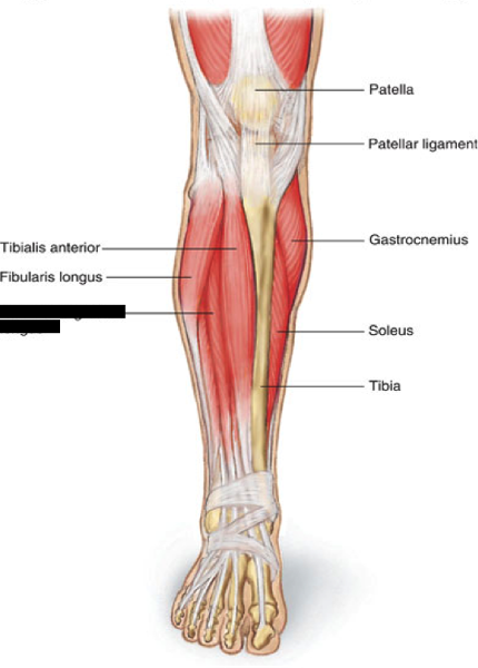
Tibialis Anterior
O: Shaft of the tibia
I: Medial cuneiform & 1st metatarsal
A: Dorsiflexion & inversion
N: Deep peroneal nerve (L4-S1)
*Most medial of the anterior leg muscles
Extensor Digitorum Longus
O: Lateral condyle of tibia
I: Distal phalanx; digits 2-5
A: Extend digits 2-5
A: Dorsiflex foot
N: Deep peroneal nerve (L4-S1)
Extensor Hallucis Longus
O: Fibula
I: Distal phalanx - digit 1
A: Extends digit 1
A: Inversion
N: Deep peroneal nerve (L4-S1)
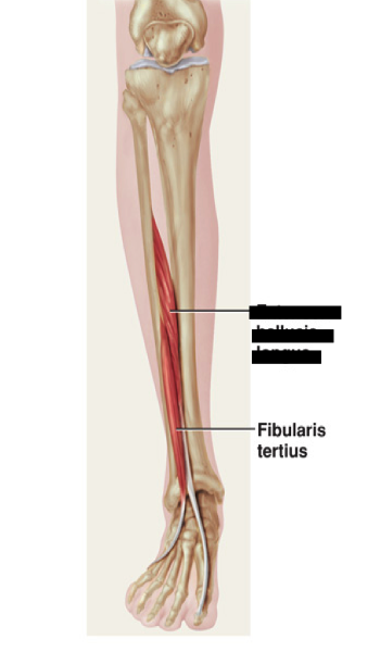
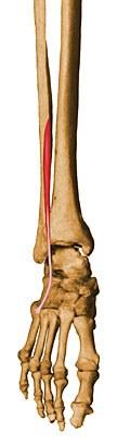
Peroneus Tertius
O: Anterior shaft of fibula
I: Base of 5th metatarsal
A: Everts & dorsiflexes foot
*Tendon crosses anterior to lateral malleolus
*Causes dorsiflexion
N: Deep peroneal nerve (L4-S1)
Peroneus Longus
O: Head & shaft of fibula
I: First metatarsal & medial cuneiform
A: Evert foot (strongest pronator)
A: Plantar flexion of the ankle
A: Tendon travels behind lateral malleolus, then under foot to medial side
N: Superficial peroneal
Peroneus Brevis
O: Lateral aspect of shaft of fibula
I: Base of 5th metatarsal
A: Eversion & plantar flexion of ankle
*tendon travels behind lateral malleolus
N: Superficial peroneal
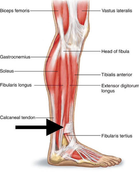
Gastrocnemius
O: Lateral & medial epicondyles of femur
I: Calcaneus via calcaneal (Achilles') tendon
A: Plantar flexion & assists knee flexion
*Important in walking/running activities
N: Tibial Nerve
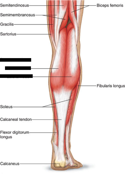
Soleus
O: Head/shaft of fibular & posterior surface of tibia
I: Calcaneus via calcaneal (Achilles') tendon
A: Plantar flex foot
N: Tibial Nerve
*Mostly lies deep to the gastrocnemius
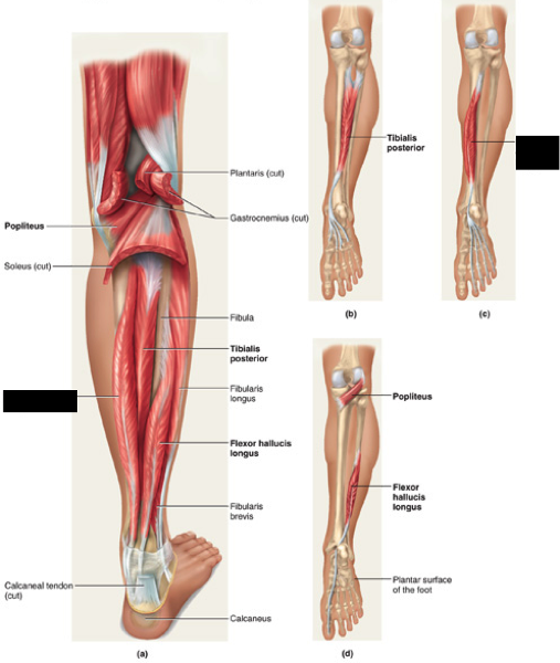
Tibialis Posterior
O: Posterior upper half of tibia & fibular
I: Lower surfaces of navicular, cuneiform, & bases of metatarsals 2-5
A: Plantar flex and invert foot
N: Tibial nerve
Flexor Digitorum Longus
O: Posterior surface of tibia
I: Distal phalanges of 2-5
A: Flex digits 2-5 & plantar flexion
N: Tibial Nerve
*Helps support the arch
Flexor Hallucis Longus
O: Middle 2/3 of posterior fibular
I: Base of distal phalanx of big toe
A: Flex digit 1, inversion, plantar flexion
N: Tibial Nerve
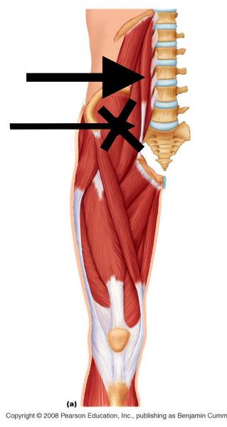
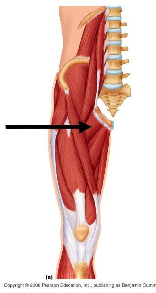
Iliopsoas
What is the name for the iliacus & psoas major together?
Anterior aspect of the hip joint, but commonly accepted as adductor group
What is the "Groin"?
Innervates the quadriceps & hip flexors
What is the Femoral Nerve?
Over activation or tightness
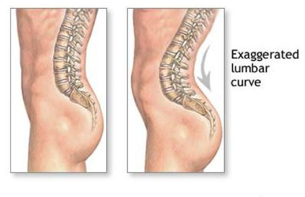
What causes this?
Medial retinaculum
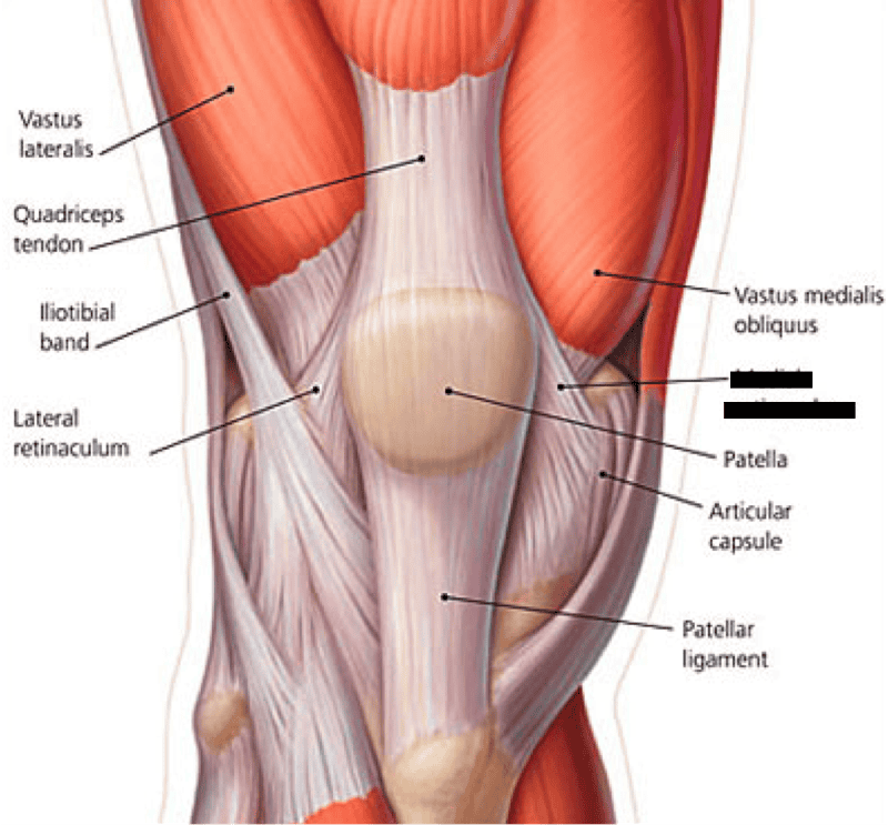
Lateral retinaculum
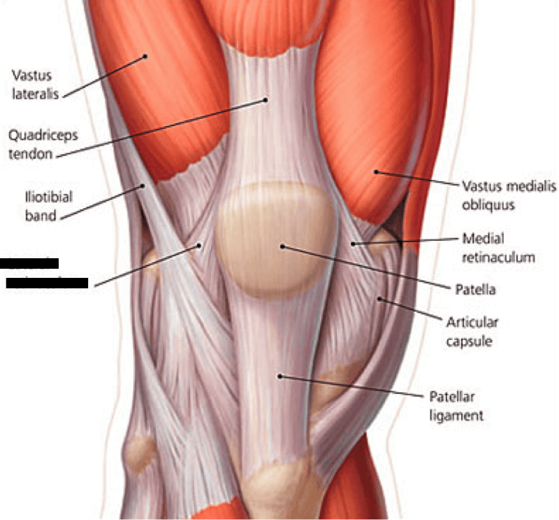
Iliotibial band
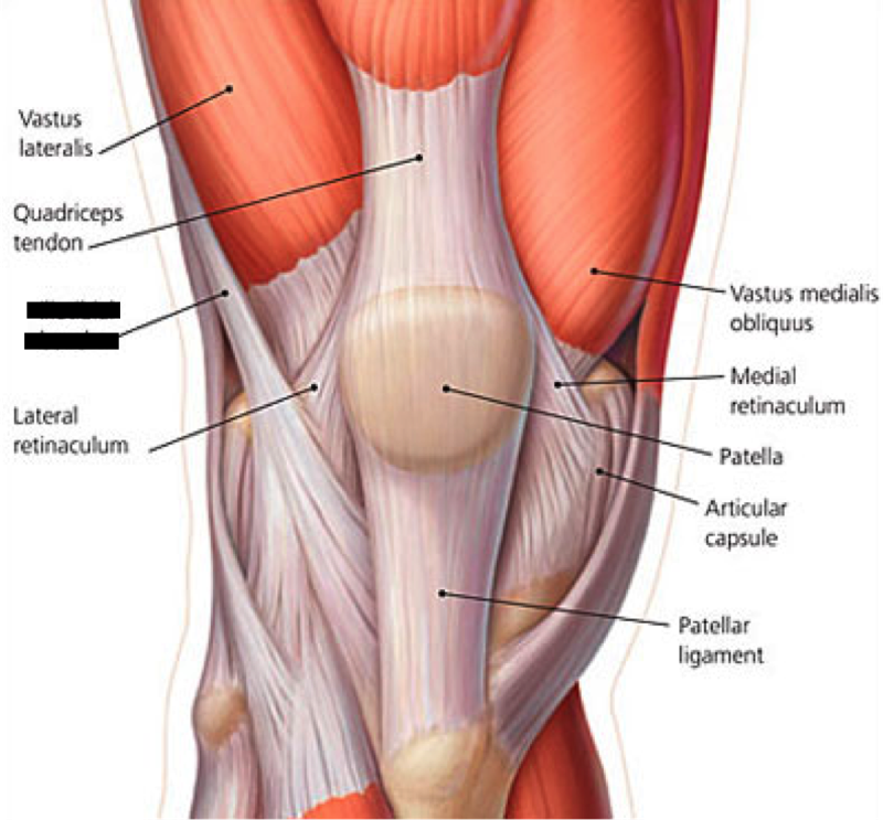
Quadriceps Tendon
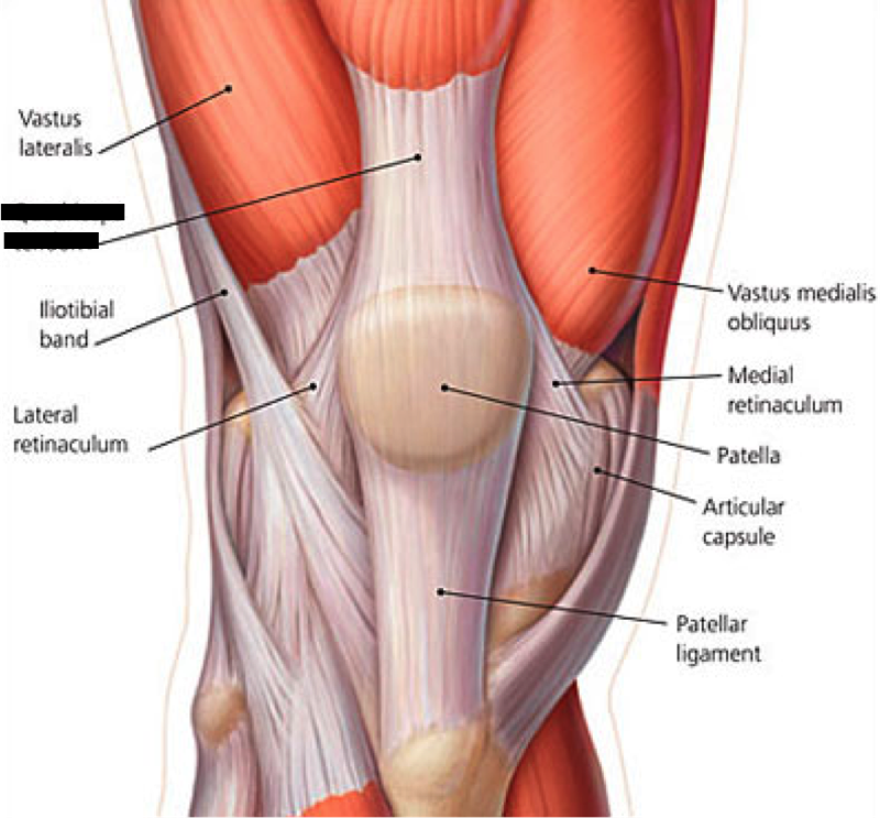
Patellar Tendon
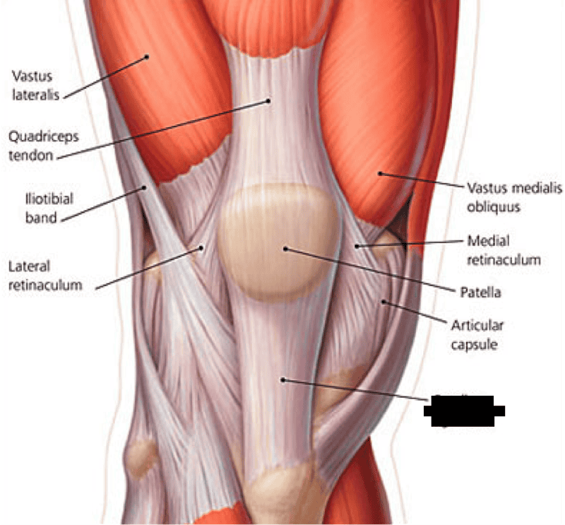
Rectus Femoris
Vastus Medialis
Vastus Intermedius
Vastus Lateralis
What is the quadriceps group?
Pectineus
Gracilis
Adductor Brevis
Adductor Longus
Adductor Magnus
What muscles are involved in a Groin strain?
Adductor Magnus
Adductor Brevis
Adductor Longus
Gracilis
Pectineus
What are the hip adductors?
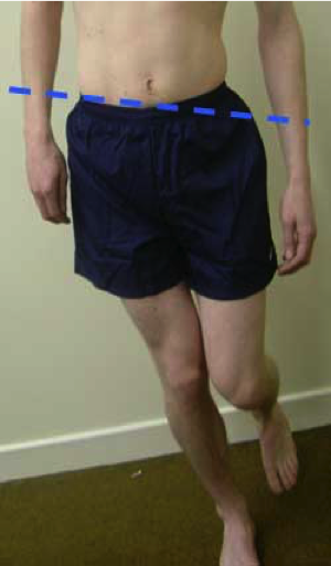
Stabilize pelvis when:
-Foot is on the ground
-They prevent the pelvis from dropping toward opposite side during walking
--Swinging leg
--Trendelenburg's Sign
What do the gluteus minimum & medium do together?
Trendelenburg’s Sign
Compression of the sciatic nerve
Radiating or shooting pain down the leg
What is sciatica?
Piriformis
Gemellus superior
Obturator internus
Obturator externus
Gemellus inferior
Quadratus femoris
What are the "Deep 6" External rotators?
Biceps Femoris (long & short head)
Semimembranosus
Semitendinosus
What are the hamstring muscles?
Deep peroneal nerve (L4-S1)
Innervation of anterior compartment
Superficial peroneal nerve (L4-S1)
Innervation of lateral compartment
Tibial nerve (L4-S3)
Innervation of deep posterior compartment
Tibial nerve (L4-S3)
Innervation of superficial posterior
Tibialis anterior
Extensor digitorum longus
Extensor hallucis longus
Peroneus tertius
Anterior compartment muscles
Peroneus longus
Peroneus brevis
Lateral compartment muscles
Gastrocnemius
Soleus
Superficial Posterior compartments
Tibialis posterior
Flexor digitorum longus
Flexor hallicis longus
Deep Posterior compartments
Fascia
What encloses a compartment?
Plantar flexors
When tendons pass behind lateral malleolus what does it make them?
Triceps surae
Another name for the calf muscle
Calcaneal tendon
Work primarily gastrocnemius because it is stretched (lengthened position) & in direct line of pull
Strengthening the Triceps surae in the standing position
Work primarily soles because gastrocnemius is relaxed (shortened position) across knee joint
Strengthening the Triceps surae in the seated position
Gluteus Maximus
Gluteus medius
Gluteus minimus
TFL
Sartorius
Piriformis (when hip is flexed)
Hip abductors
Adductor Magnus
Adductor Longus
Adductor Brevis
Pectineus
Gracilis
Psoas major
Iliacus
Gluteus Maximus (lower fibers)
Hip adductors
Semimembranosus
Semitendinosus
Gluteus Medius (anterior fibers)
Gluteus Minimus
Internal (Medial) Rotation
Biceps Femoris
Gluteus Maximus (all fibers)
Gluteus Medius (posterior fibers)
Sartorius
Piriformis
Quadratus Femoris
Obturator Internus/Externus
Gemellus Superior/Inferior
Iliopsoas
External (Lateral) Rotation
Rectus Femoris
Gluteus medius (anterior fibers)
Gluteus minimus
Add. magnus/longus/brevis (assists)
Pectineus
TFL
Sartorius
Psoas major/Iliacus
Hip Flexion
Biceps femoris
Semitendinosus
Semimebranosus
Gluteus Maximus (all fibers)
Gluteus Medius (posterior fibers)
Adductor Magnus (posterior fibers)
Hip Extension
Semitendinosus
Semimembranosus
Gracilis
Sartorius
Popliteus
Internal (Medial) Rotation
Biceps Femoris
External (Lateral) Rotation
Biceps Femoris
Semitendinosus
Semimembranosus
Gracilis
Sartorius
Gastrocnemius
Popliteus
Plantaris (weak)
Knee Flexion
Rectus Femoris
Vastus Lateralis
Vastus Medialis
Vastus Intermedius
Knee Extension
Gastrocnemius
Soleus
Tibialis Posterior
Peroneus Longus
Peroneus Brevis
Flexor Digitorum Longus (weak)
Flexor Hallucis longus (weak)
Plantaris (weak)
Plantar Flexion
Tibialis Anterior
Extensor Digitorum Longus
Extensor Hallucis Longus
Dorsiflexion
Tibialis Anterior
Tibialis Posterior
Flexor Digitorum Longus
Flexor Hallucis Longus
Extensor Hallucis Longus
Inversion
Peroneus Brevis
Peroneus Longus
Extensor Digitorum Longus
Eversion