pericardium
layer that encloses the heart
left
direction in which the heart points
visceral pericardium
layer also known as the epicardium
parietal pericardium
separated from the epicardium by the pericardial cavity
epicardium
layer that protects the heart by reducing friction
myocardium
thick middle layer of the wall of the heart; composed of smooth, cardiac muscle
endocardium
inner layer of the heart wall consisting of epithelium and connective tissue as well as some specialized muscle tissue
atria
receive blood from lungs and body
ventricles
receive blood from atria and force into body
septum
separates right and left sides of heart
tricuspid valve
between right atrium and ventricle
chordae tendinae
fibers attatched to the tricuspid valve which pull it closed when papillary muscles contract, preventing backwash of blood
papillary muscles
responsible for pulling the atrioventricular valves closed by means of the chordae tendineae
pulmonary valve
link between right ventricle and artery extending from it
bicuspid (mitral) valve
between left atrium and ventricle
aorta
largest artery in the body
aortic valve
between the left ventricle and the largest artery in the body
semilunar valves
pulmonary valve and aortic valve
skeleton of the heart
rings of dense connective tissure surrounding the pulmonary trunk and aorta
coronary arteries
first two branches of the aorta; feed the heart
agina pectoris
extreme chest pain caused by blockage (thrombus) of coronary arteries
coronary thrombosis
blood clot completely blocking a coronary artery, causes a heart attack
myocardial infarction
another name for heart attack
coronary sinus
enlarged vein from junctions of coronary veins which empty into the right atrium
systole
contraction of heart muscle
diastole
relaxation of heart muscle
cardiac cycle
atrial systole/ventricular diastole, ventricular systole/atrial diastole, brief complete diastole
functional syncytium
mass of merging cells that function as a unit
cardiac conduction system
fibers of cardiac muscle tissue which distribute impulses over the entire heart
sinoatrial node
small, elongated mass of specialized cardiac muscle tissue just beneath the epicardium in the right atrium near the opening of the superior vena cava-starts impulses
pacemaker
common name for S-A node
atrioventricular node
only normal conduction pathway between the atrial and ventricular syncytia
A-V bundle
group of fibers which receive impluse from the atrioventricular node; also known as the bundle of His
Purkinje fibers
extend from branches of A-V bundle, stimulate muscle fibers in the ventricular walls
electrocardiogram (ECG)
recording of the electrical changes that occur in the myocardium during a cardiac cycle
waves
deflection in a ECG
P wave
in an ECG corresponds to depolarization of the atrial fibers (leads to contraction
QRS complex
in ECG corresponding to depolarization of ventrical membranes, much stronger!!
T wave
in ECG last wave of cardiac cycle corresponding to repolarization
acetylcholine
decreases S-A and A-V nodal activity; leads to heart rate decrease
baroreceptors
detect changes in blood pressure
auricle
expandable extension of the atruim
coronary sulcus
groove that marks border between atria and ventricles
interatrial septum
separates the two atria
interventricular septum
separates the two ventricles
pectinate muscles
prominent muscular ridges along the inner surface of the auricle and across the adjacent anterior atrial wall
foramen ovale
penetrates interatrial septum from fifth week of embryonic development until birth
fossa ovalis
small depression of site of prior foramen ovale
trabeculae carneae
muscular ridges on the internal surface of the ventricles
how big is the heart
approximately the siz of a fist
where is the heart located
in the mediastinum between the 2nd rib and the 5th intercostal space, on the superior surface of the diaphragm, two thirds to the left of the midsternal valve , anterior to the vertibral column, posterior to the sternum
pericarditis
inflamation of the pericardium
pericardium
(peri - around cardi - heart)double walled sac the encloses the heart -
superficial fibrous pericardium
it protects, anchors, and prevents overfilling
deep two layered serous pericardium
parietal layer: lines the internal surface of the fibrous pericardium
visceral layer(epicardium): on external surface of the heart - seperated by a fluid filled pericardial cavity (decreases friction)
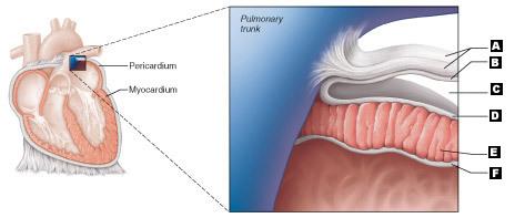
label - if you hohld your mouse over the photo it enlarges
A. Fibrous pericardium
B. Parietal layer or serous pericardium
C. Pericardial cavity
D. Epicardium
E. Myocardium
F. Endocardium
epicardium
visceral layer of the serous pericardium
describe the myocardium
(made of muscle) spiral bundles of cardiac muscle cells, fibrous skeleton of the heart: crisscrossing, interlacing layer of connective tissue
what is the function of the myocardium
anchors cardiac muscle fibers, supports great vessels and valves , limits spread of action potentials to specific parts
endocardium
(inside the heart)is continuous with endothelial lining of blood vessels
what are the four main chambers of the heart
two atria - left and right
two ventricles - left and right
internal structure of the two atria
seperated internally by the interarterial septum, coronary sulcus (atrioventricular groove) encircles the junction of the atria and ventricles , auricles increase atrial volume
internal structure of the two ventricles
seperated by hte interventricular septum, anterior and posterior interventricular sulci mark the position of the septum externally
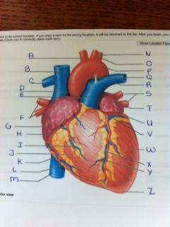
A. brachiocephalic trunk
B. superior vena cava
C. right pulmonary artery
D. ascending aorta
E. pulmonary trunk

F. right pulmonary veins
G. right atrium
H. right coronary artery
I. anterior cardiac vein
J. right ventricle

K. right marginal artery
L. small cardiac vein
M. inferior vena cava
N. left common carotid artery
O. left subclavian artery

P. aortic arch
Q. ligamentum arteriosum
R. left pulmonary artery
S. left pulmonary veins
T. auricle of the left atrium

U. circumflex artery
V. left coronary artery
W. left ventricle
X. great cardiac vein
Y. anterior interventricular artery
Z. apex
what are the recieving chambers of the heart
atria
where are the pecinate muscles found
walls of the atria
vessels entering the right atrium are
superior vena cava
inferior vena cava
coronary sinus
vessels entering the left atrium are
right and left pulmonary arteries
what are the discharging chambers of the heart
ventricles
where are the trabeculae carnae found
the ventricles of the heart
where do the papilary muscles project
into the ventricular cavities
vessels leaving the right ventricle are
pulmonary trunk
vessels leaving the left ventricle are
aorta
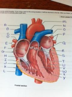
A. superior vena cava
B. right pulmonary artery
C. pulmonary trunk
D. right atrium
E. right pulmonary veins
F. fossa ovalis

G. pecinate muscles
H. tricuspid valve
I. right ventricle
J. chordae tendineae
K. trabeculae carneae
L. inferior vena cava

M. aorta
N. left pulmonary artery
O. left atrium
P. left pulmonary veins
Q. mitral (bicuspid ) valves

R. aortic valve
S. pulmonary valve
T. left ventricle
U. papilary muscles

V. interventricular septum
W. epicardium
X. myocardium
Y. endocardium
the heart is two side by side pumps, what is the right side the pump for
the pulmonary circuit - vessels that carry blood to and from the lungs
the heart is two side by side pumps, what is the left side the pump for
the systemic circuit - vessels that carry the blood to and from all body tissues
pathway of blood through the heart
Superior & Inferior Vena Cavas and coronary sinus Right atrium Tricuspid valve Right ventricle Pulmonary semilunar valve Pulmonary trunk Pulmonary arteries Lung capillaries Pulmonary veins Left atrium Bicuspid (mitral) valve Left ventricle Aortic semilunar valve Aorta to the systemic arteries systemic capillaries systemic veins superior & inferior vena cavas and coronary sinus
what occurs when blood reaches the lungs
gas exchange
are equal volumes of blood pumped to the pulmonary and systemic circuits
yes
describe the pulmonary circuit
short, low pressure circulation
describe the systemic circuit
blood encounters much resistance in the long pathways
how does the anatomy of the ventricles reflect these differences
the left ventricle is thicker than the right ventricle
the cornonary circulation is known as what? does what?
the shortest circulation in the body
is the functional blood supply of the heart
what are anastomoses
junctions - collateral routes that provide additional routes for blood delievery
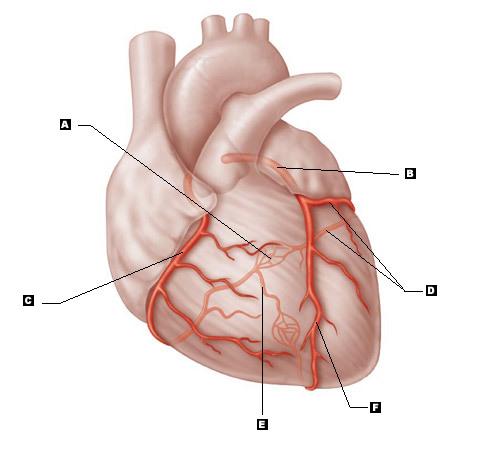
label
A. Anastomosis
B. Left coronary artery
C. Right coronary artery
D. Circumflex artery
E. Posterior interventricular artery
F. Anterior interventricular artery
the major coronary areteries are
1. right and left coronary arteries (in atrioventricular groove)
2. marginal arteries
3. circumflex arteries (wraps around the heart)
4. anterior and posterior interventricular arteries
the major cardiac veins are
1. small cardiac vein
2. anterior cardiac vein
3. middle cardiac vein
4. great cardiac veins
what is the coronary sinus
the blood pooling area into the right atrium
What is angina pectoris and what is it caused by
chest pain
thoracic pain caused by a fleeting deficiency in blood delivery to the myocardium
what is myocardial infarction and what is it caused by
heart attack
prolonged coronary blockage, areas of cell death are repaired with noncontractile scar tissue
what do heart valves do
ensure unidirectional blood flow through the heart
what is the job of the atrioventricular (AV) valves
prevent backflow into the atria when ventricles contract
tricuspid valve - right side
mitral (bicuspid) valve - left side
What do the chordae tendineae do
they anchor AV valve cusps to papillary muscles
what is the function of the semilunar (SL) valves
prevent backflow into the ventricles when the ventricles relax
aortic semilunar valve and the pulmonary semilunar valve
when the AV valves open: atrila pressure is greater than ventricular pressure - what occurs
1. blood returning to the heart fills the atria, putting pressure against the atrioventricular valves; atrioventricular valves are then forced open
2. as ventricles fill, atrioventricular valve flaps hang limply into the ventricles
3. atria contract, forcing additional blood into ventricles
when the AV valves close, atrial pressure is less than ventricular pressure - what occurs
1. the ventricles contract forcing blood against atriventricular valve cusps
2. atriventricular valves close
3. papillary muscles contract and chordae tendinae tighten, preventing valve flaps from everting into atria
what occurs when the semilunar valves open
as ventricles contract and intraventricular pressure rises, blood is pushed up against semilunar valves, forcing them to open
what occurs when the semilunar valves close
as ventricles relax and intraventricular pressure falls, blood flows back from arteries, filling the cusps of semilunar valves and forcing them to close
anatomy of cardiac muscle
cells are striated, short, fat, branched, and interconnected, the connective tissue matrix(endomysium) conects to the fibrous skeleton, t tubules are wide but less numerous, SR is simpler than in skeletal muscle, contain numerous large mitochondria (25-35 % cell volume), intercalated discs - junctions between cells which anchor cardiac cells,
what types of junctions are found in cardiac muscle
desmosomes - prevent cells from seperating during contraction
gap junctions- allow ions to pass; electrically couple adjacent cells
what does it mean that the heart behaves as a functional syncytium
that is contracts all at once
facts about cardiac muscle contraction
depolarization of the heart is rhythmic and spontaneous
•About 1% of cardiac cells have automaticity—(are self-excitable)
•Gap junctions ensure the heart contracts as a unit
•Long absolute refractory period (250 ms)
•Depolarization opens voltage-gated fast Na+channels in the sarcolemma
•Reversal of membrane potential from –90 mV to +30 mV
•Depolarization wave in T tubules causes the SR to release Ca2+
•Depolarization wave also opens slow Ca2+channels in the sarcolemma
•Ca2+surge prolongs the depolarization phase (plateau)
•Ca2+influx triggers opening of Ca2+-sensitive channels in the SR, which liberates bursts of Ca2+
•E-C coupling occurs as Ca2+binds to troponin and sliding of the filaments begins
•Duration of the AP and the contractile phase is much greater in cardiac muscle than in skeletal muscle
•Repolarization results from inactivation of Ca2+channels and opening of voltage-gated K+channels
which side of the heart are the SA and AV nodes found
the right side
Sinoatrial (SA) Nodeis also known as what
pacemaker
properties of the SA node
generates impulses about 75 times/minute (sinus rythm)
depolarizes faster than any other part of the myocardium (has to get the signal out)
sequence of electrical excitation -
1. the SA node generates impulses about 75 times a minute (depolarizes faster than any other part of the myocardium)
2. the impulses pause at the AV node (for about 0.1 second) smaller diameter fibers and fewr gap junctions, depolarizes 50 times per minute in the absence of SA node input
3. the atrioventricular bundle (bundle of His) connects the atria to the ventricles (only connection between the atria and the ventricles)
4. the bundle branches conduct the impulses through the interventricular (two pathways )septum (the right and the left bundle branches - carry the impulses toward the apex of the heart
5. the perkinje fibers depolarize the contractile cells of both ventricles (complete the pathway into the apex and ventricular walls)
in the absence of AV node how many times a minute do the AV bundles and perkinje fibers depolarize
the AV bundles and perkinje fibers depolarize only 30 times per minute in the absence of AV node imput
defects in the intrinsic conduction system of the heart may result in what?
1. arrythmias- irregular heart rythms
2. uncoordinated atrial and ventricular contractions
3. fibrilation- rapid, irregular contractions, useless for pumping blood
A defective SA node may result in what
ectopic focus - abnormal pacemaker takes over
if AV node takes over, there will be a junctional rythm (40-60 bpm)
A defective AV node may result in what
partial or total heart block, few or no impulses from SA node reach the ventricles - wont pump blood
heartbeat is modified by which system
the ANS autonomic nervous system
Where are cardiac centers located
in the medula oblongata
role of sympathetic neurons
cardioaccelaratory center inervates SA and AV nodes, heart muscles, and coronary arteries through sympathetic neurons
role of parasympathetic fibers
cardioinhibitory canter inhibits SA and Av nodes through parasympathetic fibers in the vagus nerves
what is an electrocardiogram
ECG or EKG - a composite of all the action potentials generated by nodal and contractile cells at a given time
what are the three waves of an EKG
P wave: depolarization of SA node
QRS wave: ventricular depoolarization
T wave: ventricular repolarization
explain the sequence of depolarization and repolarization of the heart related to the deflection waves of an EKG tracing
1. atrial depolarization - initiated by the SA node carries the P wave
2. with atrial depolarization complete the impulse is delayed at the AV node
3. ventricular depolarization begins at apex, causing the QRS complex - atrial repolarization occurs
4. ventricular depolarization is complete
5. ventricular repolarization begins at apex, causing the T wave
6. ventricular repolarization is complete
what are the two sounds associated with the closing of the heart valves
lub dup
when does the first sound lub occur
first sound occurs as AV valves close and signifies begining of systole
when does the second sound dup occur
second sound occurs when semilunar valves close at the begining of ventricular diastole
What are heart murmurs
abnormal heart sounds most often indicitive of valve problems
what is the cardiac cycle
all events associated with blood flow through the heart during one complete heartbeat
systole
contraction, higher pressure - ventricular contraction
diastole
relaxation - ventricles are relaxed because they are filling - lower pressure
how is blood pressure read (units of measure)
mm HG (mercury)
phase 1 of the cardiac cycle
ventricular filling - takes place in mid to late diastole
- AV valves are open
- 80% of blood passively flows into ventricles
-atrial systole occurs, delivering the remaining 20%
END DIASTOLIC VOLUME(EDV) volume of blood in each ventricle at the end of ventricular diastole
phase 2 of the cardiac cycle
ventricular systole
-atria relax and ventricles begin to contract
-risinf ventricular pressure results in closing of AV valves
-isovolumetric contraction phase (all valves are closed - breif moment)
- in ejection phase, ventricular pressure exceeds pressure in the large arteries, forcing the Semilunar valves to open = stroke vilume
stroke volume
the amount of blood ejected from the heart per beat
phase 3 of the cardiac cycle
isovolumetric relaxation occurs in early diastole
- ventricle relax
- backflow of blood in aorta and pulmonary trunk closes semilunar valves and causes breif rise in aortic pressure
-END SYSTOLIC VOLUME (ESV) volume in blood in each ventricle at the end of ventricular systole
do you want more blood at EDV or at ESV
EDV
CO =(HR) x (SV)
what is CO
what is HR
what is SV
CO: cardiac output - volume of blood pumped by each ventricle in one minute
HR: heart rate - number of beats per minute
SV: stroke volume - volume of blood pumped out by a ventricle with each beat
SV = EDV - ESV
stroke volume = end diastolic volume - end systolic volume
what are the three main factors tha taffest stroke volume
preload
contractility
afterload
preload
degree of stretch of cardiac muscle cells before they contract (Frank - Sterling law of the heart)
cardiac muscle exhibits a length- tension relationship, at rest, cardiac muscle cells are shorter than optimal length, slow heartbeat and exercise increase venous return, increased venous return distends (stretches) the ventricles and increases contraction force
contractility
contractile strength at a given muscle length, independent of muscle stretch and EDV
what do positive inotropic agents do
increase contractility,
- hormones (thyroxine, glucagon, and epinephrine)
how do negative inotropic agents decrease contractility
acidosis
increased extracellular K+
calcium channel blockers
afterload
pressure tha must be overcome for ventricles to eject blood
what increases afterload, reulting in what?
hypertension
increased ESV and reduced SV
sympathetic nervous system is activated by what
emotional or physical stressors
norepinephrin causes the pacemaker to fire more rapidly (and at the same time increases contractility)
parasympathetic nervous system opposes sympathetic effects how
acetylcholine hyperpolarizes pacemaker cells by opening K+ channels - the heart at rest exhibits vagal tone (parasympathetic)
what is the atrial (bainbridge) reflex
a sympathetic reflex initiated by increased venous return
stretch of the atrial walls stimulates the Sa node
also stimulatse atrial stretch receptors activacting sympathetic reflexes
explain heartrate and exercise
1. exercise - fright- anxiety
2. sympathetic activity increases, parasympathetic activity decreases, contractility increases and venous return increases
3. EDV (preload) increases ESV decreases
4. heart rate increases, stroke volume increases
5. cardiac output increases
chemical regulation of heart rate: hormones
epinephrine - from adrenal medula enhances heart rate and contractility
thyroxine- increases heart rate and enhances the effects of norepinephrine and epinephrine
chemical regulation of heart rate: intra and extracellular ion concentration
Ca+ and K must be maintained for normal heart function - changes in ion concentration affect heart excitability
other factors that influence heart heart (besides hormones and intra and extracellular ion concentration)
age
gender
exercise
body temperature
tachycardia
abnormally fast heart hear over 100 beats per minute
- if persistant, may lead to fibrillation
bradycardia
heart rate slower than 60 beats per minute
may result in grossly inadequate blood circulation
maybe desirable result if endurance training
congestive heart failure - what is is it? what are causes?
progressive condition where the CO is so low that blood circulation in inadequate to meet tissue needs
caused by
*coronary artherosclerosis
*persistant high blood pressure (hypertension)
*multiple myocardial infarcts
*dialated cadiomyopathy DCM)
age related changes affecting the heart
sclerosis and thickening of valve flaps
decline in cardiac reserve
fibrosis of cardiac muscle
atherosclerosis