Disease causing microorganisms are called pathogens.
Pathogenic microorganisms have special properties that allow them to invade
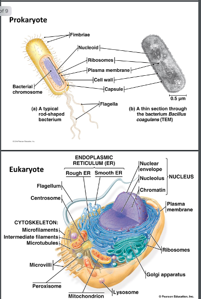
All living cells can be classified into two groups:
6.1
prokaryotes and eukaryotic based on certain structural and functional characteristics. These cells are similar in their chemical composition and chemical reactions.
Both contain nucleic acids, proteins, lipids, and carbohydrates. They have a sticky glycocalyx that surrounds them, the glue that holds the cells in place. Most bacteria are found sticking to solid surfaces, including other cells, rather than free floating. They use the same chemical reactions to metabolize food, build proteins, and store energy.
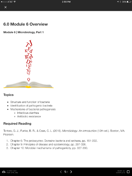
pg. 72-73.
What distinguishes prokaryotes from eukaryotes?
the structure of the cell wall, membrane and the absence of organelles.
pg. 73
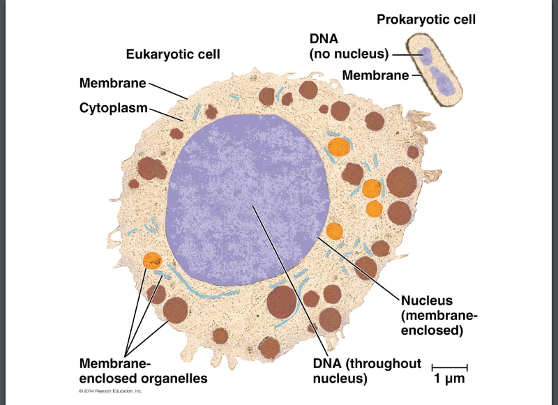
Prokaryotes
6.1
prenucleus
The DNA is usually a single circularly arranged chromosome and is not surrounded by a membrane.
DNA is not associated with histones.
Lack organelles- special structure that carry on various activities.
Their cell walls almost always contain the complex polysaccharide peptidoglycan.
They usually divide by binary fission- DNA is copied and the cell splits into 2 cells. This involves fewer structures and and processes that eukaryotic cell division.
small unicellular organisms that include Bacteria and archaea (majority are bacteria)
pg. 72
Eukaryotes
6.1
True nucleus.
The DNA is found in the cells nucleus, which is separated from the cytoplasm by a nuclear membrane, and the DNA is found in multiple chromosomes.
DNA is associated with chromosomal proteins called histones and with non histones.
Have a number of membrane enclosed organelles, including mitochondria, endoplasmic reticulum, golgi complex, lysosomes, and sometimes chloroplasts.
Cell walls when present are chemically simple.
Cell division involves mitosis, chromosomes replicate and and identical set is distributed into each of the two nuclei, cells produced are identical.
plants, animals, fungi, yeast, mold, protozoa, and algae.
pg 72-73
The two domains of prokaryotes are __________
6.1
bacteria and archaea
reproduction and adaption
6.1
bacteria reproduce quickly by binary fission and can divide every 1-3 hours
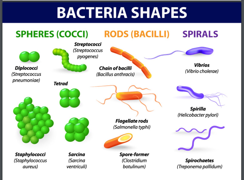
Shapes of bacteria (morphology)
6.1
coccus/cocci- spherical. berries. usually round, but can be oval, elongated, or flattened on one side. When they divide to reproduce the cells can remain attached to one another. Cocci that remain in pairs are diplococci. Those that divide and remain attached in chainlike patterns are called streptococci. Those that divide in two planes and remain in groups of four are known as tetrads. Those that divide in 3 planes and remain attached in cubelike groups of 8 are called sarcinae. Those that divide in multiple planes and form grapelike clusters or broad sheets are called staphylococci.
bacillus/bacilli- rod shaped. Little rods or walking sticks. Divide only across their short axis so there are fewer groupings of bacilli than cocci. Most bacilli appear as single rods called single bacilli. Diplobacilli appear in pairs after division. Streptobacilli occur in chains. Coccobacilli are oval and look like cocci
spiral bacteria have one or more twists, they are never straight. Bacteria that look like curved rods are called vibrios. Spirilla have a helical shape, like a corkscrew, and fairly rigid bodies, flagella used to move. Spirochetes are a group of spirals that are helical and flexible and move by means of axial filaments that resemble flagella.
pg. 74-75
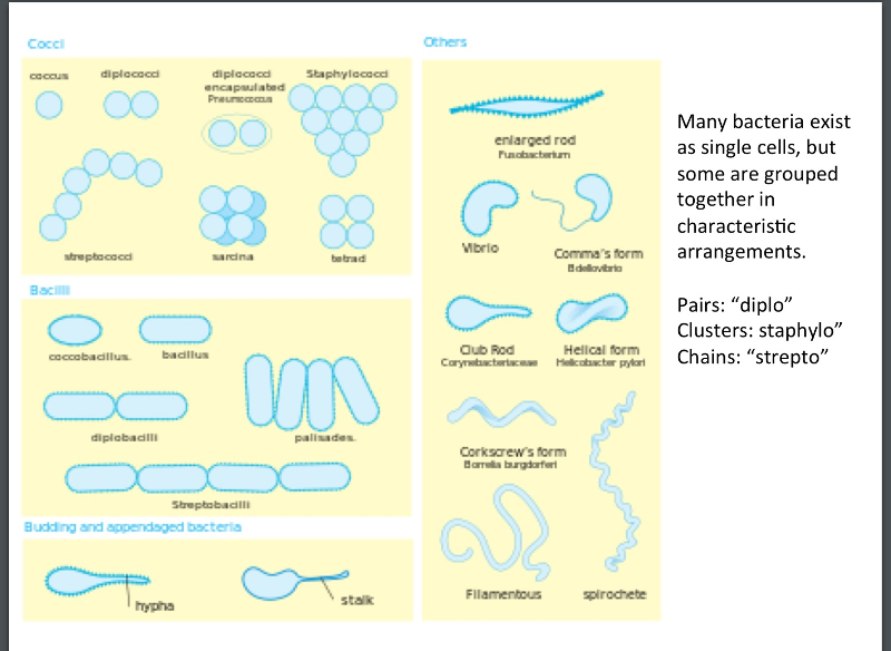
arrangement of bacterial cells:
6.1
spherical- coccus or cocci- berries. Diplococci or streptococci-division in one plane
Tetrad-division in two planes and in groups of four.
rod shaped- bacillus or bacilli -little rods or walking sticks.
Sarcinae- divide in three planes and attach in cubelike groups of eight.
staphylococci- divide in multiple planes and form grape like clusters
pg. 74
6.1
staphylococci
The prokaryotic cell: Bacteria
6.1
unicellular, multiply by binary fission, differentiated by morphology, chemical composition, nutritional requirements, biochemical activities, and source of energy.
pg 72-73
Are bacteria harmful?
6.2
<1% of the different types of bacteria make people sick.
many bacteria serve important functions in our body- bacteria in our intestines can digest fiber (plant polysaccharides) and in return produce vitamins necessary for our health.
bacteria on our skin provides protection from other harmful bacteria.
Bacteria can be harmful if they get into the wrong place or if they acquire the wrong genes.
Normal Microbiota
6.2
Microorganisms that establish more or less permanent residence (colonize) but that do not produce disease under normal conditions.
Can also be referred to as normal flora.
pg. 390
transient microbiota
6.2
may be present for several days, weeks, or months and then disappear. Microorganisms are not found throughout the entire human body but are localized in certain regions.
pg. 390 and 392.
The human microbiome project
6.2
began in 2007 to analyze microbial communities called microbiomes that live in and on the human body. Its goal is to determine the relationship between changes in the human microbiome and human health and disease. pg 390
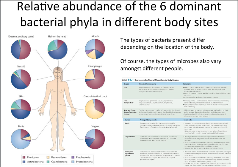
Types of bacteria present differ depending on the location of the body.
Distribution and composition of normal microbiota are determined by many factors:
6.2
- nutrients- microbes vary with respect to the types of nutrients they can use as an energy source and colonize only those site that can supply adequate nutrients. These nutrients are dead cells, food in the GI tract, secretory and excretory products of cells, and substances in body fluids. pg 391
- physical and chemical factors- temperature pH, available O2 and CO2, salinity and sunlight. pg 391
- host defenses- defenses that kill molecules, inhibit their growth, prevent adhesion and neutralize toxins.. pg. 391
- mechanical factors- age, nutritional status, diet, health status, disability, hospitalization, stress, climate, geography, hygiene, living conditons, occupation, and lifestyle. pg 391
10 times more bacteria cells than human cells. pg 390
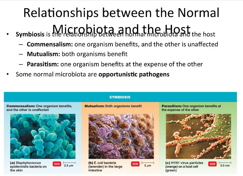
relationship between normal microbiota and the host:
6.2
Symbiosis is the relationship between normal microbiota and the host, a relationship between 2 organisms in which at least one organism is dependent on the other. Some normal microbiota are opportunistic pathogens. pg 392
commensalism: in a symbiotic relationship one organism benefits and the other is unaffected. Many of the microorganisms that make up our normal microbiota are commensals. Ie: bacteria on our skin that eat dead skin cells. pg. 392
mutualism: is the type of symbiosis that benefits both organisms. Ie: bacteria like e. coli that inhabit our intestines and eat fiber, synthesize vitamin K and some B vitamins that are abosrbed into our bloodstream and distributed for use by our body cells.. pg 393
parasitism: symbiosis that one organism benefits by deriving nutrients at the expense of the other. Many disease causing bacteria are called parasites. pg 393
Opportunistic pathogens are?
normal microbiota if they stay in the correct location on our body we can have a mutualistic relationship, but if they get into another part of our body they can become an opportunistic infection.
How does our normal microbiota help us?
6.2
It can benefit us because it prevents the growth of organisms that might actually harm us also called Microbial antagonism or competitive exclusion, a competition between microbes.
Normal microbiota protect the host by:
- The two populations compete for nutrients
- producing substances harmful to invading microbes
- affecting pH and available O2
pg 390-391
probiotics
a supplement of live microbial cultures, harmless bacteria, that repopulates your body with good bacteria and crowd out detrimental bacteria.
provides bacteria for commensalism, bacteria that can be benifical for us.
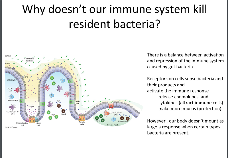
Why doesn't our immune system kill resident bacteria?
6.2
when bacteria are present all the time it promotes a tolerance. There is a balance between activation and repression of the immune system caused by gut bacteria.
Receptors on cells sense bacteria and their products and activate the immune response, release chemokines, and cytokines ( attract immune cells) make more mucous (protection)
However our body doesn't mount as large a response when certain types of bacteria are present.
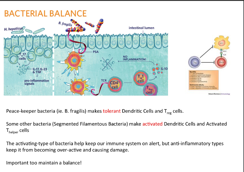
Bacterial balance
6.2
peace-keeper bacteria (i.e.: B. Fragilis) makes tolerant dentritic cells and T reg cells.
Some other bactria (segmented filamentous bacteria) make activated dendritic cells and activated T helper cells
the activating type of bacteria keep our immune system on alert, but anti-inflammatory types keep it from becoming over-active and causing damage.
Important to maintain a balance.
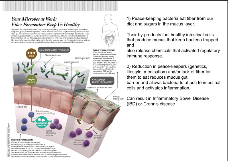
Peace keeping bacteria
6.2
eat fiber from our diet and sugars in the mucus layer.
their by-products fuel healthy intestinal cells that produce mucus that keep bacteria trapped and release chemicals that activated regulatory immune response
reduction in peace-keepers (genetics, lifestyle, medications) and /or lack of fiber for them to eat reduces mucus gut barrier and allows bacteria to attach the intestinal cells activating inflammation.
can result in inflammatory bowel disease (IBD) or crohns disease.
Normal microbiota can benefit the host by preventing the overgrowth of harmful microorganisms. This is called microbial __________.
6.2
Antagonism
Microbial antagonism - the normal microbiota can benefit the host by preventng the overgrowth of harmful microorganisms, can also be called competitive exclusion.
pg. 391.
Escherichia coli synthesizing vitamins K and B in the large intestine would be an example of which type of symbiosis?
6.2
Mutalism
pg 393
Pathology
6.3
Pathology is the scientific study of disease.
Pathology is first concerned with:
- the cause or the etiology of disease
- pathogenesis- the manner in which the disease develops.
- the structural and functional changes brought about by the disease and the effects on the body.
pg 390
Pathogen
6.3
Disease causing microorganisms are called pathogens.
pg 390
Infection
6.3
The invasion or colonization and growth of pathogens in the the body by pathogenic microorganisms.
An infection may exist in the absence of detectable disease. Patient may have the infection but not show s/s of the disease.
The presence of a microorganism in a part of the body where it is not normally found is also called an infection, and may lead to disease.
Ie: large numbers of E. Coli are normal in a healthy intestine but would cause a infection if noted in the urinary tract.
pg 390
disease
6.3
occurs when an infection results in any change from a state of health.
disease is an abnormal state in which part or all of the body is incapable of performing its normal functions.
When a microorganism overcomes the body's defenses a states of disease results.
pg 390
How do Microorganisms enter a host?
Portals of entry: many microorganisms can cause infections only when the gain access through their specific portal of entry
- Mucous membranes- easiest and most frequent portal is the Respiratory tract, by droplet or dust particles and can cause cold, pneumonia, TB, influenza and measles. Can also enter by GI- food and water and dirty hands, conjunctiva, and GU tract- STD/STI's.
- skin- unbroken skin, hair follicles, sweat glands.
- parental route- deposited directly into the tissues beneath the skin or into mucous membranes when these barriers are penetrated or injured. IE; punctures, injections, bites, cuts, wounds, surgery, cracked skin.
pg. 418
Pathogenicity
6.3
The ability to cause disease by overcoming host defenses.
pg. 418
virulence
6.3
The degree of pathogenicity.
pg 418
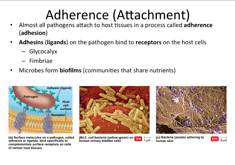
Adherance or adheasion
6.3
All pathogens have a means of attaching themselves to host tissues at their portal of entry to cause pathogenicity.
surface molecules on the pathogen called Adhesins (ligands) that bind specifically to complementary surface receptors on the cell of certain host tissues. Mannose is the most common receptor
- glycocalyx (can be glycoproteins or lipoproteins)
- pili, fimbriae, and flagella. (Frequently associated with fimbriae)
Microbes have the ability to come together in masses cling to surfaces, and take in and share available nutrients. in communities called biofilms- ie: dental plaque.
pg. 420.
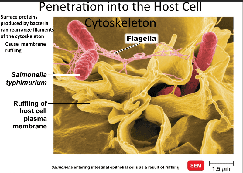
Penetration into the host cell cytoskeleton
6.3
microbes attach to host cells by adhesions, this triggers signals in the host cell that activate factors that can result in the entrance of some bacteria. The mechanism is provided by the host cytoskeleton. A major component of the cytoskeleton is a protein called actin which is used by some microbes to penetrate host cells and by others to move through and between host cells.
microbes produce surface proteins called invasins that rearrange nearby actin filaments of the cytoskeleton.
membrane ruffling is the result of disruption in the cytoskeleton of the host cell. The microbe sinks into the ruffle and is engulfed by the host cell.
pg. 423.
The easiest and MOST frequently traveled portal of entry for infectious microorganisms is the __________.
6.3
Respiratory tract
The degree of pathogenicity of an organism is known as the _________.
6.3
Virulence
E. coli is an example of what type of bacteria?
6.4
Gram-negative, bacillus
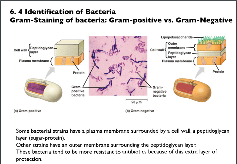
Identification of bacteria by gram stain:
6.4
Some bacterial strains have a plasma membrane surrounded by a cell wall, a peptidoglycan layer or sugar layer.
Other strains have an outer membrane surrounding the peptidoglycan layer. These bacteria tend to be more resistant to antibiotics because of this extra layer of protection.
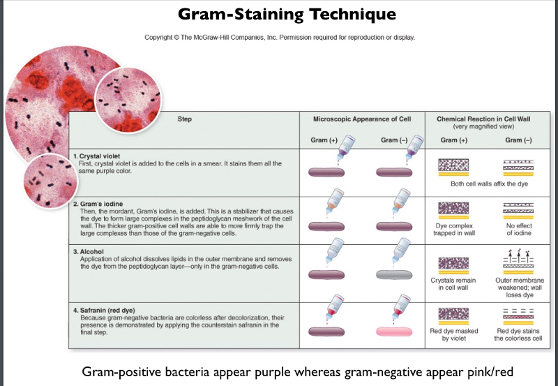
Gram staining technigue
6.4
staining means coloring the microorganisms with dye that emphasizes certain structures.
- fixed- kills the microbes and fixes them to the slide
- smear- a thin film of material containing microorganisms, passed through the flame, and stain called crystal violet is applied.
- alcholol to decolorize
- safrinin red dye and washed again.
pg. 65
Gram positive vs gram negative bacteria:
6.4
Gram positive bacteria- can be treated with antibiotics if pathogenic type bacteria.
Gram negative bacteria- generally more resistant to antibiotics due to the extra peptidoglycan plasma membrane layer.
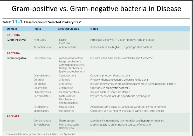
Types of gram positive bacteria are:
Separate phyla:
Firmicutes- Bacilli and Clostridia. gram + rods and cocci
Actinobacteria
pg 308
Types of gram negative bacteria are:
Proteobacteria- Alphaproteobacteria, betaproteobacteria, gammaproteobacteria, deltaproteobacteria, epsilonproteobacteria. This includes vibrio, salmonella, helicobacter and escherichia.
Cyanobacteria- oxygenic photosynthetic bacteria
chlorobi photosynthetic anoxygenic green sulfur bacteria
chlamydiae- grow only in eukaryotic host cells
plantomycetes- aquatic bacteria
bacteroidetes, flavobacteria, sphingobacteria- phylum members include opportunisttic infections.
fusobacteria- anaerobic some cause tissue necrosis and septicemia in humans.
Spirochetes- pathogens that cause syphilis and lyme disease.
pg. 291.
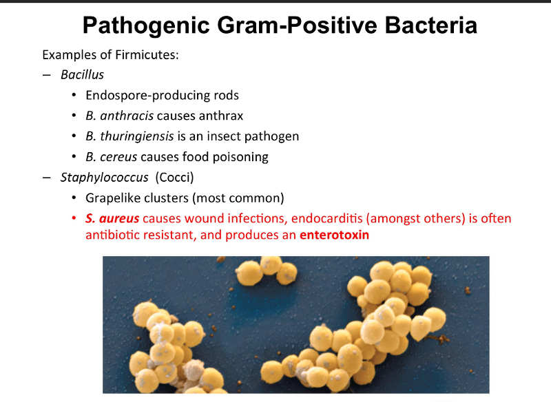
Pathogenic gram positive bacteria: Firmicutes
6.4
Will stain purple from the crystal violet.
Endospore forming bacteria -Bacillus (genus)- rod shaped bacteria and clostridium- rod shaped cells. cause Tetanus and botulism
- anthrax- used in biologic warfare
- b. Thuringiensis is an insect pathogen. Used as an insectocide for plants, sprayed on plants as a protectant from.
- B. Cereus- causes food poisoning.
-Staphylococcus (cocci) Photo shown is staph aureus. Has alot of antibiotic strains. produces endotoxins.
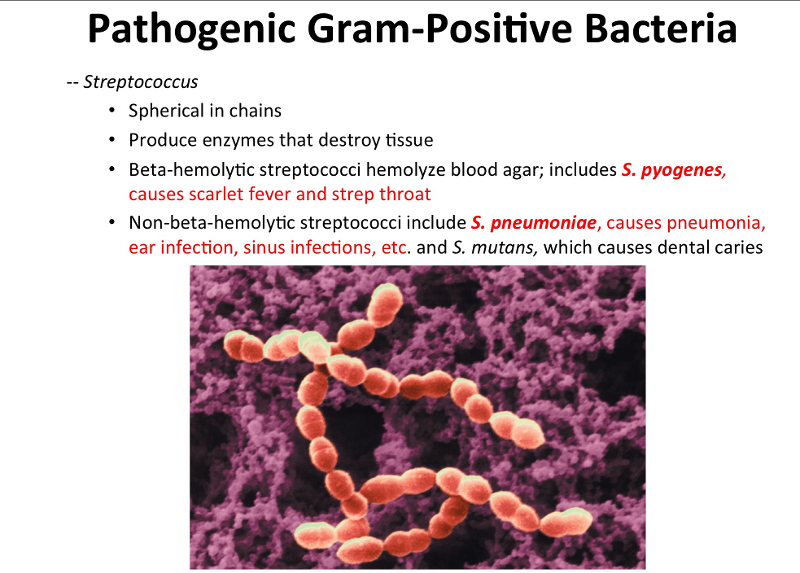
Pathogenic gram positive bacteria: Streptococcus
6.4
- spherical in chainlike form
- produce enzymes that destroy tissue- causes tissue damage.
- beta hemolytic streptococci hemolysis (lyce) blood agar; includes S. pyrogenes causes scarlet fever and strep throat.
- non-beta -hemolytic streptococci (don't lyce) include S. pneumoniae causes pneumonia, ear infection, sinus infection, etc. and S mutans which causes dental carries.
pg. 309
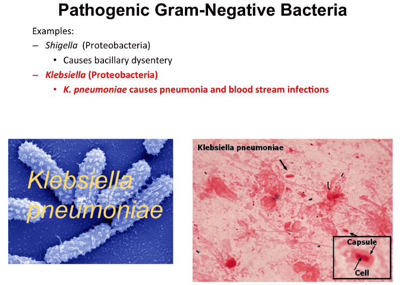
Pathogenic gram-negative bacteria:
6.4
Gram negative bacteria that have an extra plasma membrane around the outside of their cell wall. Also have a polysaccaride cell layer capsule in addition to the extra plasma membrane makes them really resistant to antibiotic treatment and can evade host immune responses as well.
proteobacteria examples:
- Shigella - causes bacillary dysentery
- klebsiella- K. Pneumoniae causes pneumonia-fluid to enter the lungs and blood stream infections or septicemia.
- Salmonella - 2400 serovars, common form of foodborne illness, salmonella typhi causes thyphoid fever.
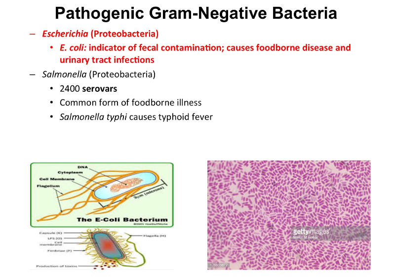
E. coli pathogen is very resistant to abx
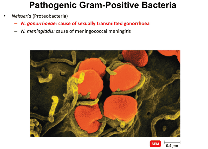
Pathogenic gram-positive bacteria proteobacteria:
6.4
- Neisseria:
- N. Gonorrhoeae: cause of sexually transmitted disease gonorrhoea
- N. meningitidis: cause of meningococcal meningitis.
- plasma membrane surrounded by the cell wall of peptidoglycan layer but they don't have extra plasma membrane
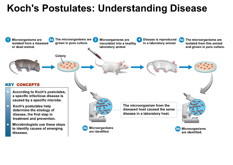
Koch's postulates:
6.4
- The same pathogen must be present in every case of the disease.
- The pathogen must be isolated from the diseased host and grown in pure culture.
- The pathogen from the pure culture must cause the disease when its inoculated into a healthy susceptible laboratory animal (reproduce the disease)
- The pathogen must be isolated from the inoculated animal and must be shown to be the original organism.
pg. 394.
Exceptions to Koch's postulates:
6.4
- Not all pathogens can cause sever disease conditions
- some pathogens cause disease only in humans
- some microbes have never been cultured
pg 394
Streptococcus pneumoniae is an example of what type of bacteria?
6.4
Gram-positive, streptococci
mechanisms of pathogenesis
6.5
When a microorganism invades a body tissue how does it cause disease or pathogenesis? it initially encounters phagocytes of the host, they destroy the invader or the pathogen will overcome the host's defenses, allowing the microorganism to damage the cell in 4 ways:
Direct damage:
- disrupts the host cell function
- uses host cell nutrients
- produces waste products
- multiplies in host cells and causes ruptures
(bacteria) Production of toxins:
- toxins- are poisonous substances that are produced by certain microorganisms- produce fever, CV problems, diarrhea, and shock
pg. 424
How do bacterial pathogens damage host cells?
1. Using the hosts nutrients: Siderphores
6.5
Iron is required for the growth of most pathogenic bacteria. The concentration of free iron in the human body is low because most of the iron is tightly bound to iron transport proteins and hemoglobin. For pathogens to obtain iron they secrete proteins called siderophores that take iron away from iron-transport proteins by binding the iron even more tightly, forms a complex and is taken up by the siderophore receptors on the bacterial surface and brought into the bacteria.
Siderophores are proteins secreted by pathogens that bind iron more tightly than host cells.
pg. 424
How do bacterial pathogens damage host cells?
2. Production of toxins
6.5
toxins are the primary factor contributing to the pathogenic properties of those microbes.
Two class types:
- exotoxins: proteins produced inside pathogenic bacteria that secreted by the bacteria outside the cell wall.
- endotoxins: lipid portions of the outer membrane of the cell wall of bacteria, are released only when bacteria die or their cell membranes break down, surface associated.
toxin genes can be spread between species via horizontal gene transfer.
424-425.
Production of toxins:
6.5
Toxigenicity- the capacity of microorganisms to produce toxins
Toxemia- the presence of toxin's in the blood.
Intoxications- caused by the presence of a toxin, not by microbial growth.
pg. 424-425.
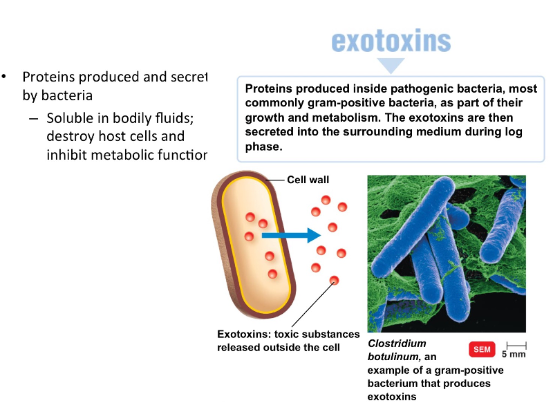
Exotoxins are proteins that are produced by bacteria and released into the extracellular environment.
6.5
- Exotoxins are proteins produced inside pathogens bacteria as part of their growth and metabolism and are secreted by the bacterium into the surrounding medium or released by lysis
- exo- means outside. This refers to the fact that exotoxins are secreted to the outside of the bacterial cells that produce them.
- most commonly gram positive bacteria, although can be gram negative.
- soluble in body fluids, can easily diffuse into the blood and are rapidly transported throughout the body.
- They are proteins and many are enzymes that catalyze certain biochemical reactions.
- they work by destroying particular parts of the host cell or by inhibiting certain metabolic functions.
- Very lethal, i.e.: clostridium botulinum- botulism, gram + bacteria.
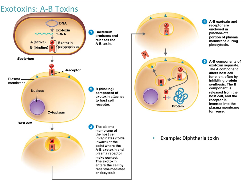
The action of an A-B Exotoxin:
6.5
They are proteins- polypeptides.
Consist of two parts: A and B, both of which are poylpeptides. Most exotoxins are A-B toxins. The part A is the active enzyme component and the part B is the binding component.
releases the A-B toxin, attaches to the host cell, taken into the host cell, the A and B come apart. The receptor B is released outside of the cell.
Diptheria is an example of a A-B toxin.
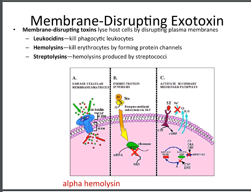
Membrane disrupting exotoxins
6.5
Cause lysis of the host cells by disrupting their plasma membranes. Some form protein channels in the plasma membrane (The cell -lysing exotoxin of Staphylococcus aureus is an example of an exotoxin that forms protein channels) others disrupt the phospholipid membrane (Clostridium perfringens).
Membrane disrupting toxins contribute to virulence by killing host cells.
- leukocidins- kill phagocytic leukocytes- immune cells, (WBC) by forming protein channels. Produced by staphylococci and streptococci.
- hemolysis- supply nutrients for bacterial cell growth, kill erythrocytes by forming protein channels.
- hemolysins Lyse RBC, produced by streptococci.
pg. 426
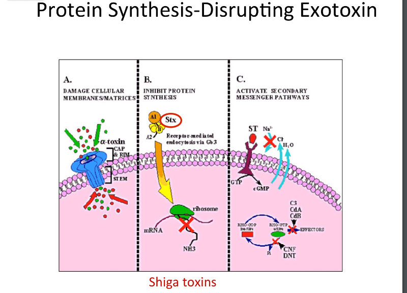
Protein synthesis disrupting exotoxin.
6.5
This toxin disrupts protein synthesis. A-B type exotoxin, the A fragment disrupts translation.
This gets into our GI tract
exotoxin known as Shigella
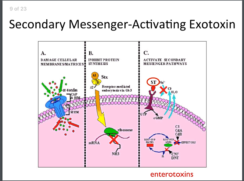
Second messenger-activating exotoxin.
last class of exotoxin:
(Binds the receptor that Ligand binds activating the G protein inside the cell to activate the series of events inside the cell.)
An exotoxin produced from e. coli. that exotoxin binds the receptor we get formation of GTP from cyclic GMP and the end result is that we end up secreting chloride ions outside of the cell, water follows and results in diarrhea.
other exotoxins: Superantigens and genotoxins:
6.5
Super antigens: cause an intense immune résponse, through a series of interaction with various immune cells. In response to superantigens enormous amounts of chemicals called cytokines are released from the host cells (T cells). Excessively high levels of cytokines causes symptoms of fever, N/V/D, shock, and death. Staphylococcus aureus- can cause food poisoning and TSS.
Genotoxins: alter the hosts DNA, damage DNA directly causing mutations, disrupting cell division and leading to cancer. helicobacter- causes breaks in DNA leading to stomach CA.
pg. 427.
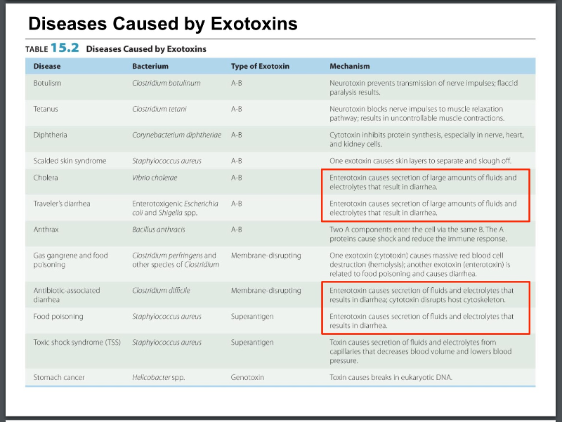
Table of diseases caused by exotoxins
6.5
Enterotoxins- in GI tract results in diarrhea
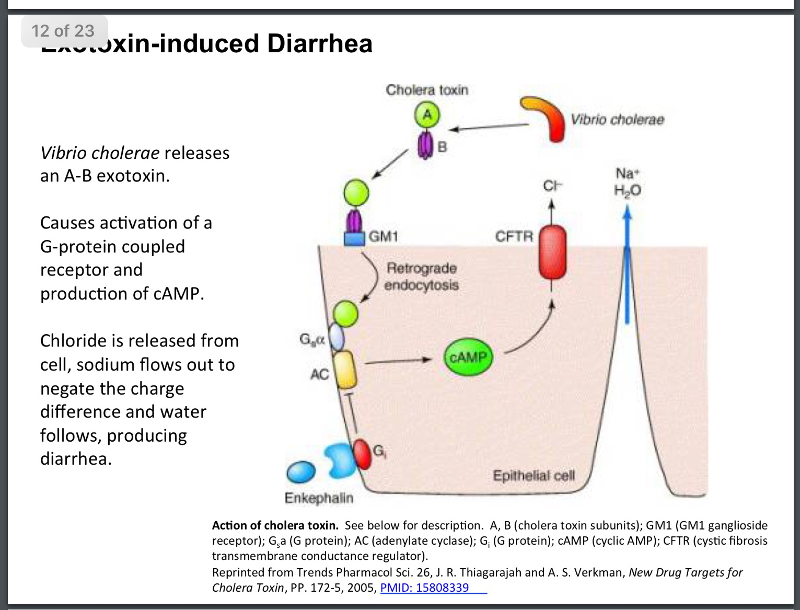
Cholera toxin- Vibrio cholera
Cholerae releases an A-B toxin.
binds and taken up into the cell
activates a second messenger cell
Chloride and water are pumped out of the cell produces diarrhea.
Can be deadly in third world countries.
Drugs used to combat this problem- encephalins- active a G protein to stop the over release of chloride
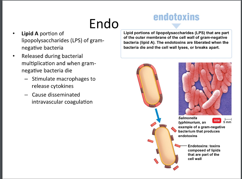
EndoToxins
6.5
Endotoxins are part of the outer portion of the cell wall of gram negative bacteria. Released during bacterial multiplication and when gram-negative bacteria dies and their cell wall lyses.
Endotoxins exert their effects by stimulating macrophages to release cytokines in very high concentrations- at these toxic levels they produce fever, chills, weakness, generalized aches, sometimes shock and death.
pg. 428
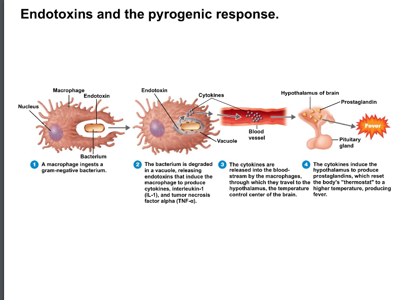
How do endotoxins produce a febrile response in a cell?
6.5
a macrophage ingests a gram negative bacteria, releases endotoxins, induce macrophages to produce cytokines ( IL1 & TNF alpha) which are released into the bloodstream, travel to the hypothalamus, and induce the hypothalamus to produce prostaglandins which reset the body's thermostat higher producing fever.
Testing for endotoxins- LAL assay
lab test uses to identify the presence of endotoxins in drugs, medical devices, and body fluids.
Limulus amebocyte lysate assay used to test for endotoxins. Blood from horseshoe crabs contain amebocytes that lyse in the presence of endotoxin producing a clot.
pg. 429
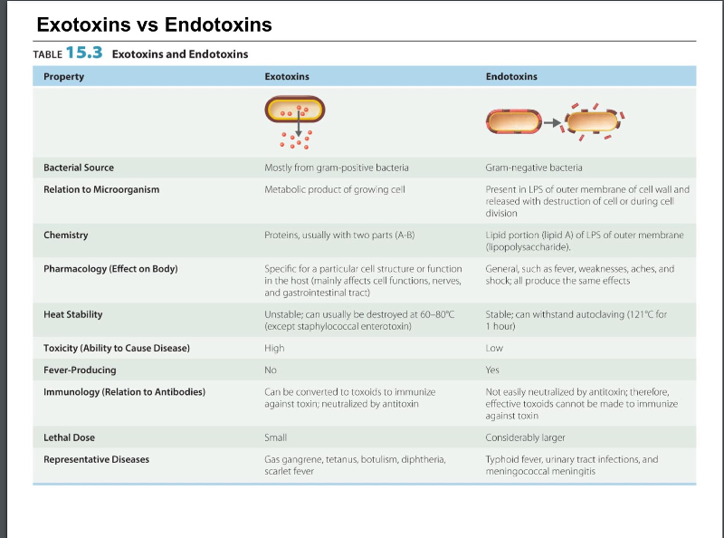
Comparison of exotoxins and endotoxins
gram negative bacteria produce endotoxins and gram positive produce exotoxins.
exotoxins have a wide range of effects
endotoxins tend to have similar effects in the host cell.
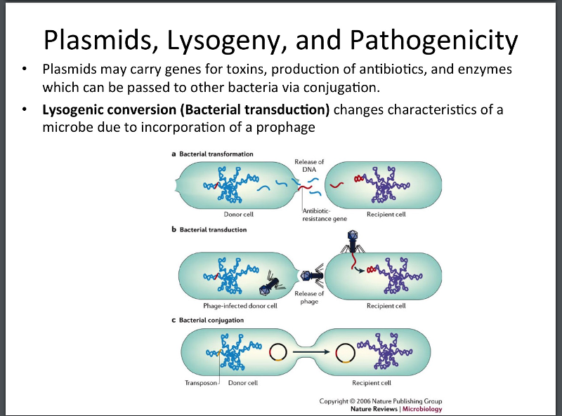
Bacterial transformation- release of DNA from one cell makes its way into a recipeint cell.
Bacterial transduction- movement of genes by viruses that infect bacteria- bacteriophage. Acquires a gene from one bacteria and when it infects another bacteria injects that genetic material
Bacterial conjugation- transmit extra chromosomal plasmids and toxins along with antibiotic resistance.
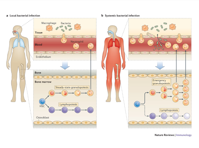
Bacterial pathogenesis can be a local infection or can be systemic if it enters the blood stream.
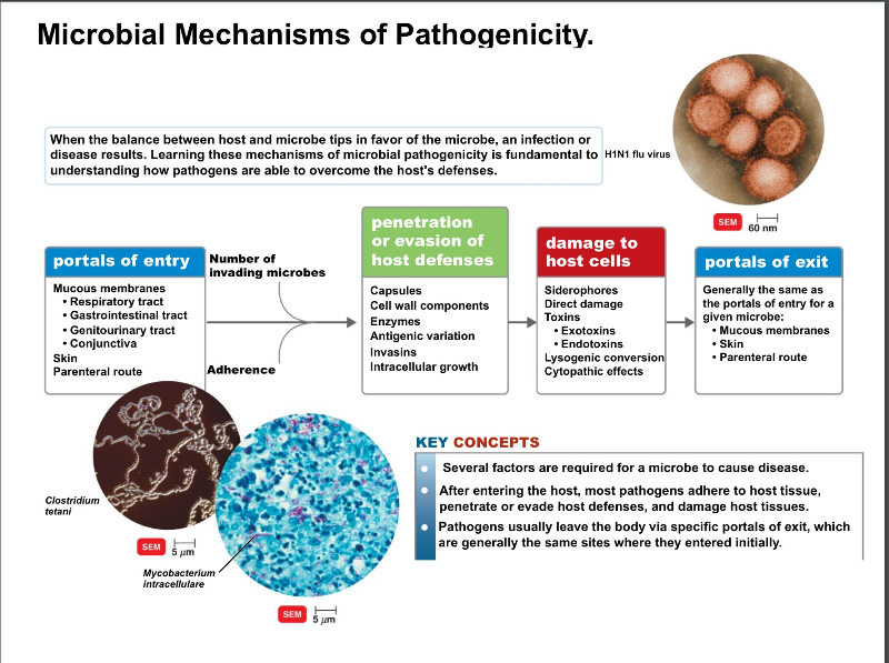
How do microbes (bacteria or viruses) evade hosts?
entry into the host cell or tissue
evasion of the host defenses
damage to the host cells either directly or by toxins
replicate and exit the body to hopefully go on and infect a new host.
The outer portion of Gram-negative cell walls contain __________.
endotoxins
Proteins secreted by pathogens that bind iron are known as __________.
Sidophores
The antibiotic revolution and antibiotic resistance.
6.6
- Abx are used to kill or inhabit the growth of bacteria
- naturally occurring, first antibiotic 1940's -penicillin from penicillium
- synthesized in the lab -arsphenamine
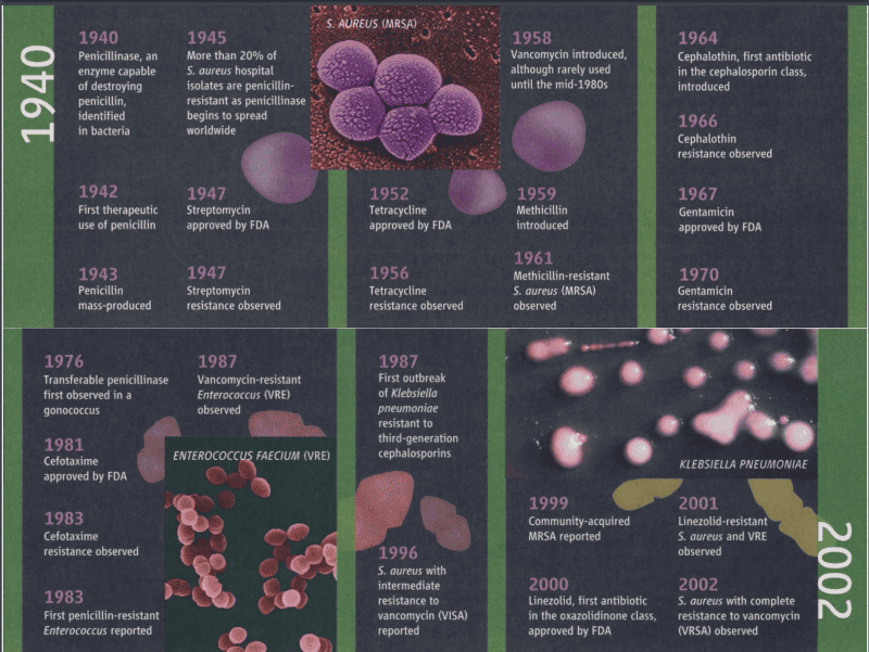
Timeline of the discovery of PCN
used in 1942, mass produced and 3 years later staph aureus shows resistance.
1947 new antibiotic is used- streptomycin. 3 years later streptomycin resistance
tetracycline, vancomycin, methicillin all produced and lead to resistance.
MRSA 1961
pathogenic bacteria with no abs to treat them.
Natural selection
The process in which individuals that have certain heritable traits survive and reproduce at a higher rate than other individuals because of those traits
over time, can increase the match between organisms and their environment.
in response to a change in environment may result in adaptation to new conditions.
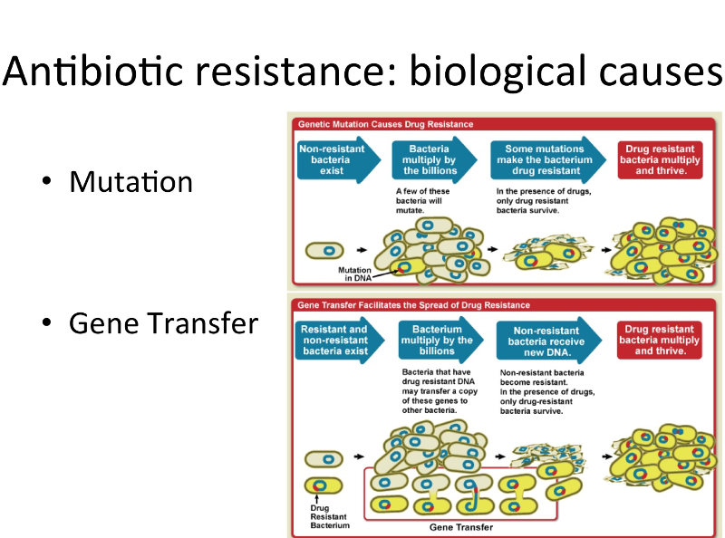
normal genetic mutations occur that make bacteria slightly different making that bacteria now resistant to abs.
gene transfer- if the bacteria has the means of transferring its genetic material it can now become resistance.
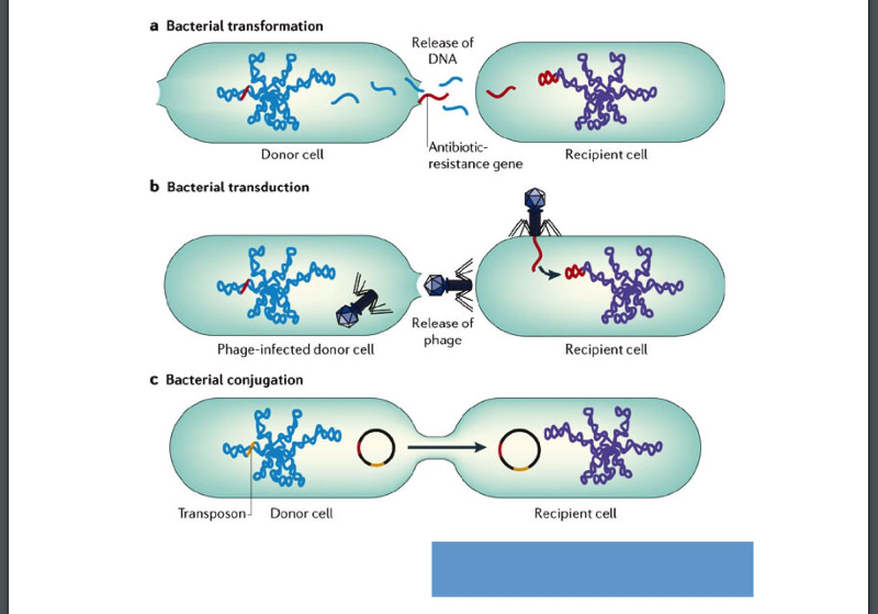
transformation- release of DNA
transduction- transfer via bacterialphage
conjugation- small plasmids are transferred to produce proteins to another bacteria.
antibiotic resistance
6.6
overview of how antibiotic resistance develops.
natural genetic variation -population of bacteria where there is few resistance to abx.
abx kill most of the bacteria, but the few left form abx resistance strains this is the result of natural mutation.
bacteria pass via gene transfer or other mechanisms to other bacteria providing resistance.
How does antibiotic resitance spread in the community?
Simply using antibiotics creates resistance. These drugs should only be used to treat infection.
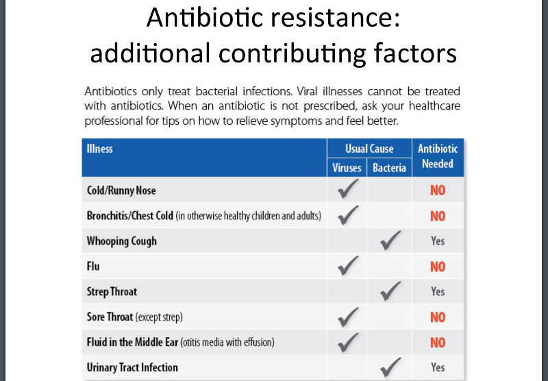
antibiotics were being misused to for bacteria vs viral infections.
abx should only be given for true bacterial infections.
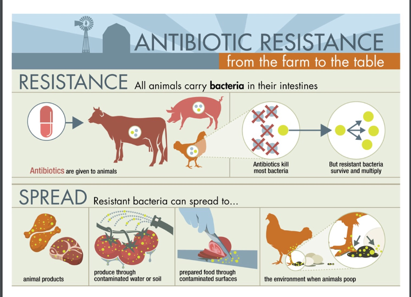
abx given to animals on the farm that then provides resistance strains in our food.
exposed to contaminated foods or from a contaminated environment.
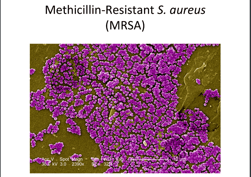
prevalent in hospitals, resistant to most abx, tends to be untreatable.
skin disease, can look like a spider bite, no obvious wound. AKA staph infections.
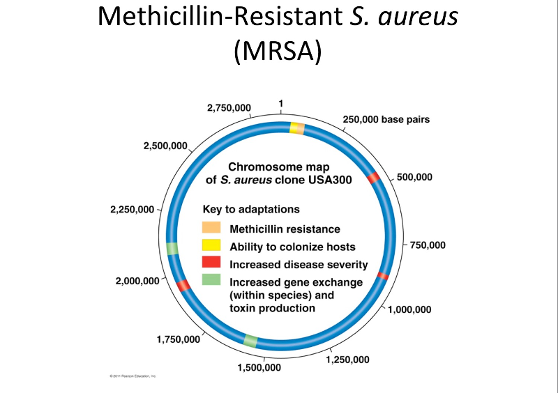
the chromosomal map of MRSA.
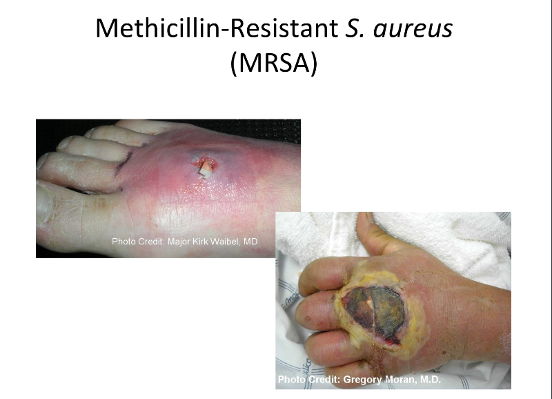
Causes huge amounts of tissue damage that is difficult to treat due to abx drug resistance.
frequent hand washing
keep wounds covered
clean linens
develop new abx
reduce the use of the ones we already have.
The removal of plasmids reduces virulence in which of the following organisms?
Streptococcus mutans
A encapsulated bacterium can be virulent because?
It resists phagocytosis and continues growing
ie: streptococcus pneumoniae and klebsiella pneumoniae produce capsules that r/t their virulence.
A drug that binds to mannose on human cells would?
Prevent the attachment of pathogenic e. Coli.