What is the function of the coronary circulation?
Provide a blood supply to the aortic arch
Provide a blood supply to the heart
Provide a blood supply to the pericardium
Provide a blood supply to the lungs
Provide a blood supply to the heart
What is the ligamentum arteriosum?
A remnant of the ductus arteriosus
A ligament attaching the aorta to the superior vena cava
A ligament securing the aorta
A remnant of the foramen ovale
A remnant of the ductus arteriosus
Which chamber of the heart exits into the pulmonary trunk?
Right ventricle
Left atrium
Left ventricle
Right atrium
Right ventricle
Identify the ear like flaps that are attached to the top chambers of the heart.
Pectinate muscles
Atrium
Auricle
Coronary sinus
Auricle
The first branch off the arch of the aorta is the brachiocephalic artery in both the sheep and the human.
True
False
True
The base of the heart is located at the bottom of the heart.
True
False
False
Identify the most muscular chamber.
Left ventricle
Right atrium
Left atrium
Right ventricle
Left ventricle
Name the inner lining of the heart.
Endocardium
Pericardium
Myocardium
Epicardium
Endocardium
Identify the valve found between the left atrium and left ventricle.
Aortic valve
Tricuspid valve
Bicuspid (mitral) valve
Pulmonary valve
Bicuspid (mitral) valve
What heart chamber pushes blood through the aortic semilunar valve?
Left ventricle
Left atrium
Right atrium
Right ventricle
Left ventricle
Name the needle like ridges of muscle lining the ventricles.
Papillary muscles
Trabeculae carneae
Chordae tendineae
Pectinate muscles
Trabeculae carneae
What fibrous structure functions to anchor the atrioventricular valves in a closed position?
Chordae tendineae
Trabeculae carneae
Moderator band
Papillary muscle
Chordae tendineae
Blood on the right never mixes with blood on the left, once the heart is fully developed.
True
False
True
Name the ridged bundles of muscle found projecting inside the right atrium.
Intercalated discs
Trabeculae carneae
Papillary muscles
Pectinate muscles
Pectinate muscles
Identify the right atrioventricular valve.
Aortic semilunar valve
Tricuspid valve
Bicuspid valve
Pulmonary valve
Tricuspid valve
Identfiy the valve located at the exit of the right ventricle.
Aortic semilunar valve
Tricuspid valve
Bicuspid valve
Pulmonary semilunar valve
Pulmonary semilunar valve
The moderator band is found on both the right and left side of the heart.
True
False
False
Oxygenated blood flows through the right side of the heart.
False
True
False
Action potentials generated by the autorhythmic cells spread to the contractile cells through what structures in the membrane?
intercalated discs
desmosomes
gap junctions
tight junctions
gap junctions
One of the changes that occurs in the pacemaker potential (unstable resting membrane potential) in the SA node (an autorhythmic cell) is a decreased efflux of what ion?
potassium
sodium
calcium
potassium
When threshold is reached at the SA node (an autorhythmic cell), what channels open causing further depolarization of the membrane?
fast sodium
fast calcium
slow calcium
potassium
fast calcium
Repolarization of an autorhythmic cell is due to the opening of which channels?
voltage-gated sodium channels
Chemically gated potassium channels
voltage-gated potassium channels
chemically gated calcium channels
voltage-gated potassium channels
In order to cause cardiac muscle contraction, the contractile cells must also depolarize. What causes the depolarization of the contractile cells?
the flow of negative ions from adjacent cells
an unstable resting membrane potential in the contractile cells
the flow of positive ions from adjacent cells
the flow of positive ions from adjacent cells
Which part of the conduction system initiates the depolarizing impulse, which spreads throughout the heart?
SA node
AV node
Purkinje fibers
AV bundle (bundle of His)
SA node
What does the ECG wave tracing represent?
contraction of the heart
electrical activity in the heart
electrical activity in the heart
What does the QRS complex represent in the ECG wave tracing?
atrial depolarization v
entricular depolarization
ventricular repolarization
atrial repolarization
ventricular depolarization
Contraction of the atria results from which wave of depolarization on the ECG tracing?
P wave
QRS complex
T wave
P wave
Which part of the intrinsic conduction system delays the impulse briefly before it moves on to the ventricles?
Purkinje fibers
SA node
bundle branches
AV bundle (bundle of His)
AV node
AV node
Isovolumetric relaxation and ventricular filling (two phases of the cardiac cycle) take place during __________.
ventricular systole
ventricular diastole
ventricular diastole
Which of the following is correct about the filling of the ventricles?
The majority of ventricular filling is caused by contraction of the atria.
Most blood flows passively into the ventricles through open AV valves.
Most blood flows passively into the ventricles through open AV valves.
Describe the pressures in the atria and ventricles that would cause the opening of the AV valves.
Pressure in the atria would be greater than the pressure in the ventricles.
Pressure in the ventricles would be greater than in the atria.
Pressures in the atria and ventricles would be equal.
Pressure in the atria would be greater than the pressure in the ventricles.
What causes the aortic semilunar valve to close?
higher ventricular pressure than aortic pressure
equal ventricular and aortic pressures
greater pressure in the aorta than in the left ventricle
greater pressure in the aorta than in the left ventricle
Put the phases of the cardiac cycle in the correct order, starting after ventricular filling.
isovolumetric contraction, ventricular ejection, isovolumetric relaxation
isovolumetric relaxation, ventricular ejection, isovolumetric contraction
ventricular ejection, isovolumetric contraction, isovolumetric relaxation
ventricular ejection, ventricular relaxation, isovolumetric contraction
isovolumetric contraction, ventricular ejection, isovolumetric relaxation
Increased pressure in the ventricles would close what valve(s)?
both semilunar and AV valves
AV valves only
semilunar valves only
AV valves only
Which of the following would increase cardiac output?
decreased calcium during contraction
parasympathetic stimulation
epinephrine
high blood pressure
epinephrine
Which of the following is NOT a factor that increases stroke volume?
increasing sympathetic
stimulation increasing afterload
increasing contractility
increasing preload
increasing afterload
Calculate the stroke volume if the end diastolic volume (EDV) is 135 mL and the end systolic volume (ESV) is 60 mL.
60 mL
75 mL
205 mL
8100 mL
75 mL
What structures connect the individual heart muscle cells?
intercalated discs
chordae tendineae
trabaculae carneae
anastomoses
intercalated discs
Which statement regarding cardiac muscle structure is accurate?
Myofibrils of cardiac muscle tissue vary in diameter and branch extensively.
Cardiac muscle cells are independent of one another both structurally and functionally.
Cardiac cells are long, cylindrical, and multinucleate.
Cardiac cells possess few mitochondria.
Myofibrils of cardiac muscle tissue vary in diameter and branch extensively.
The _________ carries oxygen-poor venous blood from above the diaphragm from areas of the upper body and extremities into the right atrium.
Superior Vena Cava
The _________ carries oxygen-poor venous blood of the coronary circulation into the right atrium.
coronary sinus
The ________ carries oxygen-poor venous blood from below the diaphragm from the areas of the lower body and extremities into the right atrium.
inferior vena cava
Art-based
Right-Sided Anatomical Features
Drag and drop the correct identification label to the box with a leader line. Answers may be used once or not at all.
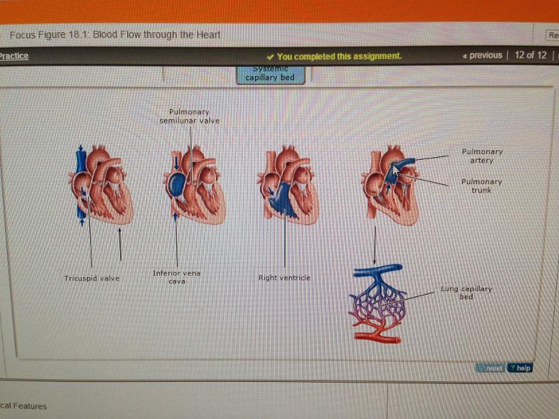
See photo
Flow through the Right Side of the Heart
Assume that blood is flowing from the coronary sinus to the lung capillaries. Place the anatomical labels in order of flow in the target boxes. Not all labels are used.
Coronary Sinus > Right Atrium > Tricuspid Valve > Right Ventricle > Pulmonary Semilunar Valve > Pulmonary Trunk > Pulmonary Arteries > Capillaries of Lungs
Part D - Oxygen-Rich Blood Returning to the Heart
Drag and drop the terms to arrange them, from left to right, in order of blood flow of oxygen-rich blood into the heart.
Lungs > Lung Capillaries > Pulmonary Veins > Left Atrium > Mitral/Bicuspid Valve > Left Ventricle > Aortic/semilunar Valve > Aorta
The structure that prevents backflow of blood into the left atrium is the _________
mitral (bicuspid) valve
The vessel that carries oxygen-rich blood to tissues is the _______
aorta
The capillaries receiving blood flow from the left side of the heart are the _________
systemic capillaries
The structure that is located anatomically between the aorta and the left ventricle is the ___________
aortic semilunar valve
Art-based Question
Part F - Conclusion: Valves in Blood Flow through the Heart
Drag and drop valve names to their correct location in the image.
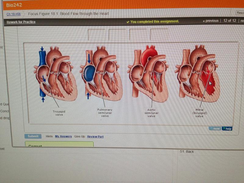
See Photo
Sort the terms into the appropriate category of either oxygen-poor or oxygen-rich.
Oxygen Poor: Superior Vena Cava, Right Atrium, Right Ventricle, Pulmonary Arteries
Oxygen Rich: Aorta, Left Ventricle, Left Atrium Pulmonary Veins
Select the correct partial path. This path is part of the complete blood flow pathway. You should be able to trace flow starting in any location.
pulmonary artery into left atrium through mitral valve to left ventricle
systemic veins returning to the left atrium and forward through the mitral valve
pulmonary trunk to pulmonary arteries to pulmonary capillaries to pulmonary veins returning to the right atrium
aorta to smaller systemic arteries to systemic capillaries to systemic veins to right atrium through the tricuspid valve
aorta to smaller systemic arteries to systemic capillaries to systemic veins to right atrium through the tricuspid valve