Interpreting an Action Potential Tracing.
Using the tracing above, drag each of the terms or values on the left to correctly complete the sentences on the right.
- 1. This neuron is most depolarized at (+30) mV.
- 2. This neuron spends approximately (2.5) msec in a hyperpolarized state.
- 3. The ion (K+) is crossing the cell's plasma membrane at 1.5 msec.
- 4. This cell reaches threshold at approximately (0.6) msec.
What does 0 mV on the Y-axis of an action potential tracing represent?
The cell's membrane is at equilibrium.
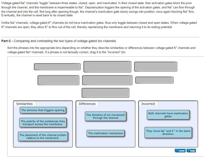
Comparing and contrasting the two types of voltage-gated ion channels
Similarities:
- The stimulus that triggers opening
- The polarity of the substances they transport across the membrane
- The placement of channel protein relative to the membrane
Comparing and contrasting the two types of voltage-gated ion channels
Differences:
- The direction of ion movement through the channel
- The inactivation mechanism
Comparing and contrasting the two types of voltage-gated ion channels
- Both channels have inactivation gates
- They move Na+ and K+ in the same direction
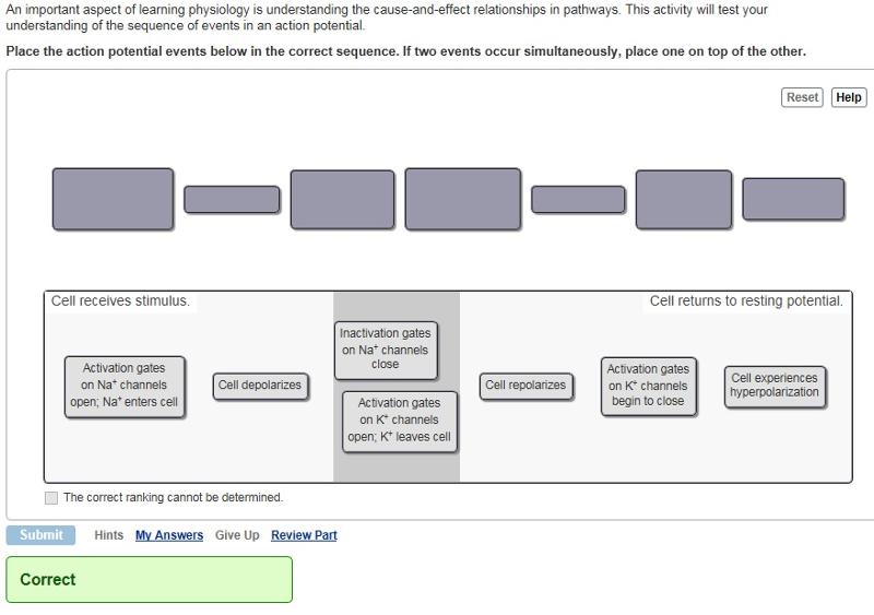
Understanding the sequence of action potential events
An important aspect of learning physiology is understanding the cause-and-effect relationships in pathways. This activity will test your understanding of the sequence of events in an action potential. Place the action potential events below in the correct sequence. If two events occur simultaneously, place one on top of the other.
1. Activation gates on NA+ channels open; Na+ enters cell.
2. Cell Depolarizes.
3. Inactivation gates on Na+ channels close
&
Activation gates on K+ channels open; K+ leaves cell
4. Cell Repolarizes.
5. Activation gates on K+ channels begin to close
6. Cell experiences hyperpolarization.
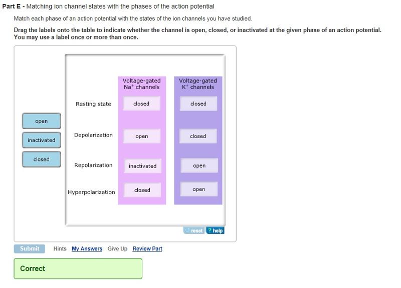
Matching ion channel states with the phases of the action potential
Match each phase of an action potential with the states of the ion channels you have studied. Drag the labels onto the table to indicate whether the channel is open, closed, or inactivated at the given phase of an action potential. You may use a label once or more than once.
Resting State: Na+ channels closed, K+ channels closed
Depolarization: Na+ open, K+ closed
Repolarization: Na+ inactivated, K+ open
Hyperpolarization: Na+ closed, K+ open
Understanding why hyperpolarization happens
Following repolarization, the neuron may become slightly hyperpolarized before it re-establishes its resting membrane potential.
Hyperpolarization is due to a difference between how the voltage-gated Na+ and K+ channels work. What is this difference?
Voltage-gated Na+ channels stop the flow of Na+ relatively quickly, while voltage-gated K+ channels are slow to close, resulting in the overshoot.
Making predictions based on your understanding
Interpreting tracings of membrane potential is an important skill in a course like this. In future chapters, you will see other instances of cells experiencing changes in membrane potential. For example, the tracing below is from a cardiac muscle fiber that is undergoing an action potential.
Based on your knowledge of action potentials in neurons, what can you conclude about how they occur in cardiac muscle fibers?
Cardiac muscle fibers depolarize more quickly and spend more time depolarized than neurons do.