Describe the functional relationship between volume changes and gas flow into and out of the lungs.(Part 1)essay
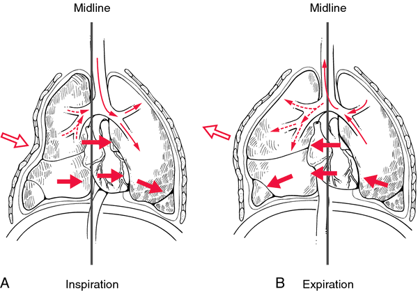
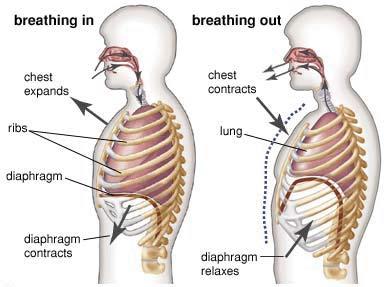
As the muscles of inspiration , the diaphragm and the external intercostal muscles contract, the thoracic cavity is expanded
inferiorly by the diaphragm pulling down as it contracts and laterally and superiorly as the external intercostal muscles cause the ribs to expand up and out. This expansion of the thoracic cavity causes the volume of the thorax to increase because it is now a larger "container" than before V(up), P(down)
Describe the functional relationship between volume changes and gas flow into and out of the lungs.(Part 2)essay
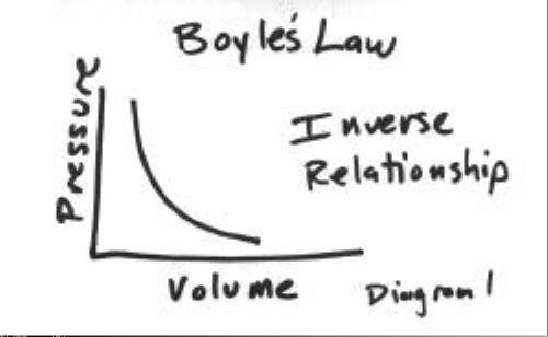
Boyle's Law, if volume increases in a given space it causes the pressure of gases in that space to decrease V(down) P (up)
Pressure in the lungs is now lower than the atmospheric pressure outside of the lungs, causing the air to be pushed into the lungs along this pressure gradient from higher to lower.
The flow of air into the lungs stops as the pressure equalizes with the atmospheric pressure. (pause)
Describe the functional relationship between volume changes and gas flow into and out of the lungs.(Part 3)essay
Expiration starts when the inspiratory muscles relax and are pulled back into their normal resting length due to the elastic properties of the muscles and tissues of the thoracic cavity.
The volume of the thoracic cavity and lungs is thus decreased as the size of the "space" in the thorax is reduced.V(down) P(up)
This decrease of volume causes the pressure in the lungs to increase to a pressure higher than the atmospheric pressure, and air is pushed out of the lungs from the higher to lower pressure gradient.
Identify the cause of common digestive system dysfunctions
Reflux
Peptic ulcers
Gallstones
Lactose intolerance
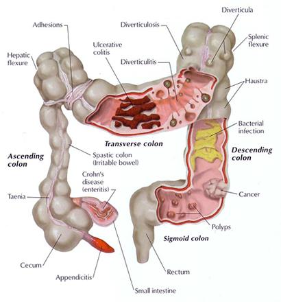
Divertticulitis
Ibs
Celiac disease
Constipation
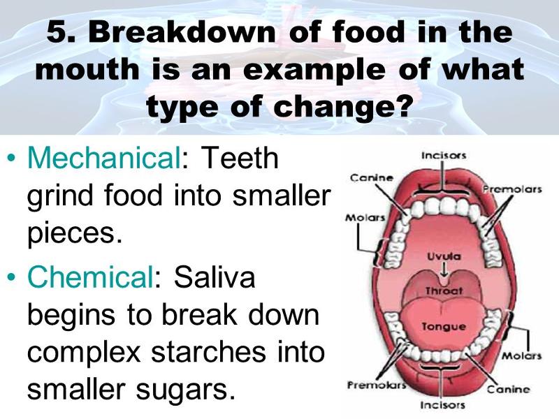
___________________is the mechanical and chemical breakdown of foods into forms that cell membranes can absorb?
Digestion
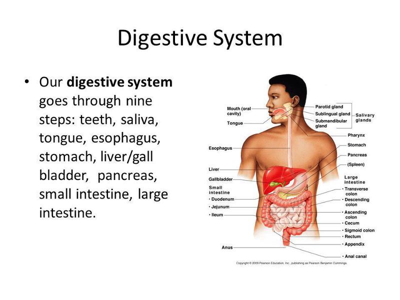
Organs of the digestive system carry out these processes, as well as ingestion, propulsion, absorption and defecation
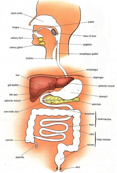
The digestive system consists of the ___________canal extending from the mouth to the ______, plus accessory ______ that empty into the alimentary canal
Alimentary
ANUS
ORGANS
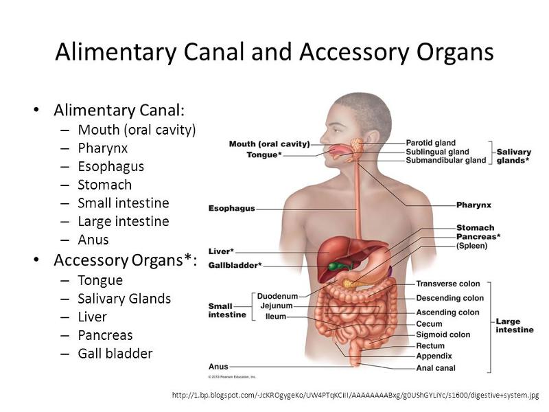
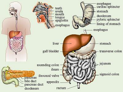
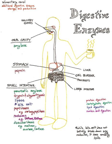
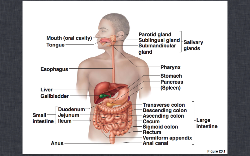
Alimentary canal (gastrointestinal or GI tract)
FUNCTION: Digests and absorbs food
SYSTEM: Mouth, pharynx, esophagus,stomach, small intestine, and large intestine
Accessory digestive organs
Teeth, tongue, gallbladder
Digestive
glands A) Salivary glands
B)
Liver
C) Pancreas
ALIMENTARY CANAL
ACCESSORY ORGANS
Salivary
glands
Secrete saliva: enzymes that initiate
breakdown
of carbs. This begins the digestion of
carbohydrates
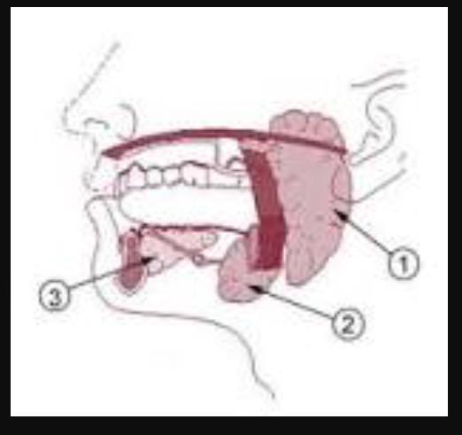
There are three pairs
of major salivary glands
Parotid
glands
Submandibular
glands
Sublingual glands
There are many minor glands scattered throughout the mucosa : t ongue, palate, and cheeks
ALIMENTARY CANAL:
STEP 1
Mouth: Mechanical breakdown of food MASTICATION begins chemical digestion of carbs
Pharynx
Connects mouth with esophagus
Esophagus pushes food to stomach
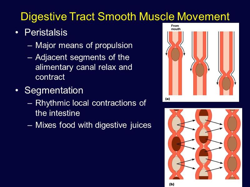
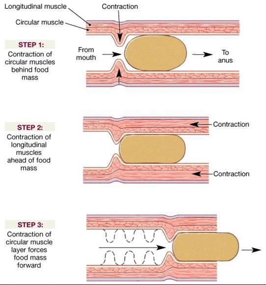
Peristalsis: Adjacent segments of
alimentary tract
organs alternately contract
and relax, which moves food along
the tract
distally
Segmentation: Nonadjacent
segments
of alimentary tract organs alternately
contract
and relax, moving the food
forward then backward. Food mixing
and
slow food propulsion occurs.
ALIMENTARY CANAL
STEP 2
Liver: Produces bile (emulsifier of fats/lipids)
Gallbladder: Stores bile and introduces it into small intestine
Pancreas: Produces and secretes pancreatic juice,
Digestive enzymes
Bicarbonate ions ----->>>> into small intestine
ALIMENTARY CANAL
STEP 3
Stomach - Secretes acid and enzymes
mixes food with
secretions to begin enzymatic
digestion of proteins
Small intestine: Mixes food with bile and pancreatic juice
FINAL enzymatic breakdown of food molecules
MAIN site of nutrient absorption
Large intestine
Absorbs water and electrolytes to form
feces
Rectum
: Regulates elimination of feces
Anus
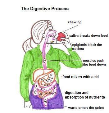
Digestive Processes
WHAT ARE Six essential activities?

Ingestion - Swallowing can be divided into
three stages:
Voluntary stage - where saliva is mixed with
chewed food
Swallowing- begins and the swallowing reflex is
triggered
Peristalsis - transports food in the esophagus to the stomach
Propulsion
-
The palate and uvula raise
The hyoid bone and
larynx elevate
The epiglottis closes off top of the
trachea
The longitudinal muscles of pharynx contract
The
inferior constrictor muscles relax and the esophagus opens; peristaltic
waves pushes food through the pharynx
Mechanical
digestion- CHEW CHURNING SEGMENTATION
Chemical
digestion- PH ENZYMES
Absorption-SM. INTESTINE/ LYMPH/ BLOOD
Defecation- LRG. INTESTINE - MOSTLY H2O AND
FECES TO ANUS
WHAT ARE THE General Characteristics of the Alimentary Canal?
The alimentary canal is a muscular tube about 8 meters long
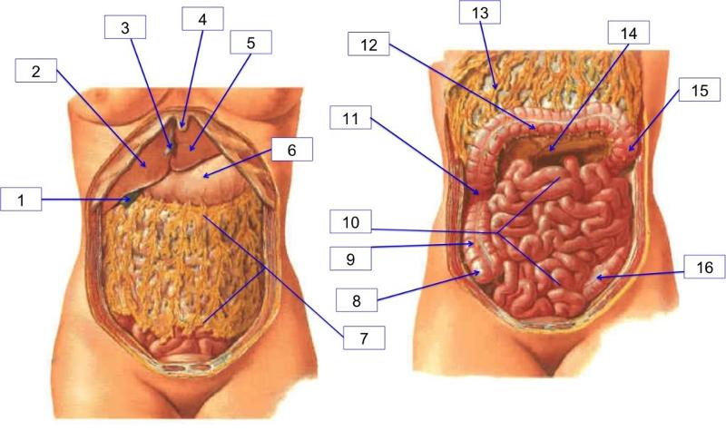
Peritoneum and Peritoneal Cavity
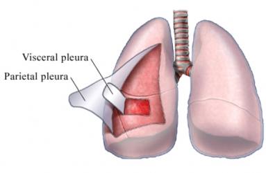
Serous membrane of
the abdominal cavity:
Visceral
peritoneum on external surface of most digestive organs
Parietal peritoneum lines the body wall
PERITONEAL CAVITY : Between the two peritoneums (Fluid lubricates mobile organs)
________________ is a double layer of peritoneum
Routes for blood vessels, lymphatics, and nerves
Holds organs in place and stores fat
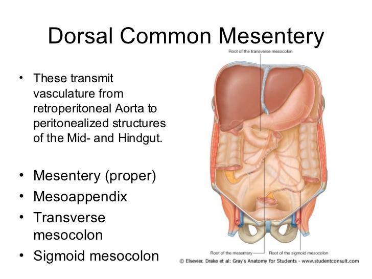
Mesentery
___________________ lie posterior to the peritoneum
___________________ are surrounded by the peritoneum.
Retroperitoneal organs
Intraperitoneal (peritoneal) organs
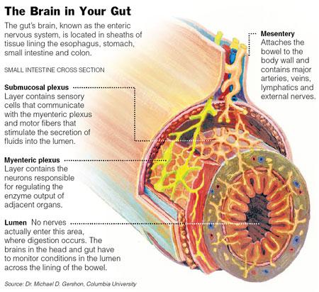
Branches of the sympathetic and parasympathetic divisions of the autonomic nervous system extensively innervate the alimentary canal, including:
____________ – controls secretions
____________ – controls gastrointestinal motility

Submucosal plexus
Myenteric plexus
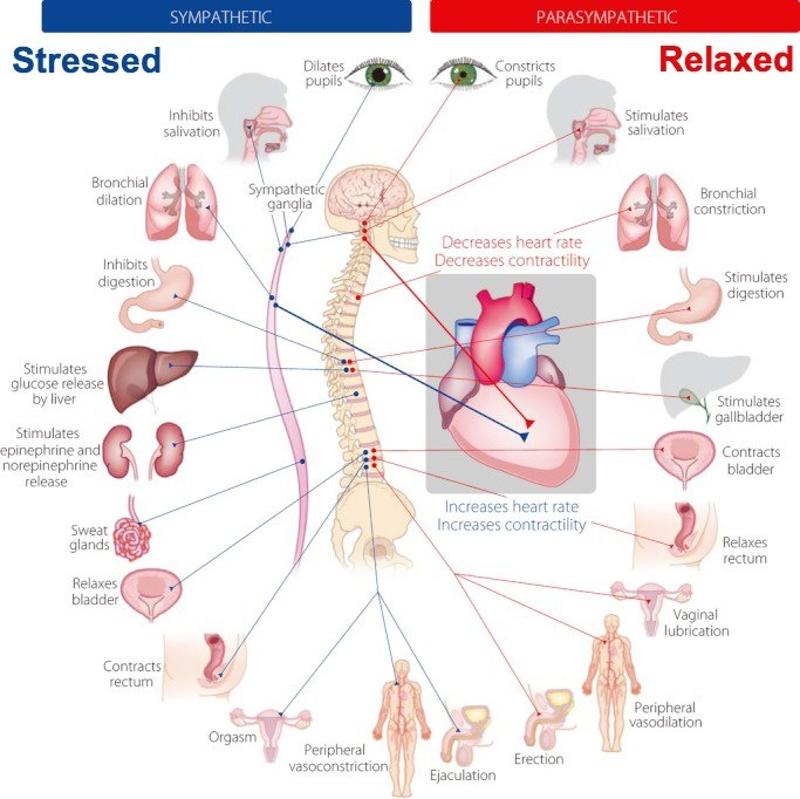
_____________ – increase activities of digestive
system
______________– inhibit certain
digestive actions

Parasympathetic impulses(FEED/BREED)
Sympathetic impulses( FIGHT OR FLIGHT)
THE ______Ingests food: Mechanically breaks up solid particles using saliva
Prepares food for chemical digestion ( Mastication)

THE MOUTH
___________ is a thick, muscular organ that occupies the floor of the mouth and nearly fills the oral cavity when the mouth is closed
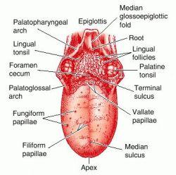
The tongue
_______________ forms the roof of the oral cavity and consists of a hard anterior part and a soft posterior part
The palate
Hardest structures in the body
There
are ____ primary (deciduous)
There are
_____secondary (permanent)
The
teeth
(deciduous) teeth
numbering 20
(permanent) teeth
numbering 32
The different salivary glands have varying proportions of two types of secretory cells
___________produce a watery fluid with a digestive enzyme
called salivary amylase
___________ secrete mucous
Serous cells
Mucous cells
__________
Secrete clear watery,
serous fluid.
Rich in salivary amylase
(CONVERTS STARCH AND GLYCOGEN= SIMPLE SUGARS )
______________
Secrete primarily
serous fluid and some mucus.
______________
Secrete primarily mucus.
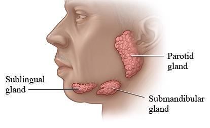
Parotid glands
Submandibular
glands
Sublingual glands
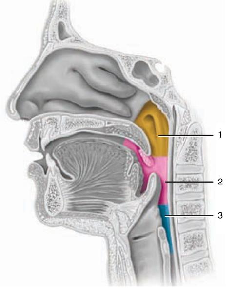
_____________is a cavity posterior to the mouth from which the
tubular esophagus leads to the stomach.
Both the _____________ and esophagus muscular walls function in swallowing.
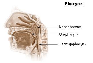
The pharynx
The pharynx can be divided into 3 parts?

Nasopharynx
Oropharynx
Laryngopharynx
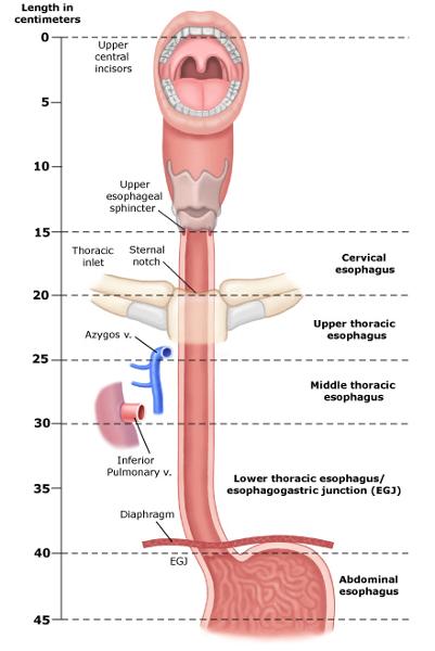
What is the Flat muscular tube from laryngopharynx to stomach,
Pierces diaphragm at esophageal hiatus
and Joins stomach at the cardiac orifice ?

Esophagus
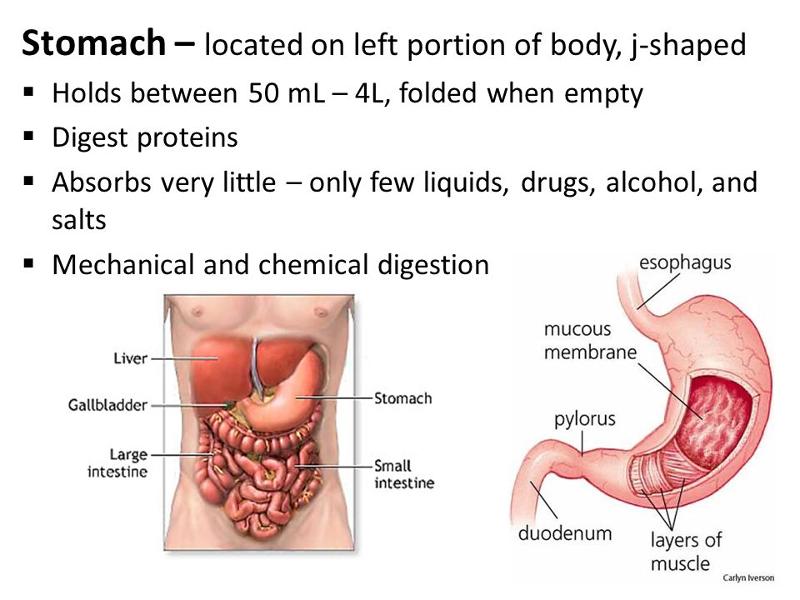
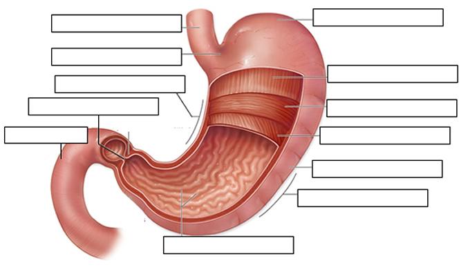
_________ is a J-shaped, pouch-like organ, about 25-30 centimeters long.It hangs inferior to the diaphragm in the upper-left portion of the abdominal cavity, has three layers of smooth muscle:
An inner
circular layer
An outer longitudinal layer
A further
inner layer of oblique fibers.
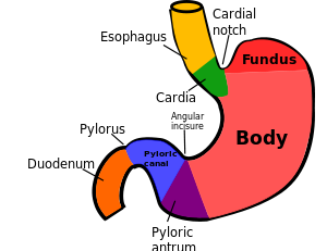
Stomach
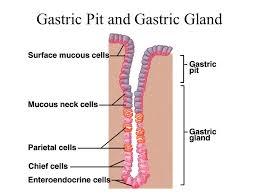
The mucous membrane of the ______ has tubular gastric glands that secrete.

STOMACH
________ IS From the chief cells
Inactive form of pepsin
___________IS From pepsinogen in the presence of hydrochloric acid Is a protein splitting enzyme
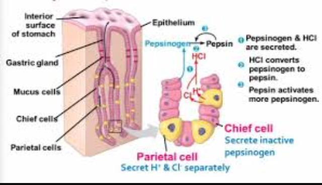
Pepsinogen
Pepsin
______ IS From the parietal cells
Needed to convert pepsinogen to pepsin
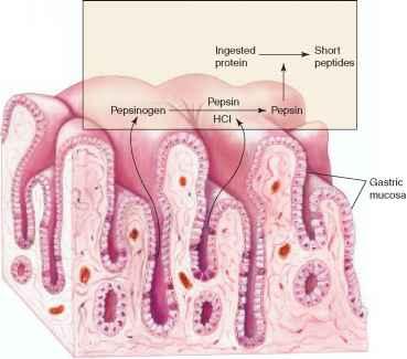
Hydrochloric acid
_________From the goblet cells and the mucous glands
Protective to stomach wall
______________From the parietal cells Is required for vitamin B12 absorption
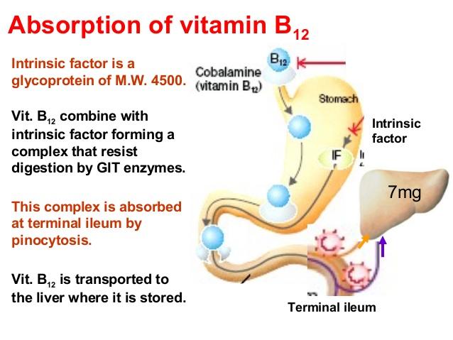
Mucus
Intrinsic factor
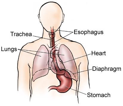
______________ (air enters) → nasal cavity → ______________ (both air and food move through) → trachea → ______________ (large tubes leading to both lungs) → lungs.
nares
pharynx
bronchus
______________ pleura; covers the surface of the lung ______________ pleura; lines the thoracic wall

PARIETAL
VISCERAL
The space in between is called the ______________ cavity and it is filled with ______________ fluid.
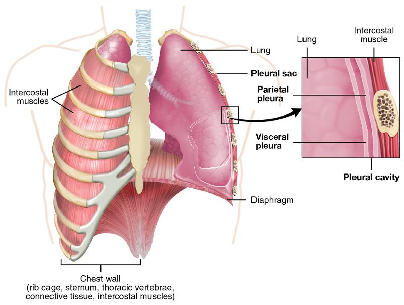
PLEURAL
SEROUS FLUID
This fluid assists breathing movements by acting as a/an ______________.
lubricant-pleural fluid
Air flows from the trachea through the ______________, ______________, and ______________ bronchi to smaller and smaller bronchi. The trachea and bronchi contain ______________ to keep the airways open. Bronchi branch into ______________, which do not contain ______________ but do contain more ______________ muscle. This allows for regulation of air flow.

main bronchus
lobar
segmental
cartilage
terminal bronchiole
cartilage
smooth muscle
Airways from the nasal cavity through the terminal bronchioles are called the ______________ zone.

conducting zone
The function of this zone is to ______________ and ______________ the air.
warm, moisten
filter
Is there gas exchange in this zone? _______
no
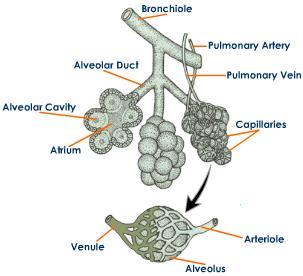
The respiratory zone contains ______________ where gas is exchanged. This zone consists of the ______________ bronchioles, ______________ ducts, and ______________ sacs.
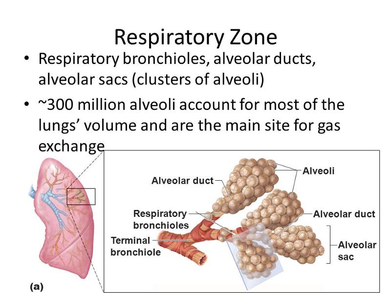
aveoli
respiratory
alveolar
alveolar
_________ From the goblet cells and the mucous glands: Protective to stomach wall
_______________ From the parietal cells. Is required for vitamin B12 absorption

Mucus
Intrinsic factor
- Parasympathetic preganglionic nerve fiber (in vagus nerve)
- Parasympathetic postganglionic impulses stimulate the release of gastric juice from gastric glands
- Impulses stimulate the release of gastric
- Gastrin stimulates gastric glands to release more gastric juice

Regulation of Gastric Secretions
O2 inhaled from environment and travels to alveoli of lungs. O2 is loaded by simple diffusion into the pulmonary capillaries and binds to hemoglobin forming oxyhemoglobin. CO2 is chemically released from the bicarbonate ion and unloaded by simple diffusion from the capillary to the alveoli to be exhaled.
O2 travels in blood to capillary bed and is released from hemoglobin unloaded by simple diffusion from the blood to the tissue. CO2 is loaded by simple diffusion into the blood from the tissue and carried in the blood as a bicarbonate ion back to the lungs.

The alveoli are the sites of the vital process of gas exchange between the air and the blood.
__________________ begin breaking down proteins, but the stomach is not well-adapted to absorb digestive products.

Gastric enzymes
Some water
Certain salts
Certain lipid-soluble drugs
Alcohol

The stomach does absorb
ALIMENTARY CANAL HAS WHAT TWO MAJOR FUNCTIONS?
- DIGEST
- ABSORB
- JEJUNUM & ILEUM
- MUSCLE TUBE @ 8 M LONG
MECHANICAL BREAKDOWN DUE TO CHEWING (MASTICATION) AND SALIVA
ENZYMES
DIGEST OF CARBS BC OF SALIVA
PAROTID - SEROUS FLUID (DIG. ENZYME AMYLAZE)
SUBMANDILAR - SEROUS / MUCUS
SUBLINGUAL - MUCUS
MOUTH
COMMON HALLWAY

PHYRYNX

MOVES FOOD THROUGH STOMACH
ESOPHAGUS
BILE- EMULSYFY FAT
WORKS WITH GALLBLADDER
STORAGE COMPARTMENT (FAT)
LARGEST INTERNAL ORGAN- CROWN UPPER RIGHT QUAD .
PRODUCES GYLCOGEN-->>GLUCOSE
CONVERTS NON CARBS
SYNTHESIZES
FORMS UREA
BREAKS DOWN RBC
REMOVES ALCOHOL
CONVERTS AMINO ACIDS
Phagocytosis of worn out RBCs and foreign substances
Removes toxins such as alcohol and certain drugs from the blood
LIVER( know 5 for test)
WASHER(CHURNING )
HC ACIDS
BREAKDOWN PROTIEN/ DIGESTION (FUNCTION)
J SHAPED POUCH
25-30 CM. @ UPPER LEFT QUADRANT
STOMACH
MIX AND BREAKDOWN
NUTRITION ABSORBTION
SML. INTESTINE
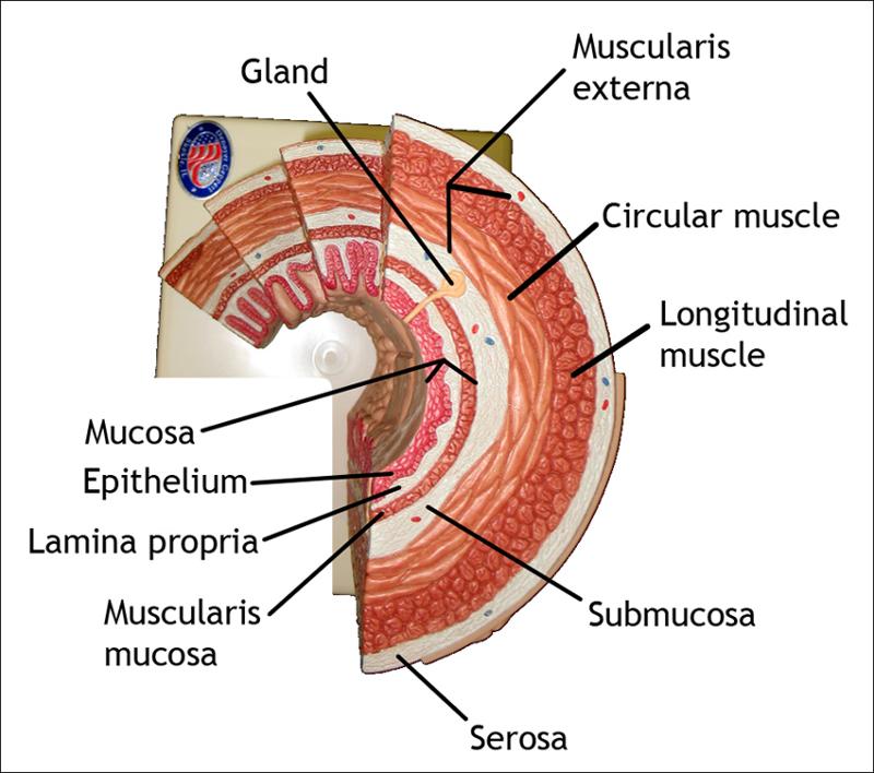
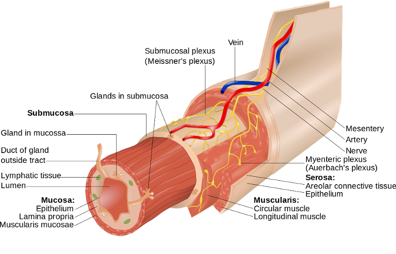
WHAT ARE THE LAYERS OF ALIMENTARY CANAL?
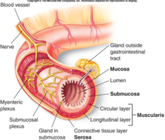
MUCOSA- CIRCULAR
SUBMUCOSA - MUCULARIS CONTROLS SECRETION
EXTERNA- LONG ( LAGITUTAL)
SEROUS- OUTER ; EPITILIAL AND MUCUS
WHAT ARE THE "INTRINSIC NERVE PLEXUSES ?
- MYENTRIC
- SUBMUCOSAL
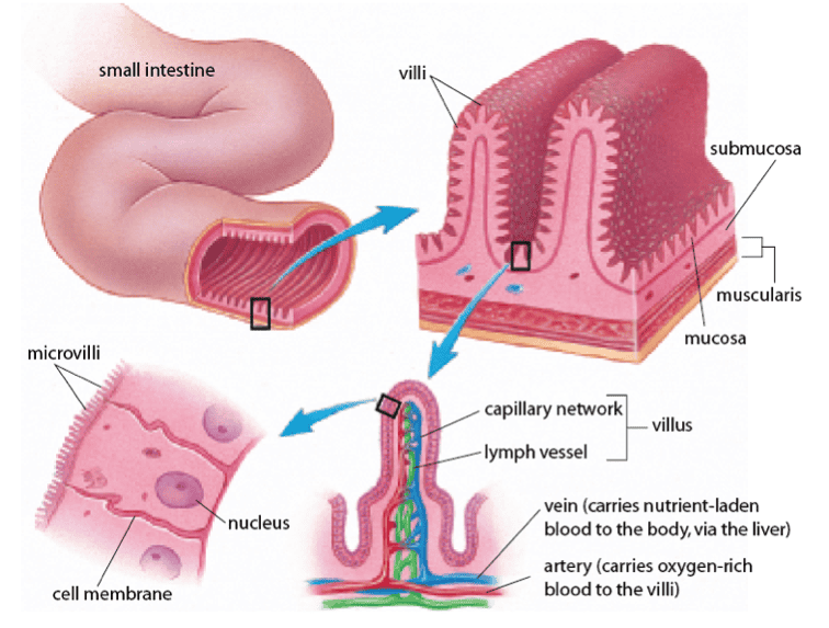
FINGER LIKE PROJECTION
VILLI
DIGESTIVE SYSTEM: ACCESSORY ARE
TEETH
TONGUE
GALLBLADDER - CARODID
DIGESTIONAL GLANDS

SALIVARY GLANDS
LIVER
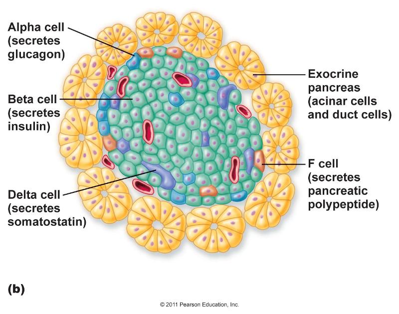
PANCREAS (ENDOCRINE) *APLHA*BETA * DELTA
FOOD

BOLLUS
__________ GOBLET ------>>> MUCOUS GLANDS (PROTECT STOMACH WALLS)
MUCUS
TOP OF STOMACH
FUNDUS
RIDGES
RUGAE
NERVE FIBER @ VAGUS NERVE
RELEASE OF GASTRIC JUICE -> GASTRIC GLAND
PARASYMPATIC
IMPULSE RELEASE OF GASTRIN

STIMULATES GASTRIC GLAND
SEE FOOD - STIMULATES TASTE/ CELL
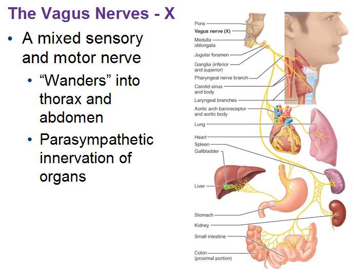
CEPHALIC (VAGUS NERVE)
ACTIVATES STRETCH RECEPTORS
FOOD CHEMICALS --->>> G CELLS
(MEDULLA ---->>> VAGUS NERVE )
GASTRIC
LOW PH
PARTIALLY : FOOD & FAT
HYPERTONIC @ DUODENUM
GASTRIC RELEASE AT BLOOD
(STOP SIGNALS)
INTESTINAL
______________ DOESNT ABSORB OF DIGEST FOOD. ABSORBS SALTS, H2O, BOOZE, LIPID SOLUABLE DRUGS.
STOMACH
DIGESTED FOOD
CHYME
DUEL FUNCTION
ENDOCRINE/ EXOCRINE
RELEASES PANCREATIC JUICES
BY DUODEM- (PANCREATIC DUCT TO DUODEM)
FAT/PROTIEN / NUCLEIC ACID
AMYLASE - SPLITS GLYCOGEN
LIPASE- BREAK DOWN TRIGLYCENIDES
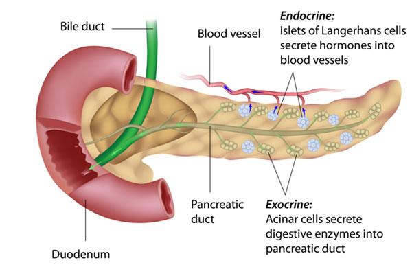
PANCREAS
EMOLSIFER / BREAKS DOWN FAT
BILE
SECRETION BY PANCREATIC JUICE
STIMULATES BILE SALTS
HEPO SPHINCTER TO RELAX
STIMULATES GALLBLADDER VIA VAGUS NERVE
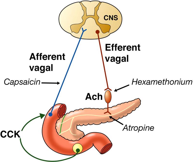
CCK
WHICH NERVE STIMULATES GALLBLADDER MAKING BILESALTS AND PANCREATIC JUICE ?

VAGUS
CYSTIC AND HEPATIC DUCT FORM THE _________ DUCT

BILE
GREEN/YELLOW
H20 AND BILE SALTS ( DETERGANT)
EMULSIFICATION AND TO BREAK DOWN FATS(DIGESTIVE ENZYME)
SALTS- ABSORB FATTY ACID (CHOLESTROL)
RECYCLED AND RETURNED TO LIVER
Bile pigments
Cholesterol
Electrolytes
BILE (SALTS)
Bile secretion is stimulated by____________________

Bile salts in enterohepatic circulation, Secretin from intestinal cells exposed to HCl and fatty chyme
Gallbladder contraction is stimulated by________________________
Cholecystokinin (CCK) from intestinal cells exposed to proteins and fat in chyme
Vagal stimulation (minor stimulus)
CCK also causes the hepatopancreatic sphincter to relax
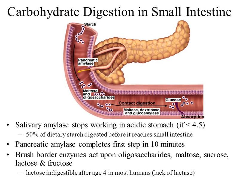
_________________is a tubular organ that extends from the pyloric sphincter to the beginning of the large intestine. It completes digestion of the nutrients in chyme, absorbs products of digestion, and transports the remaining residue to the large intestine. It consists of three parts that include. Duodenum. Jejunum. Ileum
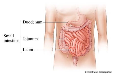
The small intestine
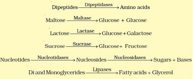
In addition to mucous-secreting goblet cells, there are many specialized mucous-secreting glands (Brunner’s glands) that secrete a thick, alkaline mucus in response to certain stimuli Enzymes in the membranes of the microvilli include________ (7)?
- Peptidase – breaks down peptides into amino acids
- Sucrase, maltase, lactase – break down : disaccharides into monosaccharides
- Lipase – breaks down fats into fatty acids and glycerol
- Enterokinase – converts trypsinogen to trypsin
- Somatostatin – hormone that inhibits acid secretion by stomach
- Cholecystokinin – hormone that inhibits gastric glands, stimulates pancreas to release enzymes in pancreatic juice, and stimulates the gallbladder to release bile
- Secretin – stimulates the pancreas to release bicarbonate ions in pancreatic juice
Regulation of small intestine secretion occurs by_______(3)?

- Mucus secretion is stimulated by the presence of chyme in the small intestine
- Distension of the intestinal wall activates nerve plexuses in the wall of the small intestine
- Parasympathetic reflexes triggering the release of intestinal enzymes
What are the steps to Absorption of the Small Intestine ?
- Villi: increase the surface area for absorption
- Small intestine absorption is so effective that very little reaches the organ’s distal end, noting that:
- Monosaccharides and amino acids absorb:
- Through facilitated diffusion and active transport
- Absorbed into blood
- Large proteins are broken down and absorbed into villi
- Fatty acids and glycerol absorb by:
- Several steps involved as noted
- Absorbed into lymph and blood
- Electrolytes and water absorb:
- Through diffusion, osmosis, and active transport
- Absorbed into blood
LARGE INTESTINE : Cecum, colon, rectum, anal, canal. UNIQUE FEATURES AND LAYERS ?
Teniae coli- Three bands of longitudinal smooth muscle in the muscularis
Haustra - Pocketlike sacs caused by the tone of the teniae coli
Epiploic appendages - Fat-filled pouches of visceral peritoneum
Functions of the Large Intestine?
- Has little or no digestive function
- Absorbs water and electrolytes
- Secretes mucus
- Houses intestinal flora
- Forms feces
- Carries out defecation
Movements of the Large Intestine?
- Movements of the large intestine are similar to those of the small intestine
- It is slower and less frequent than that of the small intestine
- Movements include:
- Mixing movements
- Peristalsis
- Mass movements usually follow meals
- The defecation reflex relaxes the internal anal sphincter and then the external anal sphincter
MOVEMENT OF FECAL MATTER
1) Distension, or stretch, of the rectal walls due to movement of feces into the rectum stimulates stretch receptors there. The receptors transmit signals along afferent fibers to spinal cord neurons.
2) A spinal reflex is initiated in which parasympathetic motor (efferent) fibers stimulate contraction of the rectal walls and relaxation of the internal ; anal sphincter
3) If it is convenient to defecate, voluntary motor neurons are inhibited, allowing the external anal sphincter to relax so that feces may pass.
Feces is composed of materials not digested or absorbed, and include?
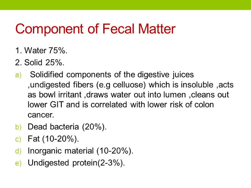
- Water
- Electrolytes
- Mucus
- Bacteria
- Bile pigments altered by bacteria provide the color
The pungent odor is produced by bacterial compounds including?
- Phenol
- Hydrogen sulfide
- Indole
- Skatole
- Ammonia
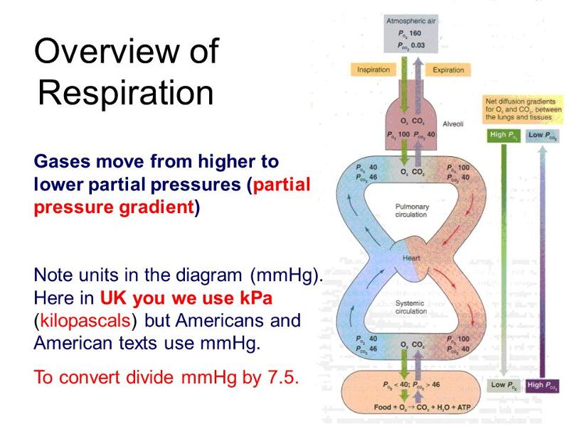
The respiratory system consists of passages that filter incoming air and transport it into the body, into the lungs, and to the many microscopic air sacs where gases are exchanged. Respiration is the process of exchanging gases between the atmosphere and body cells. Involves both the respiratory and the circulatory systems. Four processes that supply the body with O2 and dispose of CO2:_________(4) ?
- Ventilation (breathing)
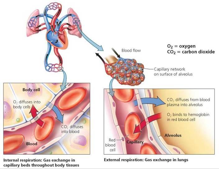
- External respiration
- Transport of gases
- Internal respiration
RESPIRATION -> CIRCULATORY
RESPIRATION Pulmonary ventilation (breathing): movement of air into and out of the lung
External respiration: O2 and CO2 exchange between the lungs and the blood (BOTH)
CIRCULATORY: Transport: O2 and CO2 in the blood. Internal respiration: O2 and CO2 exchange between systemic blood vessels and tissues
WHY DO WE BREATHE?
- Respiration occurs on a macroscopic level at the organ system
- Gas exchange, oxygen and carbon dioxide, occur at the cellular and molecular levels
- Aerobic reactions of cellular respiration allow for:
- ATP production
- Carbon dioxide generation forming carbonic acid
Organs of the Respiratory System?
Respiratory zone: site of gas exchange –respiratory bronchioles, alveolar ducts, and alveoli
Conducting zone: conduits to gas exchange sites
Includes all other respiratory structures : Respiratory muscles: diaphragm and other muscles that promote ventilation
Organs of the Respiratory System
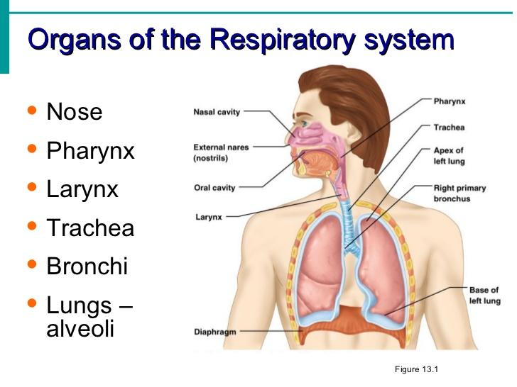
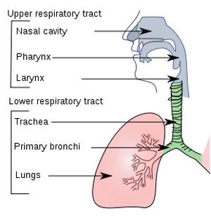
The organs of the respiratory system can be divided into two tracts:
Upper respiratory tract: The nose, Nasal cavity, Sinuses, Pharynx
Lower respiratory tract: Larynx, Trachea,Bronchial tree, Lungs
________ is an enlargement in the airway superior to the trachea and inferior to the pharynx, It is composed of a framework of muscles and cartilages bound by elastic tissue
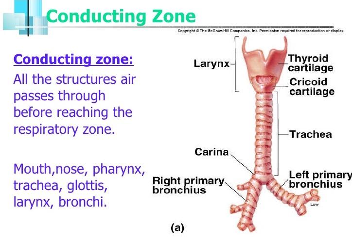
The larynx
______________ is a flexible cylindrical tube about 2.5 centimeters in diameter and 12.5 centimeters in lengthAs it extends downward anterior to the esophagus and into the thoracic cavity, it splits into the right and left primary bronchi
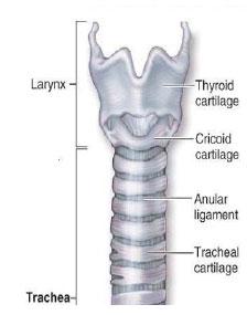
The trachea (windpipe)
___________________ consists of branched airways leading from the trachea to the microscopic air sacs in the lungs
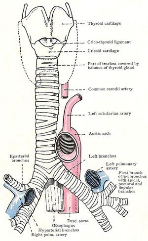
The bronchial tree
The successive divisions of the branches from the trachea to the alveoli are?
1 .Right and left primary bronchi
2.Secondary or lobar bronchi
3.Tertiary or segmental bronchi
4.Intralobular bronchioles (12-14 generations)
5.Terminal bronchioles
6.Respiratory bronchioles
7.Alveolar ducts
8.Alveolar sacs
9.Alveoli
_________________is similar to that of the trachea, but the C-shaped cartilaginous rings are replaced with cartilaginous plates where the bronchus enters the lung. These respiratory tubes become thinner and thinner, and the cell layers thin and change until the alveoli is reached. It is the alveoli that provides surface area for gas exchange is similar to that of the trachea, but the C-shaped cartilaginous rings are replaced with cartilaginous plates where the bronchus enters the lung
These respiratory tubes become thinner and thinner, and the cell layers thin and change until the alveoli is reached
It is the alveoli that provides surface area for gas exchange
The structure of the bronchus
WHAT : Surrounded by fine elastic fibers, Contain open pores that AND Connect adjacent alveoli, Allow air pressure throughout the lung to be equalized - > House alveolar macrophages that keep alveolar surfaces sterile
Alveoli
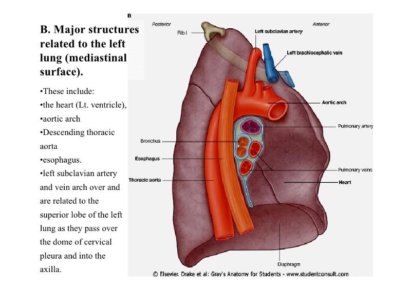
____________ are soft, spongy, cone-shaped organs in the thoracic cavity
_________has three lobes and the left lung two lobes
LEFT AND RIGHT
RIGHT
WHAT ?? Occupy all of the thoracic cavity except the mediastinum, Root: site of vascular and bronchial attachments, AND Costal surface: anterior, lateral, and posterior surfaces?
LUNGS
WHAT AM I DESCRIBING ?
- Apex: superior tip
- Base: inferior surface that rests on the diaphragm
- Hilum: on mediastinal surface; site for attachment of blood vessels, bronchi, lymphatic vessels, and nerves
- Cardiac notch ( L) concavity that accommodates the heart
LUNGS
_______ is smaller, separated into two lobes by an oblique fissure
_______ has three lobes separated by oblique and horizontal fissures
Bronchopulmonary segments (10 right, 8–9 left)
_______are the smallest subdivisions; served by bronchioles and their branches
LEFT
RIGHT
LOBULES
(low pressure, high volume)Pulmonary arteries deliver systemic venous blood
- Branch profusely, along with bronchi
- Feed into the pulmonary capillary networks
Pulmonary veins carry oxygenated blood from respiratory zones to the heart
BLOOD SUPPLY / PULMONARY CIRCULATION

Systemic circulation (high pressure, low volume)
Bronchial arteries provide oxygenated blood to lung tissue
- Arise from aorta and enter the lungs at the hilum
- Supply all lung tissue except the alveoli
Bronchial veins anastomose with pulmonary veins
Pulmonary veins carry most venous blood back to the heart
BLOOD SUPPLY/ Systemic circulation (high pressure, low volume)
- Thin, double-layered serosa
- Parietal pleura on thoracic wall and superior face of diaphragm
- Visceral pleura on external lung surface
- Pleural fluid fills the slitlike pleural cavity
Provides lubrication and surface tension
Pleurae
____________________ is the movement of air from outside of the body into the bronchial tree and the alveoli. The actions responsible for these air movements are inspiration, or inhalation, and expiration, or exhalation
Breathing or ventilation
_______ IS Atmospheric pressure (Patm)
Pressure exerted by the air surrounding the body
760 mm Hg at sea level
Respiratory pressures are described relative to Patm
Negative respiratory pressure is less than Patm
Positive respiratory pressure is greater than Patm
Zero respiratory pressure = Patm
Pressure Relationships in the Thoracic Cavity
_________________ IS (Ppul). Pressure in the alveoli, Fluctuates with breathing
AND Always eventually equalizes with Patm.
Intrapulmonary (intra-alveolar) pressure
_______ IS Pressure in the pleural cavity, Fluctuates with breathingVAND Always a negative pressure (<Patm and <Ppul)
Intrapleural pressure (Pip)
Negative Pip is caused by opposing forces–Two inward forces promote lung collapse
- Elastic recoil of lungs decreases lung size
- Surface tension of alveolar fluid reduces alveolar size
One outward force tends to enlarge the lungs
- Elasticity of the chest wall pulls the thorax outward
Intrapleural Pressure
Atmospheric pressure due to the weight of the air is the force that moves air into the lungs. At sea level, atmospheric pressure is 760 millimeters of mercury (mm Hg)Moving the plunger of a syringe causes air to move in or out . Air movements in and out of the lungs occur in much the same way
Inspiration
- The relationship between the pressure and volume of a gas
- Pressure (P) varies inversely with volume (V):
P 1 V 1 = P 2 V 2
BOYLES LAW
The forces responsible for normal resting expiration come from elastic recoil of lung tissues and from surface tension. These factors increase the intra-alveolar pressure about 1 mm Hg above atmospheric pressure forcing air out of the lungs
EXPIRATION
CO2 + H2O =
CARBONIC ACID
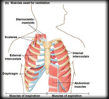
MUSCLES TO BREATHE ?
SCALENES, SCM, INTERCOSTAL (IN/EX) , DIAPHRAGM
RIBS UP, DIAPHRAGM DOWN
Alveolar ventilation rate (AVR): flow of gases into and out of the alveoli during a particular time. Dead space is normally constant, Rapid, shallow breathing decreases AVR
Alveolar Ventilation
Used to assess a person’s respiratory status
Tidal volume (TV)
Inspiratory reserve volume (IRV)
Expiratory reserve volume (ERV)–Residual volume (RV)
Respiratory Volumes
- Inspiratory capacity (IC)
- Functional residual capacity (FRC)
- Vital capacity (VC)
- Total lung capacity (TLC)
Respiratory Capacities
Air movements other than breathing are called nonrespiratory movements. They clear air passages, as in coughing and sneezing, or express emotions, as in laughing and crying
Non-respiratory Air Movements
Respiratory Disorders That Decrease Ventilation: _____________
Bronchial Asthma and Emphysema
Groups of neurons in the brainstem comprise the respiratory areas that control breathing . Impulses travel on cranial nerves and spinal nerves, causing inspiration and expiration. Respiratory areas also adjust the rate and depth of breathing. The respiratory areas include:
- Respiratory center of the medulla
- Respiratory group of the pons
Respiratory Areas
A number of factors affect breathing rate and depth including:
- Partial pressure of oxygen (Po2)
- Partial pressure of carbon dioxide (Pco2)
- Degree of stretch of lung tissue
- Emotional state
- Level of physical activity
Receptors involved include mechanoreceptors and central and peripheral chemoreceptors
FACTORS OF BREATHING
Changes in blood pH, O2 and CO2 concentration stimulates chemoreceptors
Motor impulses can travel
from the respiratory center
to the diaphragm and external intercostal muscles
Contraction of these muscles causes the lungs to expand stimulating mechanoreceptors in the lungs
Inhibitory impulses from the mechanoreceptors back to the respiratory center prevent overinflation of the lungs

Factors Affecting Breathing
The alveoli are the sites of the vital process of gas exchange between the air and the blood.
“ Describe the exchange of O2 and CO2 in the lungs and body tissues. Include the names of the body structures needed, and how O2 & CO2 are transported in the blood.”
O2 inhaled from environment and travels to alveoli of lungs. O2 is loaded by simple diffusion into the pulmonary capillaries and binds to hemoglobin forming oxyhemoglobin. CO2 is chemically released from the bicarbonate ion and unloaded by simple diffusion from the capillary to the alveoli to be exhaled.
O2 travels in blood to capillary bed and is released from hemoglobin unloaded by simple diffusion from the blood to the tissue. CO2 is loaded by simple diffusion into the blood from the tissue and carried in the blood as a bicarbonate ion back to the lungs.
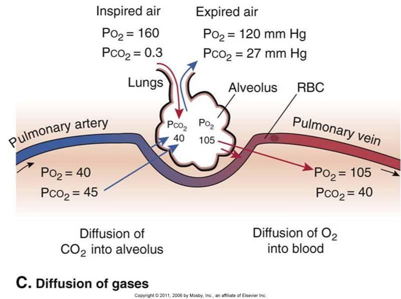
Alveolar Gas Exchanges
Part of the wall of an alveolus is made up of cells (type II cells) that secrete pulmonary surfactant . The bulk of the wall of an alveolus consists of a layer of simple squamous epithelium (type I cells) .Both of these layers make up the respiratory membrane through which gas exchange takes place
Respiratory Membrane
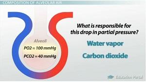
Molecules diffuse from regions where they are in higher concentration toward regions where they are in lower concentration. It is important to know the concentration gradient, In respiration, think in terms of gas partial pressures. Gases diffuse from areas of higher partial pressure to areas of lower partial pressure, The respiratory membrane is normally thin and gas exchange is rapid
Increased diffusion is favored with more surface area, shorter distance, greater solubility of gases and a steeper partial pressure gradient
Decreased diffusion occurs from decreased surface area

Diffusion Through the Respiratory Membrane
Air and food are routed into the proper channels by the ________.

larynx
The loudness of a personʹs voice depends on ________.
the force with which air rushes across the vocal folds
The walls of the alveoli are composed of two types of cells, type I and type II. The function of type II is ________.
to secrete surfactant
After the segmental (tertiary) bronchus, the next smaller branch of the respiratory passageway is (are) the ________.
terminal bronchioles
_________ O2 and CO2 between the lungs and the body cells. As the gases enter the blood, they dissolve in the plasma or chemically combine with other atoms or molecules
BLOOD TRANSPORTS
Gas Transport
Almost all oxygen carried in the blood is bound to the protein hemoglobin in the form of oxyhemoglobin. Chemical bonds between O2 and hemoglobin are relatively unstable. Oxyhemoglobin releases O2 into the body cells. About 75% of the O2 remains bound to hemoglobin in the venous blood ensuring safe CO2 levels and thereby pH
Oxygen Transport
Blood flowing through capillaries gains CO2 because the tissues have a high Pco2. The CO2 is transported to the lungs in one of three forms. WHAT ARE THEY ?
As CO2 dissolved in plasma
As part of a compound with hemoglobin
As part of a bicarbonate ion
WHERE ?
–HCO3– moves into the RBCs and binds with H+ to form H2CO3
–H2CO3 is split by carbonic anhydrase into CO2 and water
–CO2 diffuses into the alveoli
In pulmonary capillaries
Transport and Exchange of CO2
–Exemplified by chronic bronchitis and emphysema
–Irreversible decrease in the ability to force air out of the lungs
–Other common features
- History of smoking in 80% of patients
- Dyspnea: labored breathing (“air hunger”)
- Coughing and frequent pulmonary infections
- Most victims develop respiratory failure (hypoventilation) accompanied by respiratory acidosis
Chronic obstructive pulmonary disease (COPD)
–Characterized by coughing, dyspnea, wheezing, and chest tightness
–Active inflammation of the airways precedes bronchospasms
–Airway inflammation is an immune response caused by release of interleukins, production of IgE, and recruitment of inflammatory cells
–Airways thickened with inflammatory exudate magnify the effect of bronchospasms
Asthma
–Infectious disease caused by the bacterium Mycobacterium tuberculosis
–Symptoms include fever, night sweats, weight loss, a racking cough, and spitting up blood
–Treatment entails a 12-month course of antibiotics
Tuberculosis
–Leading cause of cancer deaths in North America
–90% of all cases are the result of smoking
–The three most common types
1.Squamous cell carcinoma (20–40% of cases) in bronchial epithelium
2.Adenocarcinoma (~40% of cases) originates in peripheral lung areas
3.Small cell carcinoma (~20% of cases) contains lymphocyte-like cells that originate in the primary bronchi and subsequently metastasize
Lung cancer
The smallest macroscopic subdivision of the lung is the ________.
lobule
The pleurae are vital to the integrity of the lungs because ________.
they produce a lubricating serous secretion, allowing the lungs to glide over the thorax wall during breathing
Intrapulmonary pressure is the ________.
pressure within the alveoli of the lungs
A fluid secreted into the small intestine during digestion that contains cholesterol, emulsification agents, and phospholipids is ________.
BILE
Select the statement that is true concerning primary teeth.
A) There are 27 primary teeth, and the molars are permanent.
B) There are 24 primary teeth, and no new primary teeth appear after 13 months.
C) There are 20 primary teeth, and by 24 months of age most children have all 20.
D) There are 32 primary teeth, and most children lose these teeth due to decay because they are never very strong.
20, 24 MONTH 20
Which of the following correctly describes mechanisms of CO2 transport?
- A) 7- 10% of CO2 is dissolved directly into the plasma
- B) 20% of CO2 is carried in the form of carbaminohemoglobin
- C) as bicarbonate ion in plasma
- D) attached to the heme part of hemoglobin
attached to the heme part of hemoglobin
How is the bulk of carbon dioxide carried in blood?
A) chemically combined with the amino acids of hemoglobin as carbaminohemoglobin in the red blood cells
B) as the bicarbonate ion in the plasma after first entering the red blood cells
C) as carbonic acid in the plasma
D) chemically combined with the heme portion of hemoglobin
as the bicarbonate ion in the plasma after first entering the red blood cells
The mechanical and chemical receptors that control digestive activity are located ________.
A) in the glandular tissue that lines the organ lumen
B) in the walls of the tract organs
C) in the pons and medulla
D) only in the esophagus because this is the only part of the tract that needs to change to accommodate food passage
in the walls of the tract organs
Complete the following statement using the choices below. Air moves out of the lungs when the pressure inside the lungs is
- A) less than the pressure in the atmosphere.
- B) greater than the pressure in the atmosphere.
- C) equal to the pressure in the atmosphere.
- D) greater than the intra-alveolar pressure.
greater than the pressure in the atmosphere.
When we ingest large molecules such as lipids, carbohydrates,
and proteins, they must undergo catabolic reactions whereby enzymes
split these molecules. This series of reactions is called:
a. absorption
b.
secretion
c. chemical digestion
d. mechanical digestion
chemical digestion
Which vitamin requires intrinsic factor in order to be absorbed?
- A) B12
- B) K
- C) A
- D) C
B12
The most powerful respiratory stimulus for breathing in a healthy person is ________.
A) loss of oxygen in tissues
B) increase of carbon dioxide
C) pH (acidosis)
D) pH (alkalosis)
increase of carbon dioxide
The statement, "in a mixture of gases, the total pressure is the sum of the individual partial pressures of gases in the mixture" paraphrases ________.
A) Henry's law
B) Boyle's law
C) Dalton's law
D) Charles' law
Dalton's law
The function of the hepatic portal circulation is to ________.
A) carry toxins to the venous system for disposal through the urinary tract
B) collect absorbed nutrients for metabolic processing or storage
C) distribute hormones
D) return glucose to the general circulation when blood sugar is low
collect absorbed nutrients for metabolic processing or storage
Peristaltic waves are ________.
A) segmental regions of the gastrointestinal tract
B) churning movements of the gastrointestinal tract
C) pendular movements of the gastrointestinal tract
D) waves of muscular contractions that propel contents from one point to another
waves of muscular contractions that propel contents from one point to another
Select the correct statement about oxygen transport in blood:
- A) During normal activity, a molecule of hemoglobin returning to the lungs carries one molecule of O2.
- B) During conditions of acidosis, hemoglobin is able to carry oxygen more efficiently.
- C) Increased BPG levels in the red blood cells enhance oxygen-carrying capacity.
- D) A 50% oxygen saturation level of blood returning to the lungs might indicate an activity level higher than normal.
A 50% oxygen saturation level of blood returning to the lungs might indicate an activity level higher than normal.
___ is the most common lethal genetic disease in the U.S
CYSTIC FIBROSIS
A baby is admitted to the hospital with a history of projectile vomiting after each feeding. On examination, it is found that the sphincter controlling food passage from the stomach to the duodenum is thickened and does not open readily. Because of the babyʹs loss of gastric juice, his blood probably indicates ________.
alkalosis
There are some 20 known pathogens found in the large intestine; our Ig ________ antibody-mediated response restricts them from going beyond the mucosa and causing problems.
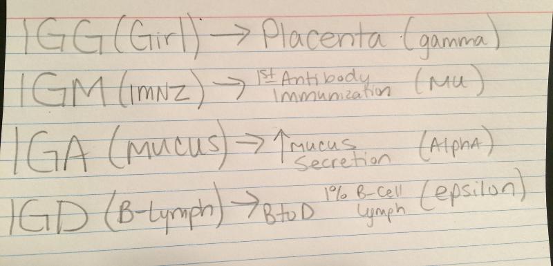
IGA
Parietal cells of the stomach produce
HYDROCLORIC ACID
The terminal portion of the small intestine is known as the small intestine is known as the ______?
Ileum
Which respiratory-associated muscles would contract if you were to blow up a balloon?
A) diaphragm would contract, external intercostals would relax
B) internal intercostals and abdominal muscles would contract
C) external intercostals would contract and diaphragm would relax
D) diaphragm contracts, internal intercostals would relax
internal intercostals and abdominal muscles would contract
Catabolism involves processes that:
a. cause a decline in circulating ketone bodies
b. mobilize fat during the post absorptive state
c. break down complex structures to simpler
ones
d. elevate glucagon levels
break down complex structures to simpler ones
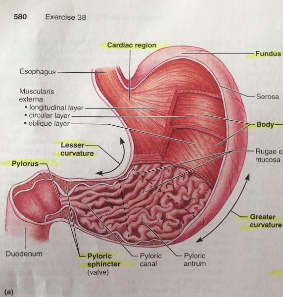
Know parts of stomach
The solutes contained in saliva include ________.
electrolytes, digestive enzyme, mucin, lysozyme, wastes, and IgA
Which of the following is not found on the right lung?
CARDIAC
NOTCH;
horizontal
fissure;
middle
lobe;
oblique
fissure
Gastrin, histamine, endorphins, serotonin, cholecystokinin,
and somatostatin are hormones or paracrines that are released
directly into the lamina propria. Which of the following cell types
synthesize and secrete these products?
parietal cells
enteroendocrine
cells
mucous neck cells
zymogenic cells
enteroendocrine cells
Paneth cells ________.
are
located next to the lacteal in a villus
secrete enzymes that kill bacteria
are more
common in the ileum than in the jejunum
are
absorptive cells in the small intestine
secrete enzymes that kill bacteria
Digestion of which of the following would be affected the most
if the liver were severely damaged?
proteins
carbohydrates
starches
lipids
lipids
Gastrin is a digestive hormone that is responsible for the
stimulation of acid secretions in the stomach. These secretions are
stimulated by the presence of ________.
simple carbohydrates and alcohols
protein and peptide fragments
fatty
acids
starches and complex carbohydrates
protein and peptide fragments
The capillaries that nourish the epithelium and absorb
digested nutrients lie in the ________.
serosa
lamina propria
muscularis mucosae
adventitia
lamina propria
If an incision has to be made in the small intestine to remove
an obstruction, the first layer of tissue to be cut is the
________.
mucosa
serosa
muscularis
externa
submucosa
serosa
Which of the following is an essential role played by large
intestine bacteria?
synthesize
vitamin K and B-complex vitamins
synthesize vitamins C and D
produce
gas
absorb bilirubin
synthesize vitamin K and B-complex vitamins
Which of these is not part of the ?
celiac artery
hepatic portal vein
superior mesenteric artery
inferior vena cava
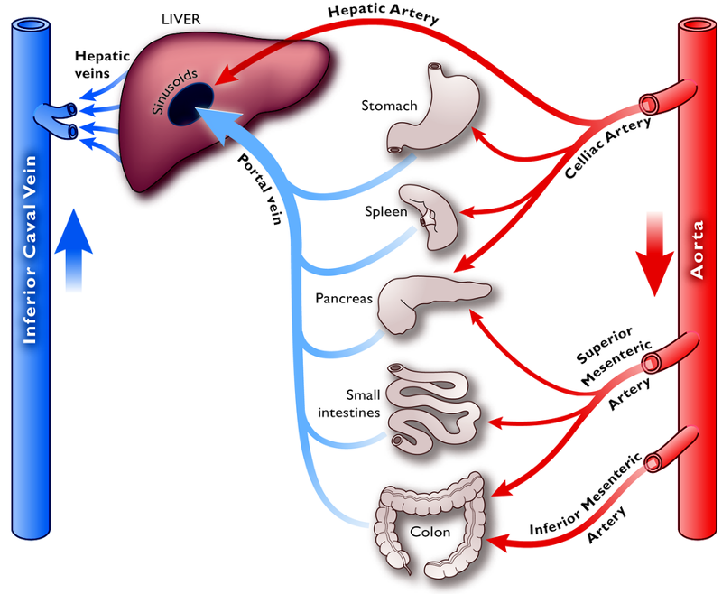
inferior vena cava
Hormones or pancreas that inhibit gastric secretion include
________.
ACh
secretin
histamine
gastrin
secretin
The ducts that deliver bile and pancreatic juice from the
liver and pancreas, respectively, unite to form the
________.
hepatopancreatic ampulla
bile canaliculus
portal vein
pancreatic acini
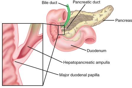
hepatopancreatic ampulla
Which hormone causes an increased output of enzyme-rich
pancreatic juice and stimulates gallbladder contraction to release
bile?
gastrin
cholecystokinin
secretin
gastric inhibitor peptide
cholecystokinin
Hepatocytes do not ________.
process nutrients
detoxify
store fat-soluble vitamins
produce digestive enzymes
produce digestive enzymes
You have just eaten french fries, buttered toast, ice cream, and whole milk. Which of the following glands would be active in helping you to digest this food?
THE PANCREAS


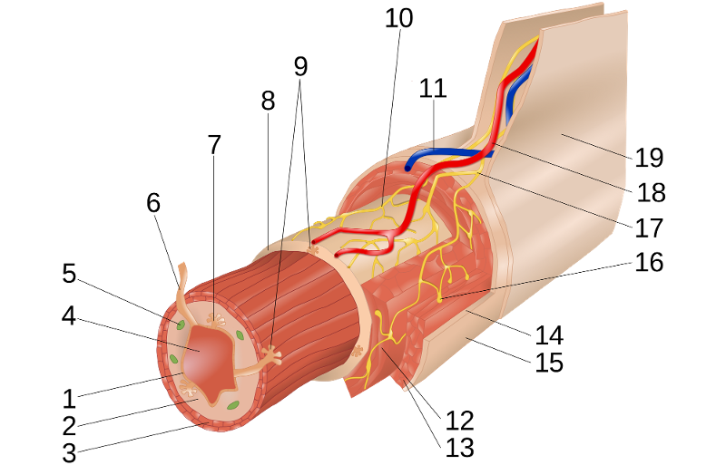
Know the layers of the alimentary canal, starting with the outermost layer

a. Serosa, Longitudinal Muscle, Circular Muscle, Submucosa, Mucosa
Where is salivary amylase released from?

a. Parotid gland
What does salivary amylase digest?
a.
Carbohydrates
b. glucose
Gastrin: What is it for?
a. Hormone secreted by the stomach that regulates gastric juice secretion by stimulating HCl production.
Secretion of Cholecystokinin(CCK) from the intestinal wall is stimulated in the presence of what foods?
a. Fatty foods and protein
Secretin: inhibits
inhibits the action of pancreatic lipase.
Peristalsis vs. Segmentation
a. Peristalsis-
occurs in stomach
b. Segmentation-
occurs in small intestine
What is the function of the large intestine?
a. Absorption of
water and electrolytes
b. Reservoir for fecal matter
What is the part of the digestive tract with the most lymph nodules and bacteria?
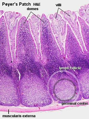
a. Ileum
What is the greater omentum formed out of?
a. Peritoneal membrane
If the liver is damaged , what is going to be harder to digest?
a. Lipids
Air moves out of the lungs when the pressure inside the lungs is ____?
a. Increased
Peristaltic waves; where are they starting?
a. Esophagus
The sheets of peritoneal membrane that hold the digestive tract in place….
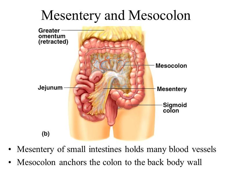
a. Mesenteries
Expiration, unlike inspiration, is a passive act. Expiration depends on 2 factors…
a . Elastic recoil
of the lungs
b. Surface tension of
alveolar fluid
What structure has the greatest surface area for gas exchange in the lungs
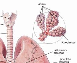
a. Alveoli
What determines the direction of respiratory gas movement?
a. Partial pressure gradient
...
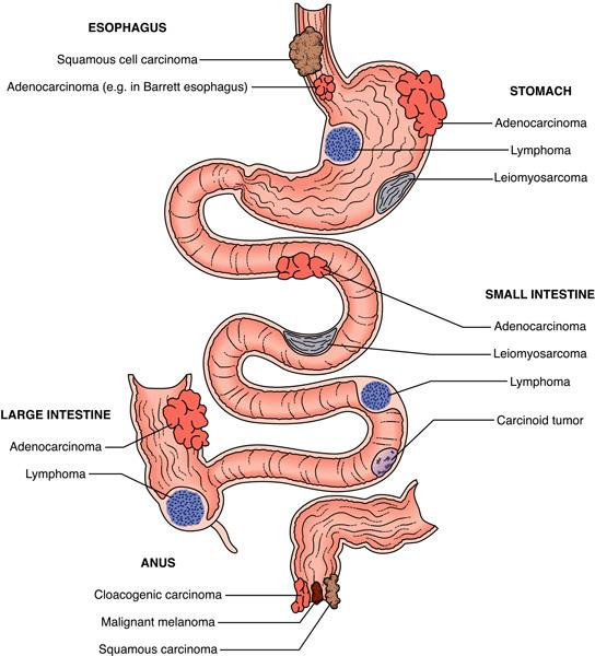
Cancers