What two structures make up the CNS?
Brain and Spinal Cord
Describe the roles of the three types of neurons in the spinal cord (sensory, motor, and interneuron). SENSORY
Sensory Neuron-
–about 10 million
–deliver information to CNS
Describe the roles of the three types of neurons in the spinal cord (sensory, motor, and interneuron). MOTOR
Motor Neuron-
–about 1/2 million
–deliver commands to peripheral effectors
Describe the roles of the three types of neurons in the spinal cord (sensory, motor, and interneuron). INTERNEURON
Interneuron-
(AKA - association neurons)
–about 20 billion
–interpret, plan, and coordinate signals in and out
Draw AND label (including the functions of the regions that you label) of a spinal cord cross section.
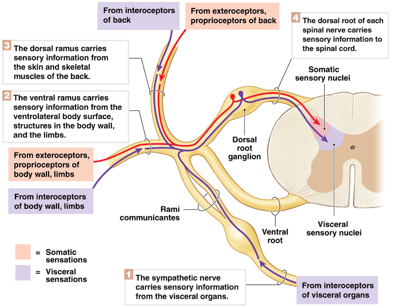
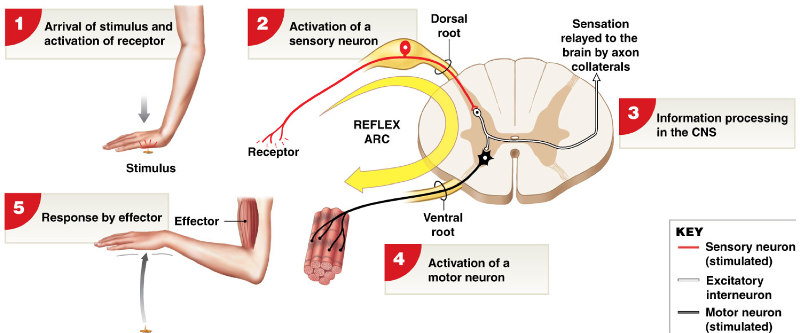
Diagram AND label the five steps that occur in a neural reflex. Make sure to keep the sensory and motor neurons straight!
Compare and contrast innate and acquired reflexes.
Innate reflexes:
- basic neural reflexes
- formed before birth
Acquired reflexes:
- rapid, automatic
- learned motor patterns
Compare and contrast somatic and visceral reflexes.
Somatic reflexes:
- involuntary control of nervous system
–superficial reflexes of skin, mucous membranes
–stretch reflexes (deep tendon reflexes) e.g., patellar reflex
Visceral reflexes (autonomic reflexes):
- control systems other than muscular system
Compare and contrast monosynaptic and polysynaptic reflexes.
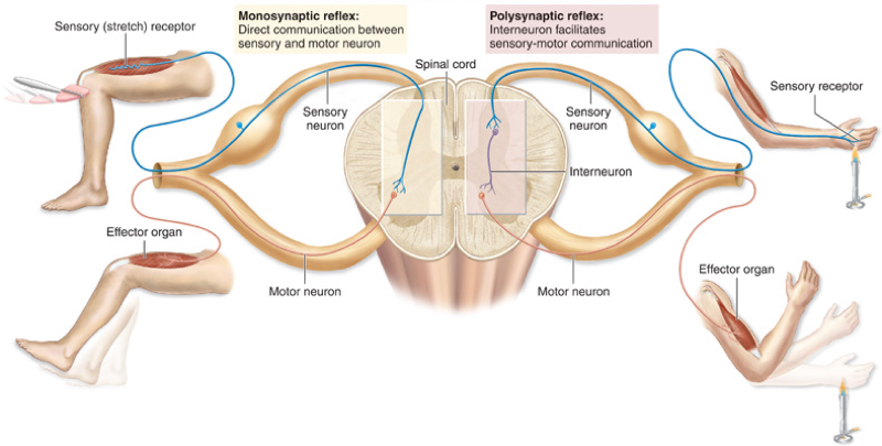
–monosynaptic - sensory neuron synapses directly onto motor neuron
–polysynaptic - at least 1 interneuron between sensory neuron and motor neuron
Compare and contrast spinal reflexes and cranial reflexes.
–spinal reflexes:
- occurs in spinal cord
–cranial reflexes:
- occurs in brain
Diagram AND label a monosynaptic spinal reflex, using the stretch reflex as an example
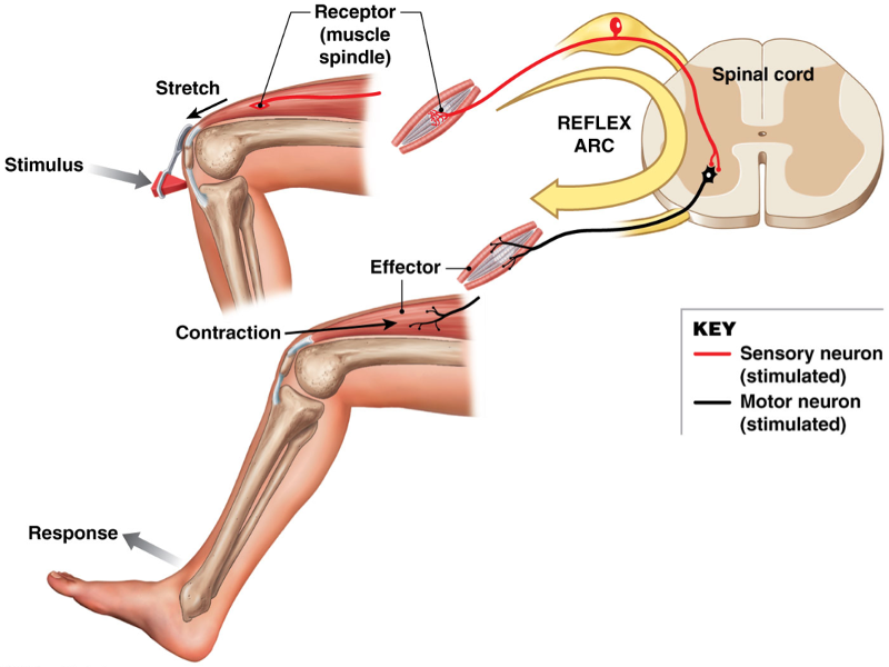
Have least delay between sensory input and motor output:–i.e. patellar reflex)
Brain cannot override the reflex
Completed in 20–40 msec
Explain the roles of intrafusal and extrafusal muscle fibers in the stretch reflex as well as the adjustment of muscles tension in postural reflexes
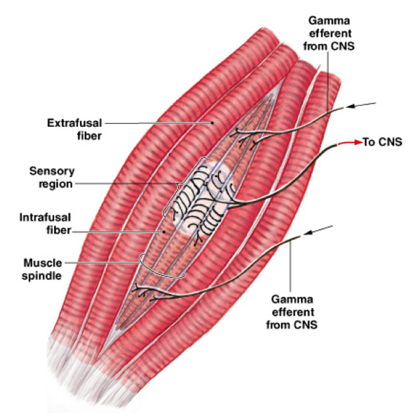
Bundles of small, specialized intrafusal muscle fibers:
–innervated by sensory and motor neurons
Surrounded by extrafusal muscle fibers:
–which maintain tone and contract muscle
Get acquainted with some of the common clinical tendon reflexes (as you will be testing these in your patients!)
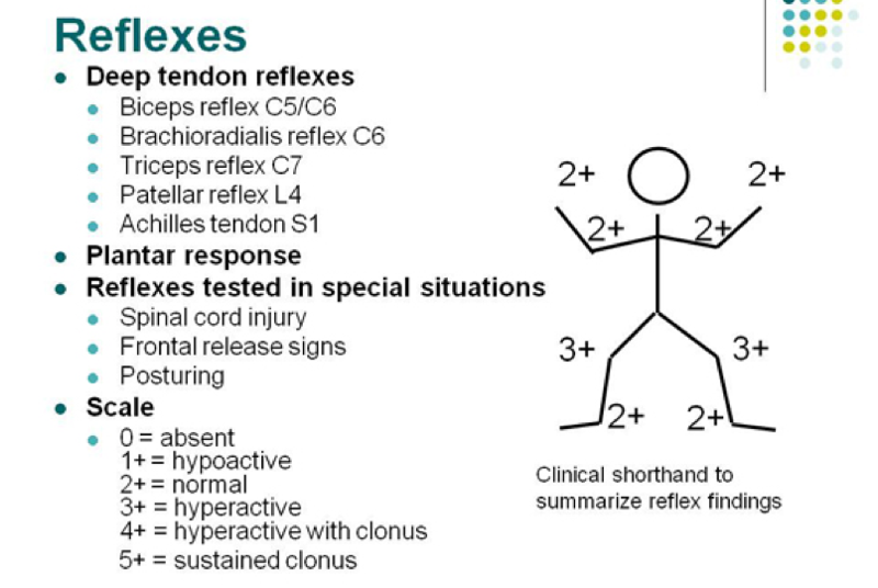
Diagram AND explain the events that occur during the flexor reflex. Make sure to include that little interneuron!
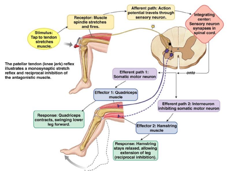
Explain the importance of reciprocal inhibition. Give an example!
For Stretch R eflex or the F lexor R eflex to work:
–the stretch reflex of antagonistic (extensor) muscle must be inhibited (reciprocal inhibition) by interneurons in spinal cord
Diagram AND label the events which occur during the tendon reflex. What would happen to us if we didn’t have this reflex?
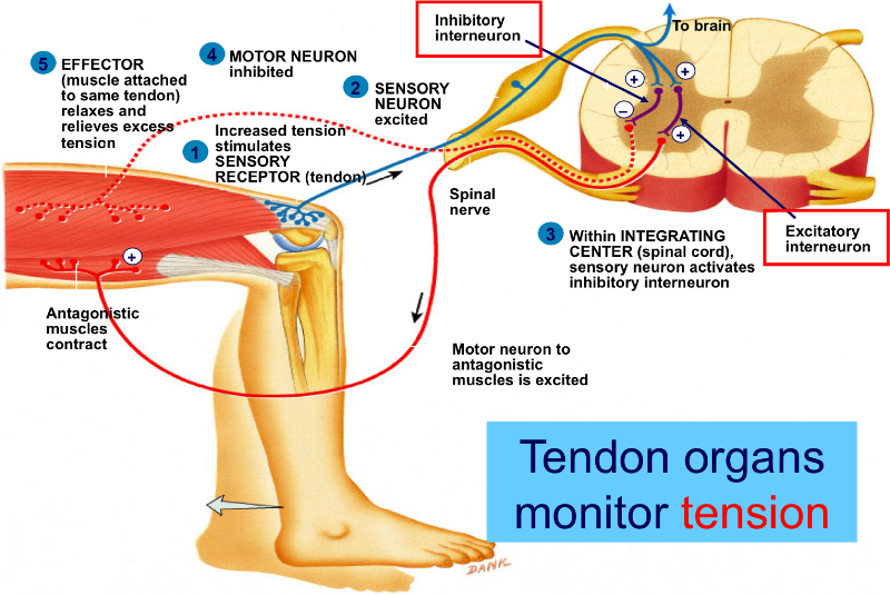
What would happen to us if we didn't have the tendon reflex?
If we did not have the tendon reflex then we would not be preventing skeletal muscles from developing too much tension, tearing or breaking tendons
What does ipsilateral mean? How about contralateral?
Ipsilateral reflex arcs:
–occur on same side of body as stimulus
–stretch, tendon, and withdrawal reflexes
Crossed extensor reflexes:
–involves a contralateral reflex arc
–occurs on side opposite stimulus
Now that you know what ipsilateral and contralateral mean, diagram an example of the crossed extensor reflex.
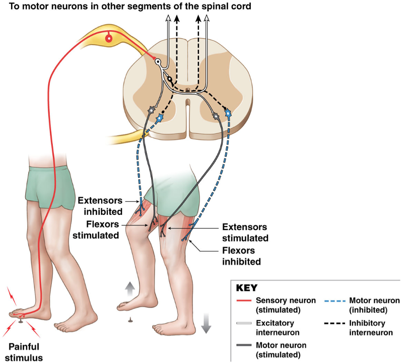
Polysynaptic Spinal Reflexes - The Crossed Extensor Reflex
Occur simultaneously, coordinated with flexor reflex
e.g., flexor reflex causes leg to pull up:
crossed extensor reflex straightens other leg
to receive body weight
Review the meanings of rostral and caudal.
Areas of the brain:
Rostral (toward forehead)
Caudal (toward cord)
What region comprises the bulk of the volume of the human brain?
–cerebrum is 83% of brain volume; cerebellum contains 50% of the neurons
Explain the difference between a gyrus and a sulcus.
–Gyri = folds; mountain
–Sulci = grooves; valley
What is a fissure in the brain?
Divides cerebral hemispheres
Explain the difference between nuclei and tracts in the brain
Nuclei = deeper masses of gray matter
Tracts = bundles of axons (white matter)
Compare and contrast grey and white matter in the brain.
Gray matter = neuron cell bodies, dendrites, and synapses
–forms cortex over cerebrum and cerebellum
–forms nuclei deep within brain
White matter = bundles of axons
–forms tracts that connect parts of brain
List the three meninges and the two spaces. Which of these three is located deepest (closest to the brain)?
Three Meninges:
Dura Mater- outermost layer- contains the Subdural space
Arachnoid Layer-contains the subarachnoid space
Pia mater-***deepest layer
Two Spaces:
Subdural Space
Subarachnoid space
Under which layer of the brain does CSF circulate?
–from choroid plexus
–through ventricles
–to central canal of spinal cord
–into subarachnoid space around the brain, spinal cord, and cauda equina
Meninges of the Brain
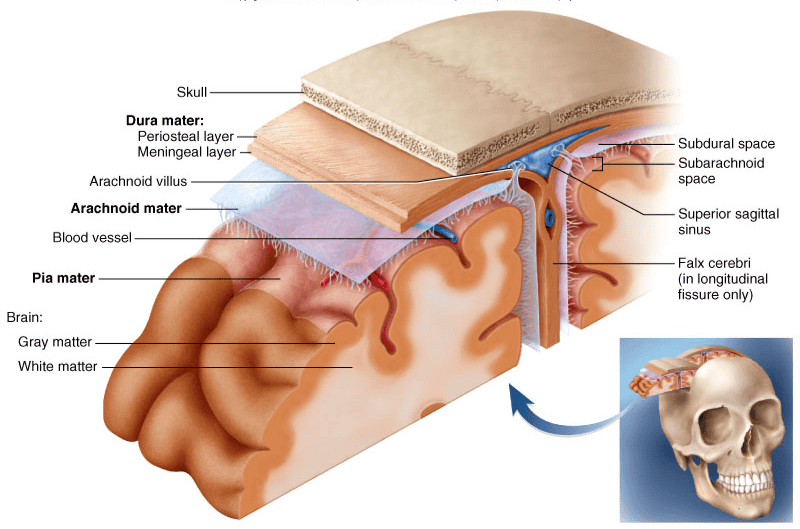
Under which of the layers in the brain are the dural sinuses located?
Dura mater
Where would a subdural hematoma occur if someone got hit in the head?
A subdural hematoma would occur in the space between the dural mater and middle layer of the meninges.
Describe the pathophysiology of meningitis? Make sure to include where you would insert a needle during a spinal tap.
- Inflammation of the meninges
- Usually a disease of infancy and childhood - between 3 months and 2 years of age
- Bacterial and virus invasion of the CNS by way of the nose and throat
- Signs include high fever, stiff neck, drowsiness and intense headache and may progress to coma
- Diagnose by examining the CSF
–lumbar puncture (spinal tap)
Label the locations of the following ventricles:
- Lateral ventricle
- Third Ventricle
- Fourth Ventrilce
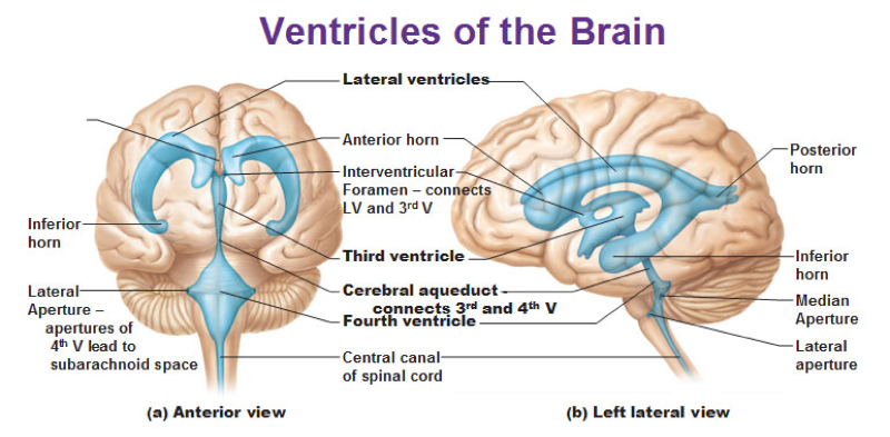
What two ventricles of the brain do each of the following connect:
- Interventricular foramen
- Cerebral aqueduct
Interventricular foramen- connects to LV and 3rdV
Cerebral aqueduct- connects to 3rd and 4th Ventricle
Predict the effects if either the interventricular foramen or the cerebral aqueduct were to become occluded.
Brain damage from build up of CSF.
– hydrocephalus
- Enlargement of the head in babies and brain damage in adults
Where is cerebrospinal fluid made, how is it circulated, and what is its purpose in the CNS? Spend some time looking at the choroid plexus!
Formed by the choroid plexus
CIRCULATES through ventricles, to central canal of spinal cord, into subarachnoid space around the brain, spinal cord, and cauda equina
Functions:
–Forms cushion for brain and other CNS organs
–Gives buoyancy to brain (which reduces weight by 97%) to prevent brain from crushing under own weight
Transports nutrients, chemical messengers, and waste
Explain the role of arachnoid villi and arachnoid granulations on the circulation of CSF.
Arachnoid villi:
–extensions of subarachnoid space
–extend through dura mater to superior sagittal sinus
Arachnoid granulations:
–large clusters of villi
–absorb CSF into venous circulation
What causes hydrocephalus?
When CSF becomes obstructed
Compare and contrast the structures involved AND the functions of both blood brain barrier and the blood CSF barrier.
BLOOD BRAIN BARRIER
BLOOD BRAIN BARRIER-
Structure: Endothelial cells stitched together by tight junctions
Very effectively protects the brain from many common bacterial infections. Thus, infections of the brain are very rare. ***ASTROCYTES
Compare and contrast the structures involved AND the functions of both blood brain barrier and the blood CSF barrier.
BLOOD CSF BARRIER
BLOOD CSF BARRIER- barrier at choroid plexus are ependymal cells joined by tight junctions
Function: Acts as barrier and produces CSF
Which portions of the brain make up the brain stem?
–Midbrain
–Pons
–Medulla oblongata
The midbrain is responsible for which functions?
Coordinates head and eye movement when we visually follow a moving object or see something out of corner of eye, even when we are not conscious of it
Coordinates head reflex movement to unexpected auditory stimulus – startle reflex
What is the major function of the pons?
Helps to maintain normal rhythm of breathing
Which portion of the brain stem adjusts the force and rate of heartbeat, vomiting, sneezing, etc?
Medulla Oblaganta
Which major region of the brain coordinates skeletal muscle contractions needed for smooth, coordinated movements of our daily lives?
Cerebellum
List the three bilaterally symmetric structures of the diencephalon.
–Thalamus
–Hypothalamus
–Epithalamus
List the three bilaterally symmetric structures of the diencephalon.
Which of these three makes up the major portion of the diencephalon?
The thalamus makes up 80% of the diencephalon
List the three bilaterally symmetric structures of the diencephalon.
Which of these three regulates hunger and fullness?
Hypothalamus
List the three bilaterally symmetric structures of the diencephalon.
Which portion serves as a gateway between the cerebral cortex and the rest of the body (where a sorting out and “editing” process occurs)?
Thalamus
List the three bilaterally symmetric structures of the diencephalon.
Which portion of the diencephalon is responsible for regulation body temperature (and does so by initiating sweating or shivering)?
Hypothalamus
List the three bilaterally symmetric structures of the diencephalon.
Which portion contains the gland responsible for the sleep/wake cycle?
Epithalamus
What are some of the structures that comprise the "limbic" system, and where in the brain are they located? What are the general functions of the limbic system?
Hippocampus –organizes sensory and cognitive information into a new memory
Amygdala- emotional memory
They are located in the cerebral hemispheres and diencephalon.
Describe the role of the basal ganglia, using Parkinson’s disease to aid in your explanation. Be specific!
With Parkinson's Disease, an inhibition of dopamine produces a balanced, restrained output of muscle-regulating signals from the basal nuclei. However with PD, neurons leading from the substantia nigra degenerate and thus do not release normal amounts of dopamine. Without the dopamine the excitatory effects of acetylcholine are not restrained, and the basal nuclei produce an excess of signals that affect voluntary muscles in several areas of the body. Overstimulation of these muscles cause rigidity and tremors of the head and limbs; an abnormal, shuffling gait: absence of relaxed arm-swinging while walking; and a forward tilting of the trunk.
What are some of the functions of the reticular formation. Make sure to describe the different “types” of sleep.
Normal Sleep: decreased activity of Ret. Form. = decreased cerebral cortex activity
Paradoxical Sleep (REM: dream sleep): impulses received by some parts of the brain but not by others.
Comatose: Ret. Form. ceases to function – cerebral cortex can’t be aroused
Compare the roles of association fibers, commissural fibers, and projection fibers of white matter. Association Fibers
Fibers that connect areas of the cerebral cortex within the SAME hemispheres of the cerebral cortex
Compare the roles of association fibers, commissural fibers, and projection fibers of white matter. Commissural Fibers
Fibers that connect one cerebral hemisphere to the other
Compare the roles of association fibers, commissural fibers, and projection fibers of white matter. Projection Fibers
Fibers that connect the cerebrum and other parts of that brain and/or spinal cord
The cerebrum is also called the
The cerebral cortex
Where is the corpus callosum located, and what is its primary function?
Corpus Callosum is a major pathway between the hemispheres.
Aids communication between cerebral areas and between cerebral cortex and CNS
The gray matter makes up which portion of the cerebrum? The white matter makes up which portion of the cerebrum?
The gray matter makes up the outer portion of the cerebrum and the white matter makes up the inner portion of the cerebrum
Locate the following of the cerebrum:
- Longitudinal fissure
- Central sulcus
- Precentral gyrus
- Postcentral gyrus
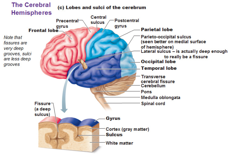
Longitudinal fissure
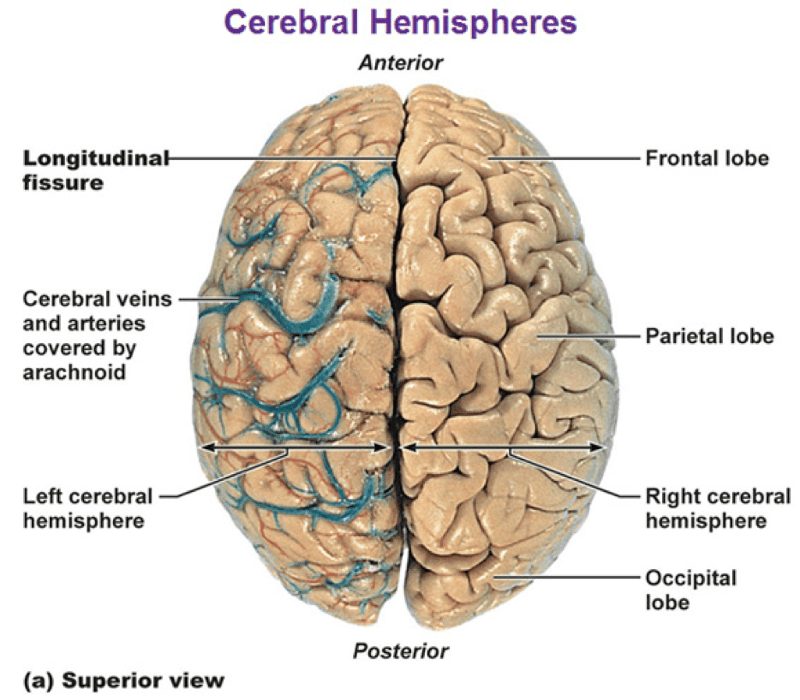
List the four major lobes of the cerebrum AND the main functions of each lobe.
Frontal Lobe
–voluntary motor functions
–planning, mood, smell and social judgment
Parietal Lobe
–receives and integrates sensory information
Occipital Lobe
–visual center of brain
Temporal Lobe
–areas for hearing, smell, learning, memory, emotional behavior
Describe the location AND functions of the sensory association areas of the cerebrum.
Interpret sensory information
Somesthetic association area (parietal lobe)
–position of limbs; location of touch or pain; shape, weight and texture of an object
Visual association area (occipital lobe)
–identify things we see
–faces recognized in temporal lobe
Auditory association area (temporal lobe)
–recall the name of a piece of music or identify a person by his voice
Describe the location AND functions of the area(s) of the cerebrum involved in motor control.
Intention to contract a muscle begins in motor association (premotor) area of frontal lobes
Precentral gyrus (primary motor area) relays signals to spinal cord
–pyramidal cells called upper motor neurons
–supply muscles of contralateral side
What does contralateral motor control refer to? Make sure to explain what decussate means!
Right hemisphere controls left side of body
Left hemisphere controls right side
Decussate is a crossing in the medulla
Why are the face and hands represented by such a large region of the cerebral cortex?
lots of sensations needed so they have larger area in cerebral cortex
Compare and contrast the locations AND functions of Wernicke and Broca’s area in the cerebral cortex.
Wernicke area posterior
–permits recognition of spoken and written language and creates plan of speech (sensory speech)
Broca area anterior
–generates motor signals for larynx, tongue, cheeks and lips
–transmits to primary motor cortex for action (Motor speech)
List AND describe the three types of aphasias covered in class. Lesion to Broca
Lesion to Broca = nonfluent aphasia
–slow speech, difficulty in choosing words
List AND describe the three types of aphasias covered in class. Lesion to Wernicke
Lesion to Wernicke = fluent aphasia
–speech normal and excessive, but makes little sense
List AND describe the three types of aphasias covered in class. Anomic aphasia
Anomic aphasia
–speech and understanding are normal but text and pictures make no sense
What is a lobotomy?
a surgical operation involving incision into the prefrontal lobe of the brain
Describe, in detail, the concept of lateralization.
Left hemisphere - categorical hemisphere
–specialized for spoken and written language, sequential and analytical reasoning (math and science), analyze data in linear way
Right hemisphere - representational hemisphere
–perceives information more holistically, perception of spatial relationships, pattern, comparison of special senses, imagination and insight, music and artistic skill
Highly correlated with handedness
–91% of people right-handed are left side dominant
Lateralization develops with age
females have more communication between hemispheres (corpus callosum thicker posteriorly)
MEMORIZE the names, numbers, functions, and whether or not each cranial nerve is sensory, motor, or both! You will thank me later for this, even though it seems like a pain in the rear right now! J
Making Separate Cards.
Predict what effects damage to each of the cranial nerves might cause. For example, a person will have trouble moving their right eye in which directions if the Oculomotor Nerve (CN III) is damaged.
Come Back *****
Use the idea of two point discrimination to explain what a receptive field is.
Area is monitored by a single receptor cell
The larger the receptive field, the more difficult it is to localize a stimulus
So, when you use the two point pair of blunt dividers, the minimum point to where they can feel both points at the same time then that is where you can locate the nerve densities. Therefore, the further apart the dividers are the more difficult it is to localize a stimulus.
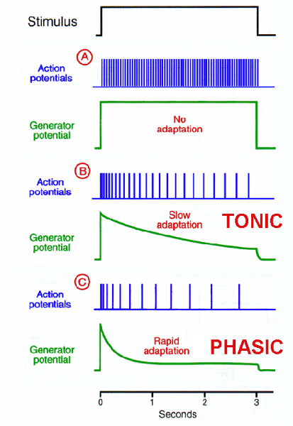
Compare and contrast tonic and phasic receptors. Draw a couple of graphs!
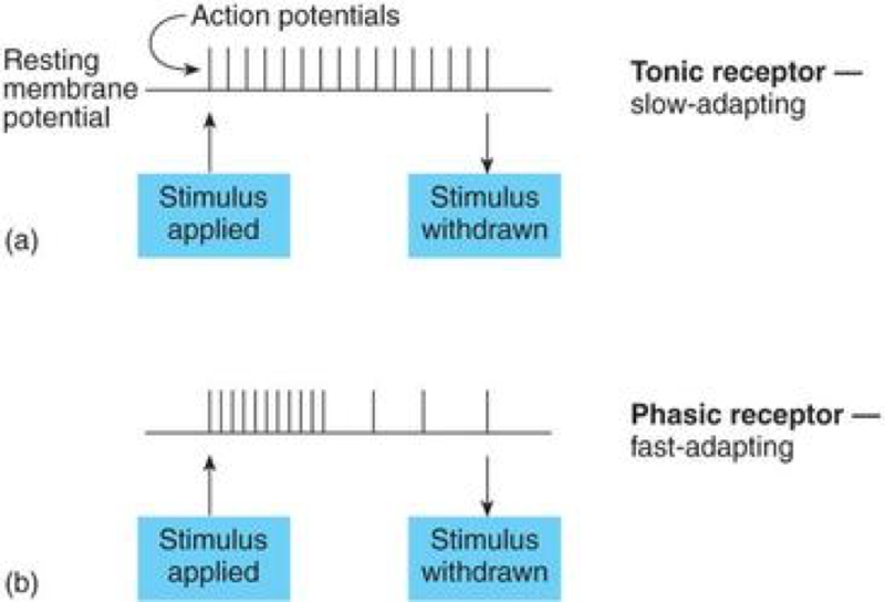
Tonic
–Are always active
Phasic
–Are normally inactive
–Become active for a short time whenever a change occurs
–Provide information about the intensity and rate of change of a stimulus
Use a graph to explain the concept of adaptation.
Pain receptors =
AKA nociceptors
Where are pain receptors found?
Are common in the:
–superficial portions of the skin
–joint capsules
–within the periostea of bones
–around the walls of blood vessels
Free nerve endings with large receptive fields
What are the two types of pain receptor fibers and what types of information do each of them carry?
Myelinated Type A Pain Fibers- Carry sensations of fast pain, or prickling pain, such as that caused by an injection or a deep cut
Type C Pain Fibers- Carry sensations of slow pain, or burning and aching pain
Where might one find thermoreceptors?
Are free nerve endings located in:
–the dermis
–skeletal muscles
–the liver
–the hypothalamus
Compare and contrast, in detail, the three classes of mechanoreceptors. Tactile receptors
Tactile receptors:
–provide the sensations of touch, pressure, and vibration
Compare and contrast, in detail, the three classes of mechanoreceptors. Baroreceptors
Baroreceptors:
–detect pressure changes in the walls of blood vessels and in portions of the digestive, reproductive, and urinary tracts
Monitor change in pressure
Consist of free nerve endings that branch within elastic tissues in wall of distensible organ (such as a blood vessel)
Compare and contrast, in detail, the three classes of mechanoreceptors. Proprioceptors
Proprioceptors:
–monitor the positions of joints and muscles
–the most structurally and functionally complex of general sensory receptors
Muscle spindles: monitor skeletal muscle length, trigger stretch reflexes
Golgi tendon organs: located at the junction between skeletal muscle and its tendon, stimulated by tension in tendon, monitor external tension developed during muscle contraction
Receptors in joint capsules: free nerve endings detect pressure, tension, and movement at the joint
Where might you find chemoreceptors? Briefly, how do they work
Located in the:
–carotid bodies:
–near the origin of the internal carotid arteries on each side of the neck
Aortic bodies:
–between the major branches of the aortic arch
–Receptors monitor Ph, carbon dioxide, and oxygen levels in arterial blood
Describe the difference in terms of location AND function of white matter and gray matter in the spinal cord.
Exterior white mater – conduction tracts
Internal gray matter - mostly cell bodies
Compare and contrast the dorsal and ventral horns of the spinal cord.
Dorsal (posterior) horns – SENSORY NEURONS!
Ventral (anterior) horns – MOTOR NEURONS!
Compare and contrast afferent and efferent neurons. Afferent
The Afferent (sensory) nervous system is all of the nerve pathways carrying signals TO the brain and/or spinal cord.
Carries toward the central nervous system.
Compare and contrast afferent and efferent neurons. Efferent
The Efferent (motor) nervous system consists of all the nerve pathways carrying signals OUT of the brain and/or spinal cord.
Carries away from the central nervous system
Draw out, in detail, the following SENSORY pathways from the spinal cord to the brain, making sure to include what type of information is relayed in each pathway:
- Posterior columns
- Anterior spinothalamic tract
- Lateral spinothalamic tract
- Spinocerebellar tract
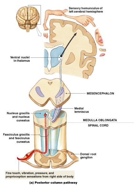
Sensory pathway- Posterior column
Carries sensations pressure, vibration, and proprioception
Sensory pathway- Anterior spinothalamic tract
Carries crude touch and pressure sensations
Sensory pathway- Lateral spinothalamic tract
Carries pain and temperature sensations
Sensory pathway- Spinocerebellar tract
conveys information to the cerebellum about limb and joint position
Diagram AND explain the differences between upper and lower motor neurons.
Upper motor neuron:
cell body lies in a CNS processing center (i.e. cerebral cortex)
- Synapses on the lower motor neuron
- Innervates a single motor unit in a skeletal muscle:
activity in upper motor neuron may facilitate or inhibit lower motor neuron
Lower motor neuron:
cell body lies in a nucleus of the brain stem or spinal cord
- Triggers a contraction in innervated muscle:
–only axon of lower motor neuron extends outside CNS
–destruction of or damage to lower motor neuron eliminates voluntary and reflex control over innervated motor unit
Where do most motor fibers decussate as they descend?
Medulla Oblaganta
Draw out the following MOTOR pathways from the brain to the spinal cord, making sure to include what type of information is relayed in each pathway:
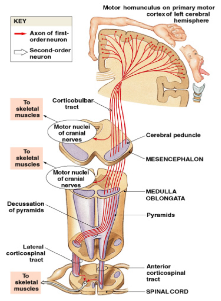
Draw out the following MOTOR pathways from the brain to the spinal cord, making sure to include what type of information is relayed in each pathway: Lateral corticospinal tract
***Lateral c orticospinal tracts (to skeletal muscles of trunk and limbs)
Draw out the following MOTOR pathways from the brain to the spinal cord, making sure to include what type of information is relayed in each pathway: Anterior corticospinal tract
...
Draw out the following MOTOR pathways from the brain to the spinal cord, making sure to include what type of information is relayed in each pathway: Corticobulbar tract
...
What are the “pyrmids” in the medulla of the brainstem?
As they descend, corticospinal tracts are visible along the ventral surface of medulla oblongata as pair of thick bands, the pyramids
Predict the effects of an upper vs. a lower motor neuron lesion in the lateral corticopsinal tract.
...
Briefly describe the components and functions of the medial and lateral pathways.
a. Medial Pathway Components - Primarily concerned with control of
muscle tone and gross movements of neck, trunk, and proximal limb
muscles
b. Lateral Pathway Components - Primarily concerned with
control of muscle tone and more precise movements of distal parts of limbs