Reasons for ultrasound exam of the neonatal head
hemorrhages around lateral ventricle
What happen in neonates under 34 weeks?
intraventricular and subependymal hemorrhages
How often do intraventricular and subependymal hemorrhages occur?
40 to 70% in neonates under 34 weeks
What is the most common ischemic lesion in a premie brain?
Periventricular leukomalacia
What is another name for Periventricular leukomalacia?
multifocal white matter necrosis
What is Periventricular leukomalacia?
when blood vessels burst, blood clots can collect in the white matter
What is Periventricular leukomalacia a predictor of?
Cerebral palsy
How often does Periventricular leukomalacia occur?
12 to 20% in infants weighing less than 2000g
Which lobe controls motor function?
frontal
Which lobe controls sensory function?
parietal
Which lobe controls audibility and olfactory function?
temporal
Which lobe controls vision function?
occipital
What controls balance?
cerebellum
Name the fontanelles
anterior
posterior
lateral
When does the anterior fontanelle close?
commonly around 6 months
What connects the 3rd and 4th ventricle?

Aqueduct of Sylvius
What connects the 4th ventricle and the spinal cord?
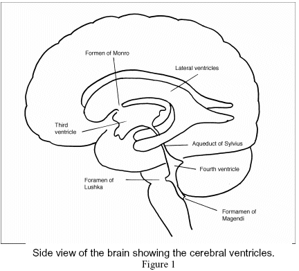
Foramen of Magendie
What connects the 4th ventricle and the subarachnoid space?
foramen of Luschka
What forms the roof of the lateral ventricles?
corpus callosum
What is below the corpus callosum and forms the medial walls of the lateral ventricles?

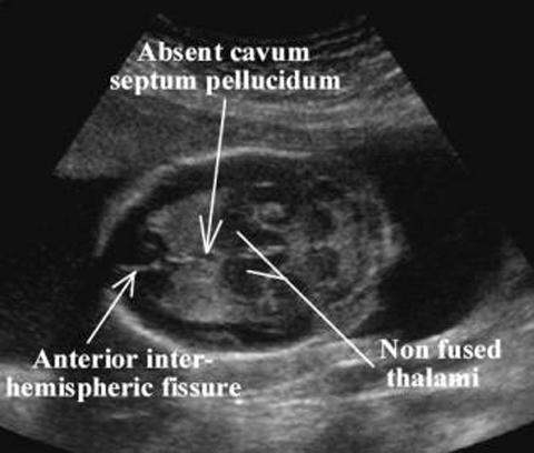
Cavum septum pellucidum
CSP
Where do all horns of the lateral ventricles merge?
trigone/atrium region
What connects the lateral ventricles and the 3rd ventricle?

Foramen of Monroe
What is the connection between the two hemispheres?
Messa intermedius
What makes up the floor of the 3rd ventricle?
hypothalamus.
What is the narrow subarachnoid space surrounding the brain and spinal cord?
Cistern
What is the largest of the cisterns?
cisterna magna
What does the narrow subarachnoid space surrounding the brain and spinal cord contain?
CSF
The _______, along with the cavum septum pellucidum (CSP) is a persistence of the embryological fluid-filled space between the leaflets of the septum pellucidum
Cavum Vergae
What forms the lateral borders of the frontal horns?
caudate nucleus
What does the caudate nucleus consist of?
head, body and tail
Where is a common site for hemorrhage on the caudate nucleus?
head
What does the brain stem consist of?
midbrain
pons
medulla oblongata
What is the cerebral peduncles responsible for?
communication between cerebellum and the sensory nerves
thalami and cerebellum
How is the midbrain divided?
into two cerebral peduncles
Thalami > cerebellum > sensory & balance
...
Explain the circle of willis.
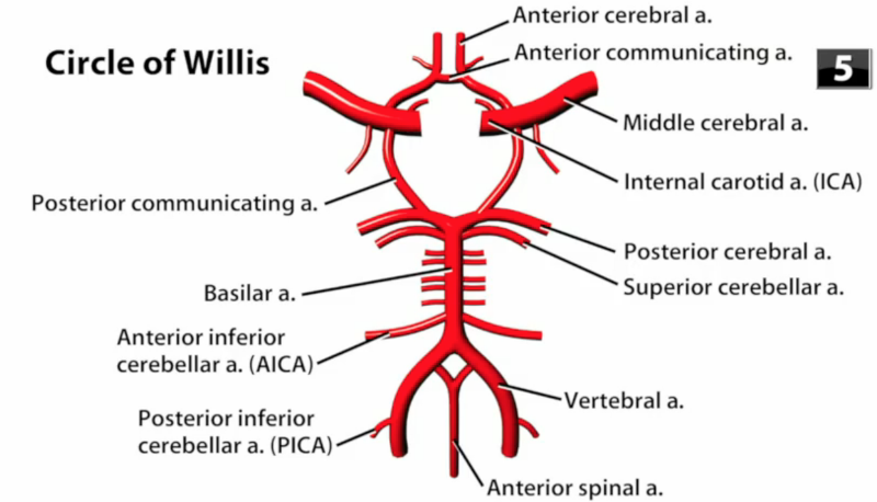
the two vertebral arteries come together to form the basilar artery
the basilar divides posterior cerebral artery
the internal carotid artery turns into the middle cerebral artery
the posterior communicating artery connects the posterior cerebral and the middle cerebral arteries
the anterior cerebral artery branches off the middle cerebral artery
the anterior communicating artery connects the two anterior cerebral arteries
Where does the middle cerebral artery extend?
into the sylvian fissure Y
Why is ultrasound the exam of choice when there is an open fontanelle?
portable
inexpensive
non-invasive
requires no sedation
Which transducer should be used for a neonate head scan?
5 MHz to 10 MHz phased array is optimal
small linear array may also be used
Where is the notch pointing during a neonate head scan?
toward the patients nose
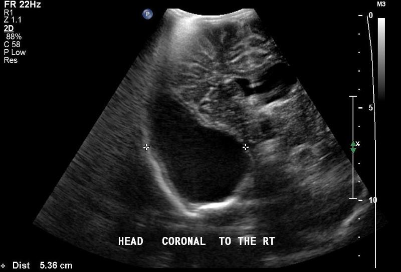
In what is the orientation of a sagittal view image of a neonate head scan?
top - superior - superficial
bottom - inferior - deep
right - anterior
left - posterior
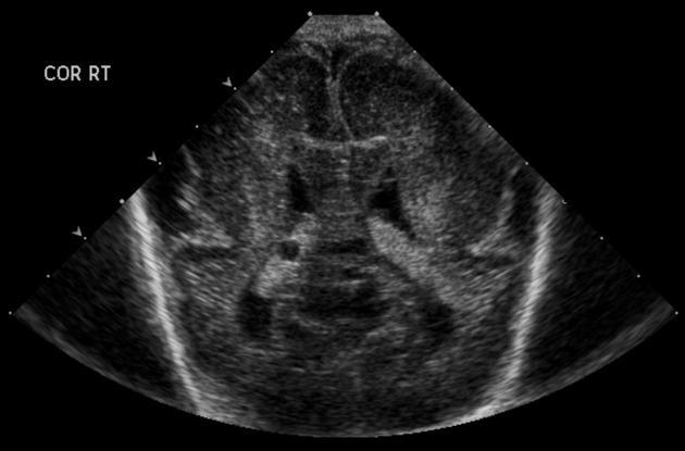
In what is the orientation of a coronal view image of a neonate head scan?
top - superior - superficial
bottom - inferior - deep
right - left
left - right
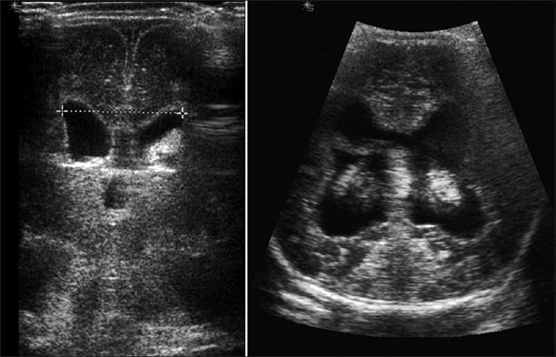
Explain the coronal view image of a neonate head scan.
slowly angle through the coronal plane
beginning rostrally at the frontal lobes angle posteriorly
What is the patient prep for a neonatal cranial exam?
The infant should be disturbed as little as possible. The exam should be done in the neonatal unit. Transducer gel should be body temperature.
How do you explain the cranial exam?
If the parents or guardians are available the sonographer should explain the exam is being performed to visualize the structures of the cranium for possible defects or problems that might arise in the premature infant.
How should the patient dress for the exam?
Keep the infant as warm as possible.
What transducer should be used for an infant weighing less than 1500 grams or less than 32 weeks?
7.5 MHz sector is a good choice but a curve linear transducer may also be used.
What transducer should be used when examining a full term infant?
3 – 5 MHz sector is a good choice but a curve linear transducer may also be used.
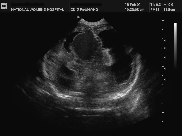
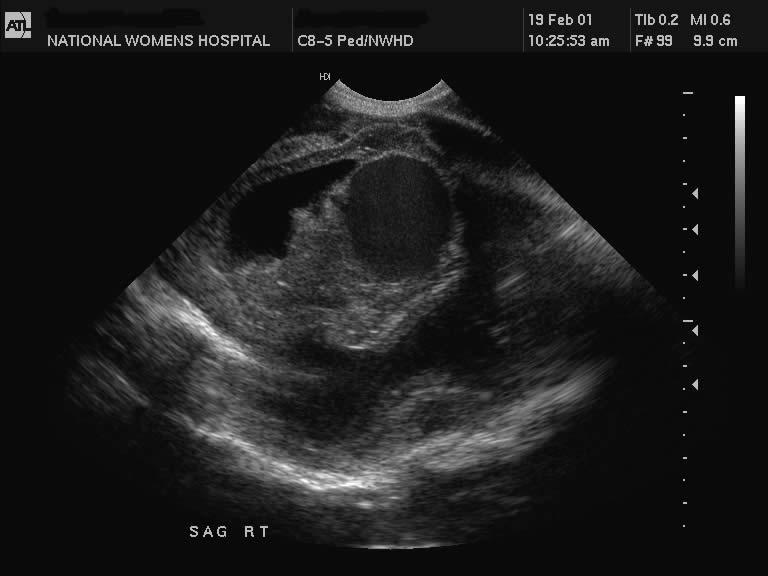
Explain the cranial procedure.
Begin doing a full sweep through the anterior fontanelle. Starting perpendicularly with a coronal view, slowly angle the transducer toward the face, scanning through the anterior horns and through the frontal lobes. Then slowly angle toward the occipital lobes and the posterior portion of the cranium and back to perpendicular. Change to a sagittal view, once again beginning perpendicular sweep the transducer toward the right lateral horn and through the temporal lobe and back to perpendicular. Repeat on the left side through the left lateral horn and temporal lobe. Finish back at a perpendicular position.
Look for:
- Hemorrhage
- Defects
- Size
- Vascularity
- Fluid
- Masses
After scanning through the brain start taking images.
Document the normal anatomy and any pathology found, including measurements and vascularity if indicated
What medical history is pertinent to a Neonate cranial exam?
Sex
Gestational Age
Weight
Family History
Anomalies previously found
What is the patient position during a neonate cranial exam?
Supine with the head facing up
Prone with head facing the side may be used as necessary
What are the scan planes for a neonatal cranial exam?
Coronal & Sagittal
Explain the coronal plane in a neonatal cranial exam?
Begin perpendicular to the anterior fontanelle, angle the transducer toward the face return midline and sweep the posterior.
Explain the sagittal plane in a neonatal cranial exam?
Begin perpendicular to the anterior fontanelle, Angle the transducer to the right return midline and sweep the left
What are the techniques used for a neonatal cranial exam?
Scanning a baby while they sleep is easier on the patient and easier for scanning
Explain the neonatal anatomy
After the fourth week after conception the neural tube separates into three main structures that will form the brain. These structures include the prosencephalon (forebrain) , mesencephalon (midbrain) and rhombencephalon (hindbrain).
The forebrain consists of the cerebrum, thalamus, hypothalamus and the diencephalon. The midbrain will develop into ventricles and the cerebral peduncles. The hindbrain will develop into the cerebellum and the brain stem including the medulla oblongata and pons
What is the purpose of the ventricles?
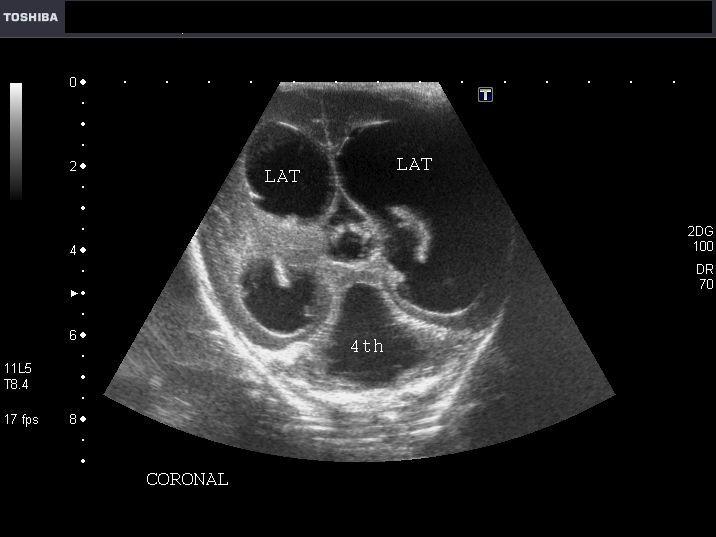
Four ventricles circulate cerebral spinal fluid around the brain.
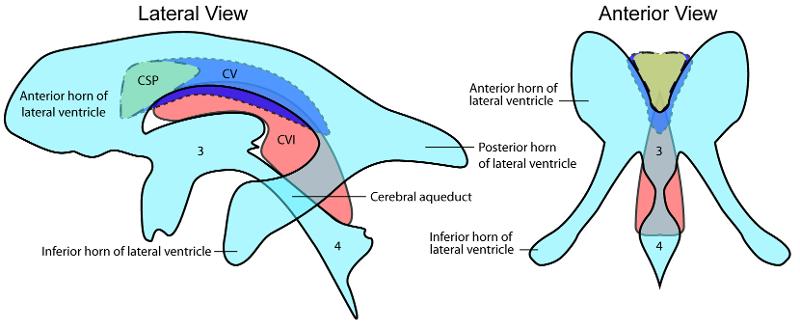
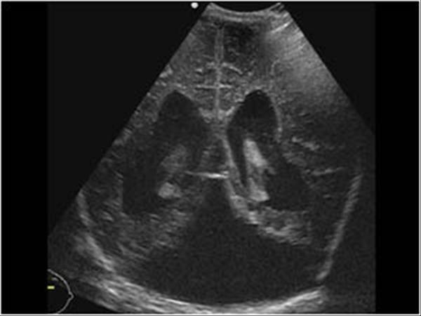
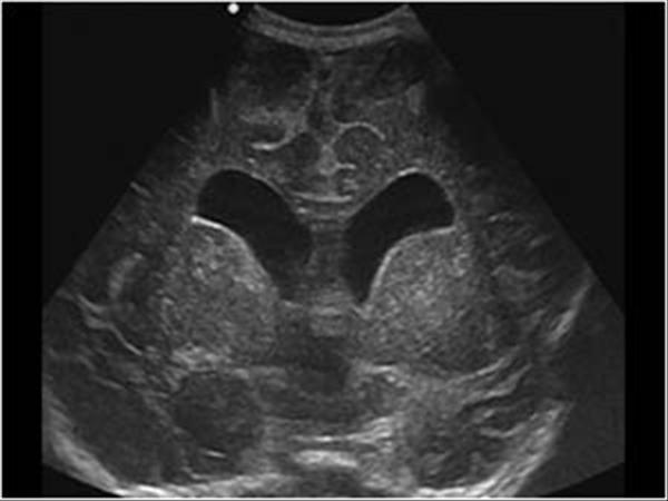
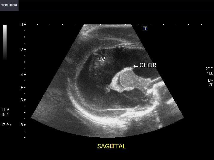
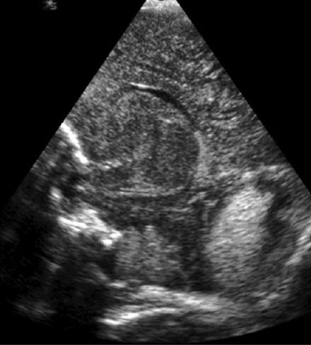
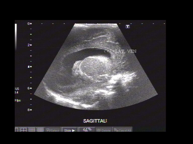
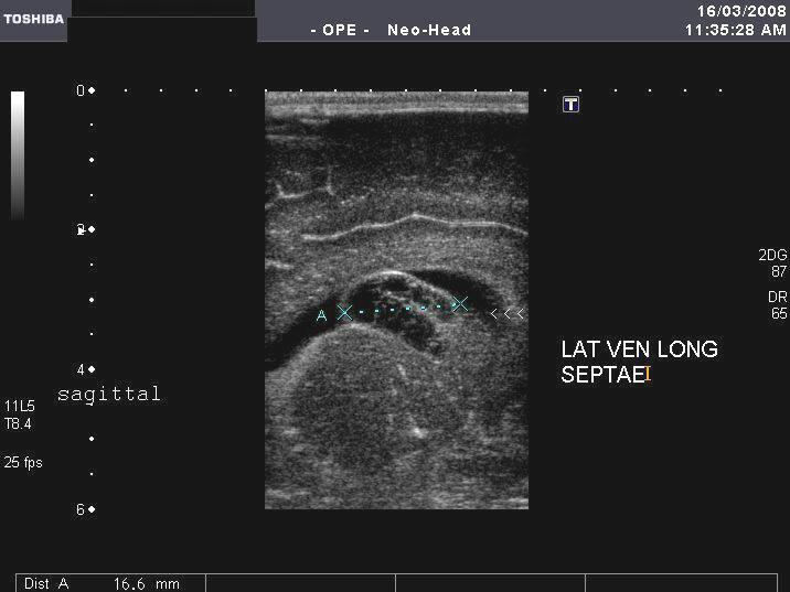
Explain the anatomy of the lateral ventricles.
divided into the frontal, occipital and temporal temporal horns. All horns converge at the trigone region. They are filled with cerebral spinal fluid.
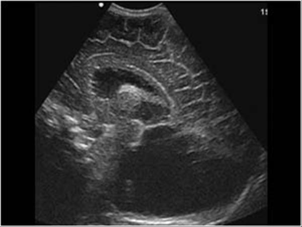
Explain the anatomy of the third ventricles.
The third ventricle is a midline structure containing cerebral spinal fluid. It is connected with the lateral ventricles by the foramen of Monroe and to the forth ventricle by the aqueduct of sylvius.
Explain the anatomy of the fourth ventricles.
The fourth ventricle is also filled with cerebral spinal fluid.
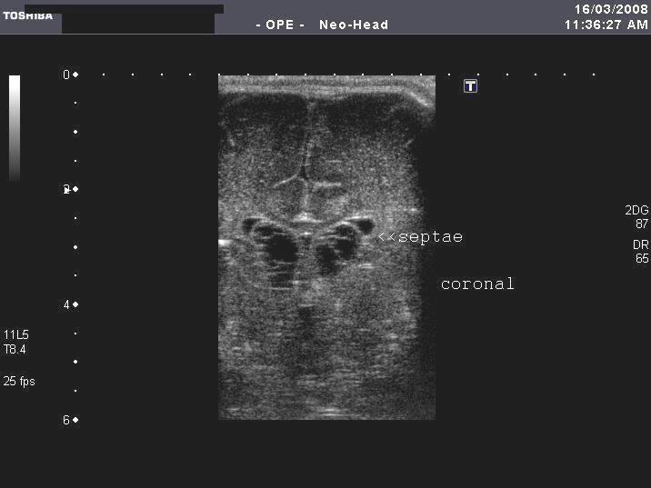
Explain the anatomy of the Corpus Callosum.
Lies midline and consists of connective fibers connecting the two hemispheres. It creates the roof of the lateral ventricles and sits superior to the cavum septum.
Explain the anatomy of the Cavum Septum Perdiculum and Vergae.
A midline structure that creates the floor of the corpus callosum. It is filled with cerebral spinal fluid. It sits between the lateral ventricles. It is present at birth but closes around 4 months.
Explain the anatomy of the Thalamus.
The thalamus is an egg-shaped structure. It sits within the 3rd
Explain the anatomy of the Cerebellum.
The cerebellum takes up most of the posterior fossa. The connection between each lobe is called the vermis.
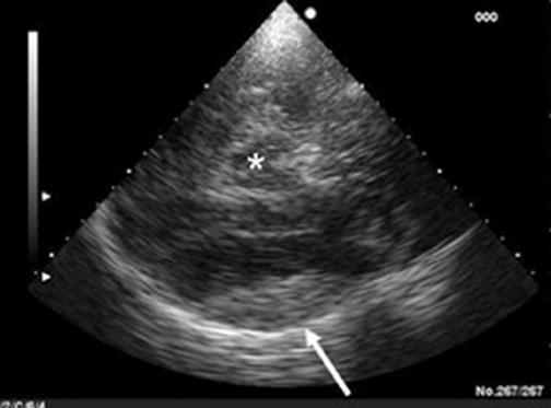
Explain the anatomy of the Cisterna Magna.
Lies posteroinferior to the cerebellum.
Explain the anatomy of the Choroid Plexus.
Consists of two curved structures that wrap around the thalamus. It is responsible for absorbing cerebral spinal fluid.
Explain the anatomy of the Aqueduct of Sylvius.
channels that connects the 3rd and 4th ventricle
Explain the anatomy of the Foramen of Monroe.
channels connecting the lateral ventricle with the 3rd ventricle
Explain the anatomy of the Brain Stem.
Consists of the midbrain, pons and medulla oblongata. This structure connects the spinal cord to the brain.
Explain the anatomy of the Interhemispheric Fissure.
A midline structure where the falx lies between the two hemispheres.
Explain the anatomy of the Massa Intermedia.
A pea-shaped structure within the 3rd
Explain the anatomy of the Hippocampal Gyrus (Choroid Fissure).
spiral-like fold covering the temporal horns.
Explain the anatomy of the Cerebral Peduncles.
Column like structures connected to the pons and the thalamus
Explain the anatomy of the Sulci.
fissures of the brain that separate the folds.
Explain the anatomy of the Tentorium.
echogenic covering that separates the cerebellum from the cerebral. It is part of the dura mater.
Explain the anatomy of the Sylvian Fissure.
A fissure located laterally between the temporal and frontal lobes. The middle cerebral artery lies in this fissure.
Explain the anatomy of the Caudate Nucleus.
located within the concavity of the lateral angles of each ventricle.
Explain the anatomy of the Germinal Matrix/Caudothalmic Groove.
A vascular network located near the caudate nucleus. This is a common site for hemorrhage in the neonate infant.
Explain the anatomy of the Quadrigeminal Plate.
Immediately superior to the superior aspect of the tentorium.
What are appropriate reasons for a neonate cranial exam?
Evaluate the cranial anatomy of a neonate infant for pathology including: anomalies, intracranial hemorrhage, and ventricular dilation
Premature Delivery
Abnormal posturing
Low birth weight
Seizers
Apnea
coma
What are the required coronal images?
Anterior: orbits
Anterior: anterior horns and lateral ventricles
Middle: lateral ventricles, cavum septum pellucidum, 3rd ventricle, and corpus callosum
Posterior: ambient wings of the cisterna magnum
Posterior: tentorium and cisterna magnum
Posterior: choroid plexus
Posterior: glomus of choroids
Posterior: occipital lobe
What are the required sagittal images?
Midline: cavum septum pellucidum, corpus callosum, 3rd ventricle and foramen of Monroe, aquaduct of slyvius, 4th ventricle, tentorium, cisterna magna
Left Thalamus
Left Caudothalamic groove
Left Lateral ventricle: anterior, body, and occipital (temporal is hydrocephalic
Left Angle slightly lateral from lateral ventricle to show the white matter
Left very lateral: Sylvain fissure/ middle cerebral artery
Repeat on left side
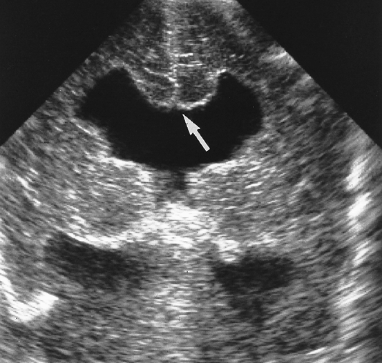
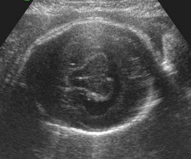
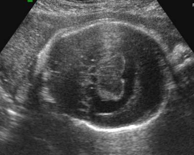
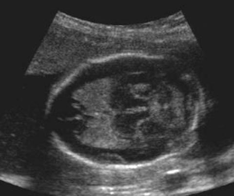
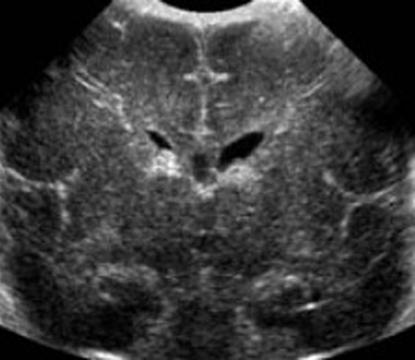
What is the sonographic appearance of the ventricles?
Two Lateral Ventricles: filled with cerebral spinal fluid and appear as echogenic slits.
Third Ventricle: Midline structure, echogenic walls and the center appears anechoic.
Forth Ventricle: Midline structure inferior to the third ventricle, echogenic walls and the center appears anechoic.
What is the sonographic appearance of the Corpus Callosum?
midline, echogenic structure, midgray and has medium to low level echoes.
What is the sonographic appearance of the Cavum Septum Perdiculum and Vergae?
appears anechoic, fluid filled structure. Anterior to the corpus callosum
What is the sonographic appearance of the Thalamus?
midline, echogenic structure within the 3rd ventricle
What is the sonographic appearance of the Cerebellum?
vermis is echogenic and the surrounding parenchyma appears midgray
What is the sonographic appearance of the Cisterna Magna?
fluid filled, anechoic structure lays posteroinferior to the cerebellum
What is the sonographic appearance of the Choroid Plexus?
echogenic structures that wrap around the thalamus
What is the sonographic appearance of the Aqueduct of Sylvius?
rarely seen during ultrasound unless dilated
What is the sonographic appearance of the Foramen of Monroe?
channels connecting the lateral ventricle with the 3rd ventricle
What is the sonographic appearance of the Brain Stem?
appears midgray with low echogenicity
What is the sonographic appearance of the Interhemispheric Fissure?
echogenic area separating the two hemispheres
What is the sonographic appearance of the Massa Intermedia?
mid-gray, best seen with ventricular dilatation
What is the sonographic appearance of the Hippocampal Gyrus (Choroid Fissure)?
echogenic, spiral-like fold coving the temporal horn.
What is the sonographic appearance of the Cerebral Peduncles?
Column like structures connected to the pons and the thalamus
What is the sonographic appearance of the Sulci?
echogenic spider- like fissures of the brain that separate the folds.
What is the sonographic appearance of the Tentorium?
echogenic covering that separates the cerebellum from the cerebral.
What is the sonographic appearance of the Sylvian Fissure?
A fissure located laterally between the temporal and frontal lobes. It appears as a Y shape. The middle cerebral artery lies in this fissure.
What is the sonographic appearance of the Caudate Nucleus?
appears midgrey
What is the sonographic appearance of the Germinal Matrix/Caudothalmic Groove?
small echogenic area at the junction of the caudate and the thalamus . hemorrhages are common here.
What is the sonographic appearance of the Quadrigeminal Plate?
echogenic area superior to the tentorium.
What is Arnold-Chiari Malformations?
Congenital anomaly associated with spina bifida. The brain stem and cerebellum are pulled toward the spinal cord, secondary hydocephus develops.
What is the sonographic appearance of Arnold-Chiari Malformations?
Small posterior fossa
Myelomeningocele decompression of the ventricle
Small cerebellum
Absence of the cisterna magna
4th ventricle in low position
Absence of the septum pellucidum
Widening of the 3rd ventricle
Cerebellar tonsil herniation into enlarged foramen magna
Displacement of pons and medulla
Elongation of 4th ventricle
Enlarged massa intermedia
3rd ventricle slightly larger
Small anterior horns
Enlargement of posterior horns
Wide interhemispheric fissure
Small posterior fossa
Low tentorium
Hydrocephalus
What are the presenting symptoms of Arnold-Chiari Malformations?
40 to 75% aqueductal stenosis
What is Agenesis of the Corpus Callosum?
Partial or complete agenesis is often seen with heterotopias and polymicrogyria.
What is the sonographic appearance of Agenesis of the Corpus Callosum?
Narrow frontal horns
Separation of the anterior horns
Widening of the occipital horns
Widening of the 3rd ventricles
3rd ventricles have pointed upper corners (bat-wings)
What are the presenting symptoms of Agenesis of the Corpus Callosum?

poor muscle tone
porencephaly
hydrocephalus
microgyria
Arnold-Chiari
fusion of the hemispheres.
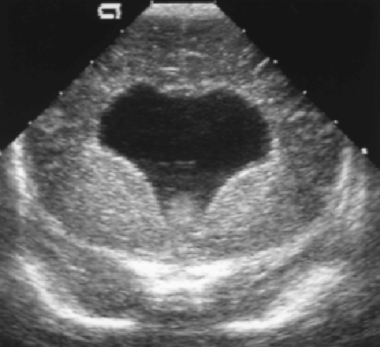
What is Dandy-Walker Malformation?
A congenital anomaly in which a 4th ventricle cyst occupies the cerebellar space.
What is the sonographic appearance of Dandy-Walker Malformation?
Hydroplasia of cerebellar vermis
Enlarged 4th ventricle
Cysts in posterior fossa
Small brain stem
Hydrocephalus
Atresia of the Luschka and Magendie
4th ventricle communicates directly to cysts
Obstruction above and below 4th ventricle
Absence of Corpus Callosum
What are the presenting symptoms of Dandy-Walker Malformation?
hydrocephalus
agenesis of the corpus callosum
encephalocele
holoprosencephaly
microcephaly
infundibular hamartomas
brain stem lipomas.
What is Holoprosencephaly?

Complex abnormality from failure of cleavage of the prosencephalon
*Must obtain modified coronal studies of the whole frontal lobe to determine if frontal horns are present
What is Alobar Holoprosencephaly?

The most severe form of Holoprosencephaly
What is the sonographic appearance of Alobar Holoprosencephaly?

Single midline ventricle
Thin, primitive cerebral cortex
Fused Thalami and hemispheres , hyper echogenic choroid plexus
Absent corpus callosum & interhemiphermic fissure, 3rd ventricle
Large dorsal cyst
What are the presenting symptoms of Alobar Holoprosencephaly?
Multiple facial abnormalities
- cebocephaly
- cyclopia
- ethmocephaly
What is Semilobar Holoprosencephaly?

Characterized by the abnormal storage and collection of glycogen in the tissue of the liver and kidneys.
What is the sonographic appearance of Semilobar Holoprosencephaly?
Single ventricle
More brain parenchyma present
Posterior faux and interhemispheric fissure
Splenium and genu seen midline
3rd ventricle is small
What are the presenting symptoms of Semilobar Holoprosencephaly?
Mild facial
- hypotelorism
- cleft lip
What is lobar Holoprosencephaly?
Least severe form
What is the sonographic appearance of lobar Holoprosencephaly?
Nearly complete separation of hemispheres
Faux and interhemispheric fissure development
Some front lobe fusion
Absent septum pellucidum
Anterior horns fused
Occipital horns separation
3rd ventricle separates the thalami
Absent genu
What are the presenting symptoms of lobar Holoprosencephaly?
mild facial abnormalities
What is Ischemic Lesions: ?

Lesions in the midline that induce malformations of the telencephalon
What is the sonographic appearance of Ischemic Lesions: ?
Single ventricle cavity
Absent corpus callosum
* Presence frontal horns helps differentiate between Holoprosencephaly
What are the presenting symptoms of Ischemic Lesions: ?

Gastrointestinal bleeding
blood in the stools
vomiting of blood
Encephalopathy
What is Porencephalic?
Also known as porencephaly, is a cyst filled with cerebral spinal fluid. May be caused by hemorrhage , infarction, trauma, inflammation of the nervous system.
What is the sonographic appearance of Porencephalic?
Cyst without mass
Reduction of hemisphere
Midline shift
Contralateral ventricular enlargement
What is Hydranencephaly?
Brain development is destroyed and preplaced with cerebral spinal fluid. May be caused by bilateral occlusion of the internal carotid
What is the sonographic appearance of Hydranencephaly?
Midbrain, basal ganglia, choroid plexus, and thalamus sparing
Presence of cerebral spinal fluid
Absent Doppler flow in carotid
Possible absent falx cerebri
What is Congenital Hydrocephalus?

An imbalance of production and absorption of cerebral spinal fluid. There are three types: obstruction to outflow, decreased absorption, or rarely overproduction.
What is the sonographic appearance of Congenital Hydrocephalus?
Blunting of the lateral angles of the lateral ventricle
Widening of the ventricle system
Rare choroid plexus cyst
What are the presenting symptoms of Congenital Hydrocephalus?
enlargement of head, bulging fontanelles
What is Obstructive Hydrocephalus?

interference of cerebral flow
What is the sonographic appearance of Obstructive Hydrocephalus?
Enlargement of proximal ventricle cavity
What is Communicating Hydrocephalus?
pathway of cerebral fluid are open but there is a decrease in absorption. The ventricular system becomes uniformly distended. The most common cause is aquaduct stenosis.
What is the sonographic appearance of Communicating Hydrocephalus?
Narrow aqueduct of Slyvius may be replaced by small network
Widening of lateral ventricles
Normal size 4th ventricle
Small posterior fossa
Cerebellum displaced posteriorly
Absent cisterna magna
What is Subarachnoid Cysts?
lined by arachnoid tissue and contain cerebral spinal fluid.
What is the sonographic appearance of Subarachnoid Cysts?

Normal vermis (Not Dandy- Walker)
Sonolucent
Verify with color Doppler
Interhemispheric cysts
Suprasellar cysts
Cerebral convexity cysts
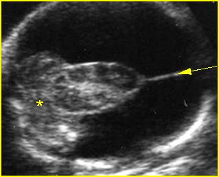
What is Choroid Plexus cysts?
A hepatic cyst is usually a solitary, non-parasitic cyst of the liver. solitary or multiple.
What is the sonographic appearance of Choroid Plexus cysts?

Common and single
Well-defined
Anechoic mass
Unilateral
Left larger than right
4 to 7 mm
*Rare Multiple cysts larger than 10 mm associated with trisomy 18
What is Subependymal cysts?
Discrete in the lining of the ventricle. Commonly the result of sequel of germinal matrix
What is the sonographic appearance of Subependymal cysts?

Smooth walled
Spherical
Located in lateral ventricle
What is Galenic Venous Malformation?
Dilation of the vein of Galen, caused by vascular malformation off the posterior cerebral.
What is the sonographic appearance of Galenic Venous Malformation?
Anechoic cystic
Between lateral ventricles
Calcification
Hydrocephaly with possible thrombus
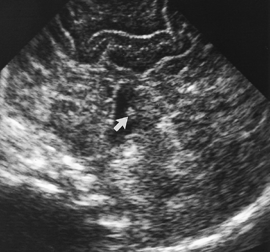
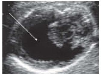
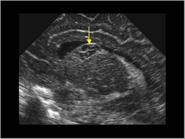
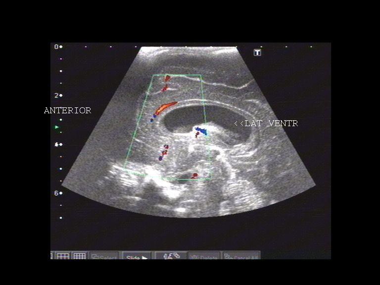
What is Subependymal-Intraventricular Hemorrhages?
Most common hemorrhage in preterm, affecting 30 to 50% of infants born before 32 weeks. The most common site is the interthalmic groove. Can cause obstruction in the choroid plexus.
What is the sonographic appearance of Subependymal-Intraventricular Hemorrhages?

Fluid
Echogenic structure in white matter
Become cystic and are absorbed leaving cavity
What is Intraparenchymal Hemorrhages?

Complicate Subependymal hemorrhage, meaning brain tissue has been
What is Intracerebellar Hemorrhages?
Difficult to find in live infants. Primmary: intracerebellar, venous infarction, traumatic laceration – resulting from occipital diastasis, extension to the cerebellum of a large SHE-IVH
What is the sonographic appearance of Intracerebellar Hemorrhages?

Echogenic structures
Within the less echogenic cerebellar parenchyma
Become cystic with time leaving cavity lesions
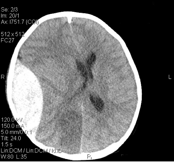
What is Epidural Hemorrhages?

Better diagnosed with CT. located peripherally along the surface of the brain.
What is the sonographic appearance of Epidural Hemorrhages?
Nonechogenic spaces
Between the echogenic calvarium and the cortex
What are the presenting symptoms of Epidural Hemorrhages?

persistent
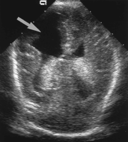
What is Periventricular Leukomalacia?
Also called Multifocal White Matter Necrosis, most common ischemic lesion in the immature brain. Highly echogenic areas in the white matter.
What is the sonographic appearance of Periventricular Leukomalacia?
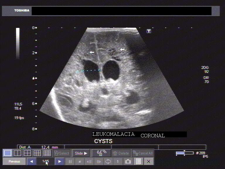
echolucencies in white matter
What are the presenting symptoms of Periventricular Leukomalacia?
Associated with Cerebral Palsy
What is Focal Brain Necrosis?

occur within large arteries. Early on cause destruction of cerebral tissue and leave cavitary lesions. These lesions correspond to cerebral infarction.
What is the sonographic appearance of Focal Brain Necrosis?
Echogenic localized lesions
Sonolucencies appear after few days
What is Ventriculitis?
A common complication of purulent meningitis. This is probably caused by infection spreading to the choroid plexus. Stents and other objects placed the body can cause this infection. Can lead to hydrocephalon
What is the sonographic appearance of Ventriculitis?
Thin septations extending from lateral ventricles.
Septations thicken