respiration
(A) The general term for the set of physiological mechanism used to carry oxygen from the atmospheric air to the mitochondria of every cell in the body; this involves bulk transport (ventilation, circulation) and diffusion (in alveoli, across the alveolar membrane into the erythrocytes and from the capillary beds through the interstitial fluid and into the cell cytoplasm and ultimately to the mitochondria); it is generally divided into three stages: (1) ventilation - the process of inhaling and exhaling, i.e., breathing; (2) external respiration - the diffusion of gases between the alveolar air and the blood; and (3) internal respiration - the diffusion of gases between the blood and the cells. (B) The oxidative process occurring within living cells by which the chemical energy of organic molecules is released in a series of metabolic steps, some occurring in the cytoplasm and some within the mitochondria, involving the consumption of oxygen and the liberation of carbon dioxide and water. aka - aerobic respiration
pulmonary ventilation
The process of exchange of air between the lungs and the atmosphere; it is sometimes divided into two components: (1) pulmonary ventilation - the total exchange of air, usually measured in liters/minute, and (2) alveolar ventilation - the effective ventilation of the alveoli in which gas exchange with the blood actually takes place; the movement of air is accomplished by the muscles of respiration under the autonomic control of the respiratory center in the medulla.
external respiration = alveolar gas exchange = pulmonary gas exchange
The interchange by diffusion (through the alveolar walls) along concentration gradients of oxygen and carbon dioxide between the alveolar air and the blood (plasma and suspended erythrocytes).
internal respiration = tissue gas exchange
The interchange by diffusion along concentration gradients of oxygen and carbon dioxide between the cells of the body and the interstitial fluid surrounding them, which in one sense is a process of nutrition.
cellular respiration
The oxidative process occurring within living cells by which the chemical energy of organic molecules is released in a series of metabolic steps with the production of ATP, some occurring in the cytoplasm and some within the mitochondria, involving the consumption of oxygen and the liberation of carbon dioxide and water; it is generally divided into two stages: (1) anaerobic glycolysis in the cytoplasm followed by (2) the citric acid cycle and oxidative phosphorylation at the electron transport chain in the mitochondria. aka - aerobic respiration
bulk flow
The movement of volumes of water and the solutes (unrestricted by size or charge) dissolved in that water under the influence of hydrostatic or osmotic pressures between cells in the tissues; the movement occurs rapidly through intercellular clefts (not across or through cell membranes); it is the major means of transport across capillary walls.
diffusion
The spontaneous intermingling of molecules or the particles of two or more substances as a result of random thermal motion within a solution (gas or liquid); it is the major means of transport for uncharged molecules across or through cell membranes; it is a form of passive transport which does not require additional ATP expenditure; the rate is dependent on the concentration gradient and the surface area available. aka - simple diffusion, free diffusion
upper respiratory system = conducting portion
The system of airways for ventilating the lungs, consisting of the nasal and oral cavities, the pharynx, the larynx, and the trachea which divides into a series of ever-smaller branches (about 23 anatomical divisions are recognized): the main bronchi, lobar bronchi, segmental bronchi, and so on, to the smallest bronchioles which do not have alveoli, the terminal bronchioles; all these branches have smooth muscle in their walls and are lined with bronchial epithelium, either pseudostratified columnar, columnar, or cuboidal; although the base airway diameter decreases with branching, the overall or total cross-sectional diameter increases tremendously so that peripheral resistance to air flow decreases as the air moves closer to the alveoli; the volume of the air in this system of airways is not available for actual gas exchange, so it has been referred to as the anatomic dead space (normally that is ~0.15 L, i.e., ~30% of a tidal volume).
lower respiratory system = respiratory portion
That portion of lung tissue where external respiration, actual gas exchange, takes place; all included structures have alveoli present in them; the structures involved include 2-5 "generations" or anatomical divisions lined by cuboidal epithelium with smooth muscle in their walls termed respiratory bronchioles, though minimal gas exchange occurs through their walls, then the final branches from the respiratory bronchioles, which have no muscle in their walls, and are lined by a squamous epithelium, the alveolar ducts, and, finally, the alveolar sacs and individual alveoli, which are lined by alveolar type I and type II squamous cells
List the anatomical and physiological defenses the body uses to provide clean "sterile" air to the respiratory membranes of the lungs.
(1) the conducting portion of the airways are lined with mucous membranes:
(a) mucous traps dust particles and microbes
(b) mucous contains lysozyme and other non-specific resistance factors
(c) mucous contains IgA class antibodies for immune resistance
(d) much of the mucous lining is ciliated (trachea to smaller bronchioles) and ciliary motion sweeps trapped dust particles and microbes upward (the "mucociliary elevator") to be swallowed and sterilized in the acid bath of the stomach
(e) beneath the mucous membranes lining the conducting portion of airways are collections of lymphatic tissue, e.g., the tonsils, to provide immune defensive cells to prevent infection
(2) the respiratory portion of the airways where gas exchange occurs are patrolled by alveolar macrophages = "dust cells" which will phagocytize and attempt to destroy any dust particles or microbes which reach this location
Sketch and label: A frontal section of the thorax illustrating the organs, membranes, and (cavities) spaces involved in ventilation.
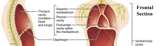
The Frontal Section is on the right. To answer the question you need only name the lungs, diaphragm and intercostal muscles, parietal and visceral pleural membranes and the pleural cavities. See below. The more complete diagram of body cavities is provided at the bottom as a reminder of the larger relationships.
external nares
The pair of anterior openings of the nasal cavity on the inferior surface of the nose to the outside of the body; the entryway for air required in respiration. aka - nostrils, nasal openings
internal nares = choanae
The pair of posterior openings of the nasal cavity into the (naso)pharynx, above the palate of the mouth; a part of the passageway for air required in respiration.
nasal cavity
The chamber lying between the base of the cranium and the roof of the mouth and extending from the nose to the pharynx and connecting with the paranasal sinuses of the skull; it is lined by a stratified squamous epithelium and moistened by secretions of mucous glands and the lacrimal glands (connected via the lacrimal canal); passage of air through this chamber warms and humidifies the air before it reaches the alveoli of the lungs and also exposes the olfactory epithelium at the roof of the cavity to chemicals which can be sensed as smells.
nasal septum
The vertical dividing wall which runs down the middle of the nasal cavity so that there are normally two subdivisons to the cavity, each ending in a nare; it consists of the bony "perpendicular" plates of the ethmoid and vomer bones.
(nasal) vestibule
In general, a cavity, chamber, or channel which leads to or is an entrance to another cavity; in the anterior part of the nasal cavity, especially the part enclosed by cartilage; this is the area where the external nares are guarded by nasal hairs; a part of the passageway for air required in ventilation.
nasal meatuses
Three connected air passages in each side of the nasal cavity formed by the projection of the conchae: the inferior nasal meatus lies below the inferior concha; the middle nasal meatus lies between the middle and inferior conchae; the superior nasal meatus lies between the superior and middle conchae.
nasal conchae
Three curled or shell-shaped plates of bone on each side of the nasal cavity which are covered by moist nasal mucosa and define the air passageways (nasal meatuses); the superior and middle conchae are formed by projections of the ethmoid bone; the inferior concha, which is the largest, is a distinct bone articulating with the lateral wall of the nasal cavity.
rhinoplasty
Plastic surgery of the nose to correct deformity or to replace lost tissue; tissue may be transplanted from the patient's cheek, forehead, arm, etc., or even from another person. nickname - nose job
pharynx
The "overlapping" cone-shaped section of the respiratory tree and the alimentary canal which extends from the mouth and nasal cavities to the larynx, where it becomes continuous with the esophagus; it is lined by a stratified squamous epithelium and moistened by secretions of mucous glands and the salivary glands; its walls consist of skeletal muscle and MALT lymphatic tissue, e.g., the tonsils and lingual tonsils; it is subdivided into the nasopharynx, oropharynx, and laryngopharynx.
nasopharynx
The superior part of the pharynx above the soft palate which is continuous with the nasal passages at the posterior nares; a part of the airway for ventilation of breathing gases.
oropharynx
The middle part of the pharynx below the soft palate and above the base of the tongue which is directly posterior to the oral cavity; the space where the food passageway and the airway intersect or overlap.
laryngopharynx
The inferior part of the pharynx below and behind the base of the tongue where the opening to the larynx is guarded by the epiglottis and behind the larynx where it becomes continuous with the esophagus at approximately the same point that the larynx joins the trachea; the part of the passageway for ingested foods and liquids to pass into the esophagus.
fauces
The opening or passageway from the back of the mouth to the pharynx, an anatomical landmark bounded by the soft palate, the base of the tongue, and the palatine arches where the palatine tonsils are located.
stratified squamous epithelium
A membranous tissue composed of two or more layers of cells, those cells at the apical surface being markedly flattened and possibly keratinized, separated by very little intercellular substance and forming the covering of many internal and external surfaces of the body and its organs; e.g., the skin, the lining of the oral and nasal cavities, the pharynx, the esophagus, the anus, etc.
larynx
The expanded region of the respiratory tract between the pharynx and the trachea, having walls of hyaline cartilage and various skeletal muscles; it is attached to the hyoid bone; it is connected with the pharynx by an opening, the glottis, which is protected by a lid-like epiglottis; it contains the elastic vocal cords which are enveloped in folds of mucous membrane and are the source of the vocal tone in speech. nickname - voice box
larynx diagram
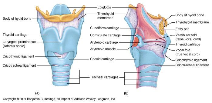
thyroid cartilage
The largest cartilage (hyaline cartilage) covered with a non-keratinized stratified squamous epithelium in the framework of the human larynx; it is attached to the hyoid bone; it is shield-shaped and has two broad processes which join anteriorly to form the protuberance on the front of the neck nicknamed the "Adam's apple" (which is more prominent in males), and is articulated below to the ring-like cricoid cartilage.
epiglottis
The thin elastic cartilaginous lid-like structure (elastic cartilage) located at the root of the tongue which folds posteriorly over to close the glottis to prevent food and liquid from entering the larynx and trachea while food or drink is passing through the pharynx during the act of swallowing or in preparation for a cough or sneeze; its movement is controlled by the Autonomic Nervous System.
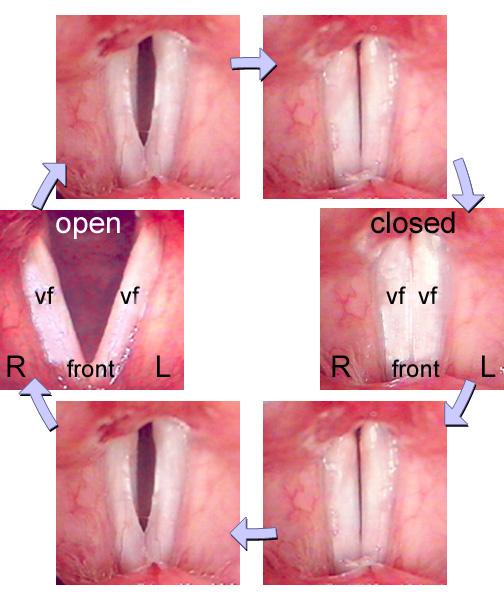
glottis
The opening between the vocal cords at the upper part of the larynx and the vocal apparatus of the larynx; it is protected by the epiglottis, which folds posteriorly over to close it to prevent food and liquid from entering the larynx and trachea while food or drink is passing through the pharynx during the act of swallowing.
cricoid cartilage
The second largest and lower most cartilage (hyaline cartilage) in the framework of the human larynx; it is narrow in front and high behind, where, being positioned within the thyroid cartilage, it is surmounted by the two arytenoid cartilages, from which the vocal cords pass forward to be attached together to the front of the thyroid cartilage. [Note: Adjacent to the cricoid cartilage and the first tracheal ring is the cricothyroid membrane, a site used for rapid emergency airway access (cricothyroidotomy).]
arytenoid cartilages
The pair of small pyramidal cartilages (hyaline cartilage) which articulate with the cricoid cartilage; the vocal cords and several skeletal muscles are attached to them; their movement produces tension or relaxation of the vocal cords resulting in a variety of sound pitches.
corniculate cartilages
The pair of small conical nodules of elastic cartilage which surmount the apex of each arytenoid cartilage.
cuneiform cartilages
The pair of small nonarticulating rods of elastic cartilage which lie in the internal mucosal wall of the the larynx somewhat superior to the corniculate cartilages.
vestibular folds
The pair of ridges of the mucous membrane stretching across the laryngeal cavity from the angle of the thyroid cartilage to the arytenoid cartilage; the enclose a space called the "false glottis;" they serve to protect the vocal folds beneath them from aspirated materials; they contribute to the closure of the glottis during swallowing or in preparation for a cough or sneeze. nickname - false vocal cords
vocal folds
The sharp edges of a pair of ridges of the mucous membrane overlying the vocal ligament and stretching along the laryngeal wall from the the angle between the lamina of the thyroid cartilage to the vocal process of the arytenoid cartilage; the movement of skeletal muscles attached to the arytenoid cartilages produces tension or relaxation of these tissue edges which then vibrate in a stream of expired air resulting in a variety of sound pitches; their movement is controlled by the Somatic Nervous System, especially the speech centers. nickname - true vocal cords
Diagram: vocal folds in action
laryngitis
Inflammation of the mucous membrane of the larynx, characterized by dryness and soreness of the throat, hoarseness or loss of voice, coughing and dysphagia (difficulty in swallowing).
1/2 List: The cartilages of the larynx, their functions,
Cartilages of the Larynx: epiglottis (epiglottal cartilage) lid-like structure located at the root of the tongue which folds posteriorly over to close the glottis to prevent food and liquid from entering the larynx and trachea while food or drink is passing through the pharynx during the act of swallowing or in preparation for a cough or sneeze.
thyroid cartilage
cricoid cartilage
corniculate cartilages
cuneiform cartilages form the framework of the larynx
arytenoid cartilages attachment points for the vocal cords and several skeletal muscles; their movement produces tension or relaxation of the vocal cords resulting in a variety of sound pitches
2/2 List: the other parts of the larynx involved in voice production.
Other Parts of the Larynx
vocal folds vibrate in the ventilation airstream resulting in a variety of sound pitches
skeletal muscles attached to the arytenoid cartilages produce tension or relaxation of the vocal cords resulting in a variety of sound pitches
trachealis muscle
A thin sheet of smooth muscle running the length of the posterior wall of the trachea, spanning the space between the open ends of the C-shaped cartilage rings which keep the trachea open; its contraction narrows the tracheal diameter (opening) to increase air velocity during a cough or sneeze
carina
The angle made between the two primary bronchi when they diverge at the tracheal bifurcation; it is richly innervated with sensory nerve endings to respond to the arrival of any aspirated material by initiating a cough reflex; it may be visualised as a ridge within the bronchial tree when using a bronchoscope.
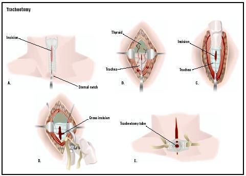
tracheotomy
A surgical operation which creates an opening into the trachea with a tube inserted to provide a passage for air; performed when the pharynx is obstructed by edema or cancer or other causes.
Diagram: Tracheotomy
endotracheal tube (intubation)
A procedure by which a flexible plastic tube is inserted through the mouth to go down into the trachea; the clinician inserts the tube with the help of a laryngoscope (an instrument which permits one to see down into the trachea, and even see the vocal cords); the purpose of endotracheal intubation is to permit air to pass freely to and from the lungs in order to ventilate the lungs; endotracheal tubes can be connected to ventilator machines to provide artificial positive pressure ventilation.
bronchial tree
The bronchi together with all their branches which deliver air to the alveoli of the lungs. The sequence of passages is as follows: trachea to main/R & L/primary bronchi (to lungs) to secondary/lobar bronchi (to lobes of lungs) to tertiary / segmental bronchi (to bronchopulmonary segment) to interlobular bronchi (to lobules) to primary/lobular bronchioles (no more cartilage supports) which branch to terminal bronchioles which branch to form respiratory bronchioles (to alveolar sacs) to alveolar ducts (to alveoli).
Diagram: respiratory tree
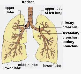
primary bronchi
The thin-walled, cartilaginous tubes, the first branches (right and left) of the trachea, which carry air to the lungs; their walls consist of a pseudostratified columnar ciliated epithelium, a loose fibrous connective tissue submucosa with submucosal glands, O-shaped hyaline cartilage rings, and smooth muscle.
lobar bronchi
The thin-walled, cartilaginous tubes, the first branches of the primary bronchi, which carry air to the individual lobes of the lungs; their walls consist of a pseudostratified columnar ciliated epithelium, a loose fibrous connective tissue submucosa thrown into irregular folds with submucosal glands, O-shaped hyaline cartilage rings, and smooth muscle; as the diameter of the bronchi decreases the size of the cartilage cross-sections also decreases.
segmental bronchi = secondary bronchi
The thin-walled, cartilaginous tubes, the first branches of the lobar bronchi, which carry air to the ~ten bronchopulmonary segments of each lung; their walls consist of a pseudostratified columnar ciliated epithelium, a loose elastic connective tissue submucosa thrown into irregular folds with submucosal glands, O-shaped hyaline cartilage rings, some diffuse lymphatic tissue and smooth muscle; as the diameter of the bronchi decreases the size of the cartilage cross-sections also decreases.
tertiary bronchi
The thin-walled, cartilaginous tubes, the first branches of the the segmental bronchi, which carry air to the lobules of each lung; their walls consist of a pseudostratified columnar ciliated epithelium, a loose elastic connective tissue submucosa thrown into irregular folds with submucosal glands, small O-shaped hyaline cartilage rings, some diffuse lymphatic tissue and smooth muscle; as the diameter of the bronchi decreases the size of the cartilage cross-sections also decreases.
bronchioles
The fine airways which constitute the passages produced by the 11th to 17th divisions of the bronchi within the lung parenchyma; they have luminal diameters from 0.5 mm to 0.2mm; they have no submucosal glands or hyaline cartilage rings within their walls which are lined by a a pseudostratified columnar ciliated epithelium; as the diameter of the bronchioles decrease, the lining epithelium changes to a simple columnar ciliated epithelium and then to a cuboidal ciliated epithelium; they have a prominent outer wall layer of smooth muscle cells capable of constricting the bronchiolar lumen in response to parasympathetic (vagal) inputs; sympathetic fibers dilate the bronchioles.
terminal bronchioles
The final divisions of the bronchioles which are lined by a cuboidal ciliated epithelium and only occasional smooth muscle fibers are observed in their walls; they carry air toward the alveoli.
respiratory bronchioles
The final divisions of the terminal bronchioles which are lined by a cuboidal ciliated epithelium and only occasional smooth muscle fibers are observed in their walls; their walls are interrupted by the presence of occasional alveolar sacs where gas exchange takes place; they divide into the alveolar ducts, which are the smallest branches of the bronchial tree; they carry air toward the alveoli.
ciliated pseudostratified columnar epithelium
The lining membrane of the majority of the branches of the bronchial tree, consisting of a tissue sheet which gives a superficial appearance of being in several layers because the cell nuclei are at different levels, but in which all cells reach the basement membrane, hence it is classed as a simple epithelium; on its apical surface is a brush border of hairlike projections which are the cell organelles known as cilia which are microtubluar motile structures which beat in a oar-like fashion to propel mucus secretions upward through the bronchial tree to the pharynx where they can be swallowed or otherwise expelled.
bronchoscopy
An examination used for inspection of the interior of the tracheo-bronchial tree, performance of endobronchial diagnostic tests, taking of specimens for biopsy and culture and removal of foreign bodies using a thin, flexible instrument (bronchoscope) which is a flexible plastic tube that can be inserted through the mouth to go down into the airways and which has optical devices and various accessories to perform the actions listed above.
List: In correct sequence the structures of the conducting portion of the respiratory system through which a gas molecule passes during inspiration.
nose/mouth → pharynx → larynx → trachea → primary bronchi → lobar bronchi → segmental bronchi = secondary bronchi → tertiary bronchi → bronchioles → terminal bronchioles → [ respiratory bronchioles* → alveolar sacs* → alveoli* -- *Note: these last three portions of the airway are not technically a part of the "conducting portion" of the airway because actual respiratory gas exchange occurs in these locations.]
Identify: The type of epithelial tissue lining each part of the conducting portion of the respiratory system and explain why this type of epithelium promotes the functions of its respective part.
1. nose/mouth → pharynx → larynx moist mucous membrane composed of non-keratinized stratified squamous epithelium provides protective barrier to separate underlying tissues from non-sterile air; contributes to the warming and humidifying of inspired air
2. trachea → primary bronchi → lobar bronchi → segmental bronchi = secondary bronchi → tertiary bronchi → bronchioles moist mucous membrane composed of pseudostratified ciliated columnar epithelium provides protective barrier to separate underlying tissues from non-sterile air; contributes to the warming and humidifying of inspired air; mucous secretions trap inspired microbes and dust particles; mucociliary elevator sweeps trapped microbes and dust particles upward to the pharynx to be swallowed and eliminated
3. bronchioles → terminal bronchioles moist mucous membrane composed of ciliated simple columnar/cuboidal/ in transition to unciliated squamous epithelium provides protective barrier to separate underlying tissues from non-sterile air; contributes to the warming and humidifying of inspired air; mucous secretions trap inspired microbes and dust particles; mucociliary elevator sweeps trapped microbes and dust particles upward to the pharynx to be swallowed and eliminated
4. respiratory bronchioles moist simple squamous epithelium provides a surface for gas exchange with air which should be clean and nearly sterile at this location
Identify: The structural divisions of the lung supplied by a primary bronchus, lobar bronchus, secondary bronchus, tertiary bronchus, and a terminal bronchiole.
*primary bronchus, entire lung
*primary bronchus, entire lung
*segmental bronchi = secondary bronchus, bronchopulmonary segment
*tertiary bronchus, lobule
*terminal bronchiole, respiratory bronchiole
pleura
The thin serous membrane, a simple squamous lining, which secretes pleural fluid and envelops each lung and folds back to make a lining for each pleural cavity.
parietal pleura
That portion of the thin serous membrane, a simple squamous lining, which secretes pleural fluid and makes a lining for the thoracic wall of the pleural cavity.
visceral pleura
That portion of the thin serous membrane, a simple squamous lining, which secretes pleural fluid and envelops the lung within the pleural cavity.
pleural cavity
A subdivision of the thoracic cavity; the potential space which lies in between the visceral pleura and the parietal pleura and contains a small amount of lubricating serous fluid, the pleural fluid.
base
The broad inferior surface of the lung which faces the diaphragm.
apex
The tip or pointed superior end of the lung.
costal surface
`The broad lateral surface of the lung which faces the ribs and the thoracic wall of the pleural cavity.
mediastinal surface
The broad medial surface of the lung which faces the mediastinal wall of the pleural cavity; it is the location of the hilus of the lung.
hilus
The depression or fissure where vessels, nerves, and a primary bronchus enters each lung on its mediastinal surface.
cardiac notch
The lateral deflection of the anterior border of the left lung which is produced to accommodate the space taken up by the heart.
oblique fissure
The deep groove, roughly parallel to the sixth rib, which divides the lung on both sides into upper and lower lobes; along this surface, visceral pleura apposes visceral pleura; both surfaces are smooth and separated by a layer of lubricant fluid which allows individual lobes to move freely with respect to one another.
horizontal fissure
A recessed groove in the parenchyma of the right superior lobe of the lung which divides the right-sided volume of lung tissue above the oblique fissure into the right superior lobe, above the fissure, and the right middle lobe, below the fissure; along this surface, visceral pleura apposes visceral pleura; both surfaces are smooth and separated by a layer of lubricant fluid which allows individual lobes to move freely with respect to one another.
lobe
The anatomical term for a major division of an organ; there is usually more than one lobe making up the object; lobes may be separated by clefts or fibrous septa; examples of organs with lobes include the brain and lung; air is delivered to each lobe of the lung by a secondary bronchus.
bronchopulmonary segment
The anatomical term for a major subdivision of the lobes of each lung; they are separated by fibrous septa; air is delivered to each bronchopulmonary segment of the lung by a tertiary bronchus; in humans, each lung consists of ~10 bronchopulmonary segments.
lobule
A subdivision of an organ; a lobe is often divided up into many lobules; examples of organs with lobules include the lung, liver and breast; air is delivered to each lobule of the lung by an interlobular bronchus.
alveolar duct
The terminal branch(es) of each respiratory bronchiole in the lungs, lined by a simple squamous epithelium, which deliver inspired air to the alveolar sacs.
alveolar sac
A cluster of alveoli located at the end of each alveolar duct in the lungs; each of the alveoli is a tiny, thin-walled, capillary-rich sac, lined by a simple squamous epithelium, where the exchange (diffusion) of oxygen and carbon dioxide (and other less important gases) takes place.
alveolus
A tiny, thin-walled, capillary-rich sac in the lungs, lined by a simple squamous epithelium, where the exchange of oxygen and carbon dioxide (and other less important gases) takes place; the approximately 300 million alveoli have a total cross-sectional area of 50- 70 m2, which equals ~40 times the surface area of the skin. aka - air sac (2) A tooth socket in the jawbone.
type I alveolar cell
The squamous epithelial cell forming the alveolar wall and through whose cell membranes and cytoplasm the exchange of oxygen and carbon dioxide (and other less important gases) takes place; collectively they cover 95% of the alveolar surface area.
type II alveolar cell
The cuboidal cell found as a minor component of the alveolar wall, they are scattered among the type I cells and account for only 5% of the alveolar surface; they have three main functions: (1) secretion of surfactant, (2) control of alveolar fluid levels by water recapture using active sodium transport to return excess alveolar surface water to the interstitial fluid, and (3) the stem or progenitor cell which can proliferate to replace both type I and type II cells after injury.
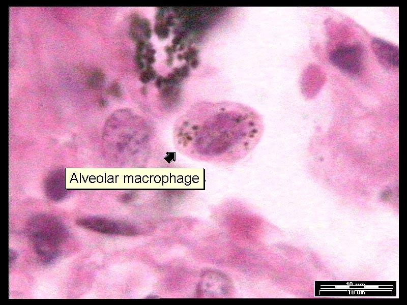
alveolar macrophage = dust cell
The large mobile cell, derived from the monocyte, occurring in low numbers within the connective tissue of the alveolar walls and, more commonly, patrolling on the walls of the alveoli, which ingests by phagocytosis any foreign particles and infectious microorganisms which reach the alveoli; they may also transport indigestable materials such as soot or silica dust to the lymph nodes of the lungs for storage.
Diagram: alveolar macrophage
alveolar-capillary membrane = respiratory membrane
The collective term for all the structural components through which respiratory gases must diffuse in order to be exchanged between the air in the alveoli and the blood; this consists of the cell membranes and cytoplasm of the type I alveolar cells, the alveolar epithelial basement membrane, the very limited amount of loose fibrous connective tissue present, if any, beneath it, the capillary basement membrane, and the cell membranes and cytoplasm of the capillary endothelial cells.
alveolar wall
The anatomical term for all the structural components through which respiratory gases must diffuse in order to be exchanged between the air in the alveoli and the blood; this consists primarily of the type I alveolar cells (a minority of the type II alveolar cells and alveolar macrophages are also present), the alveolar epithelial basement membrane, the very limited amount of loose fibrous connective tissue present, if any, beneath it, the capillary basement membrane, the capillary endothelial cells, and the blood passing through the capillaries.
epithelial basement membrane
The thin microscopic network of fibrous proteins synthesized by the basal layer of any epithelial tissue which forms a physical foundation and a physiological barrier between the epithelial tissue and the underlying connective tissue.
capillary basement membrane
The thin microscopic network of fibrous proteins synthesized by the endothelial cells of capillaries which forms a physical foundation and a physiological barrier between the endothelial cells and the underlying tissue.
endothelial cell
Any of the simple squamous cells which form a single layer and line the walls of the heart, blood vessels, and lymphatic vessels; this tissue arises from mesoderm.
simple squamous epithelium
Tissues with a high degree of cellularity and limited extracellular material, connected with specialized contact structures such as desmsomes, organized in a single layer in which the cells present are flattened (cells wider than they are tall with reference to the basement membrane), which have no direct blood supply and which are derived from embryonic ectoderm or endoderm.
surface tension
That property, due to unbalanced molecular cohesive forces, which exists in the surface film of all liquids and tends to bring the contained volume into a form having the least superficial area and where the surface tends to contract and has properties resembling those of a stretched elastic membrane; the thickness of this film, amounting to less than a thousandth of a millimeter, is considered to equal the radius of the sphere of molecular action, that is, the greatest distance at which there is cohesion between two particles; particles lying below this film, being equally acted on from all sides, are in equilibrium as to forces of cohesion, but those in the film are on the whole attracted inward, and tension results.
List: The four factors that affect pulmonary gas exchange (external respiration) across the respiratory membrane.
(1) the surface area, thickness, and other structural components of the moist respiratory membranes available for gas diffusion
(2) the partial pressures of the gases oxygen and carbon dioxide in the alveloar air
(3) the gas solubilities (solubility coefficients) of the gases oxygen and carbon dioxide
(4) the physiological relationship between ventilation (bulk air movements into and out of the alveoli) and perfusion (bulk flow of blood through the alveolar capillary beds)
List: The five cell types found in the walls of an alveolus and a function for each cell.
*type I alveolar cell form the alveolar wall (95%) and permit gas exchange
*type II alveolar cell (1) secretion of surfactant, (2) control of alveolar fluid levels by water recapture using active sodium transport to return excess alveolar surface water to the interstitial fluid, and (3) the stem or progenitor cell which can proliferate to replace both type I and type II cells after injury
*alveolar macrophage = dust cell phagocytosize any foreign particles and infectious microorganisms which reach the alveoli; they may also transport indigestable materials such as soot or silica dust to the lymph nodes of the lungs for storage
*capillary endothelial cell form the capillary wall and permit gas exchange
*erythrocyte = RBC essential for oxygen transport and, to a lesser degree, carbon dioxide transport within the blood
Sketch and label: The respiratory membrane. Describe the kinds of cells that make up this membrane and their functions.

Diagram: mechanics of breathing
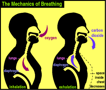
inspiration
The act of drawing in, inhaling, air into the lungs, using the external intercostals and, if necessary, the diaphragm, to increase the volume of the pleural cavities to create a favorable negative pressure gradient; this action is normally under autonomic control by means of the inspiratory area of the medullary rhythmicity area.
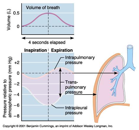
intrapleural pressure
The pressure (i.e., the force applied uniformly against the pleural cavity walls) within the pleural cavity, which, in life, is always negative; this negative force acts as a suction to keep the lungs inflated; the negative intrapleural pressure is due to the surface tension of the alveolar fluid, the elasticity of the lungs, and the elasticity of thoracic wall; changes in the intrapleural pressure contribute to the movement of air during ventilation; the intrapleural pressure between breathes is less than atmospheric pressure by about 4 mm Hg. aka - intra-thoracic pressure
intra-alveolar pressure
The pressure (i.e., the force applied uniformly against the alveolar walls) within the alveoli; when this pressure is less than atmospheric pressure, air will move into the alveoli; when this pressure is greater than atmospheric pressure, air will move out of the alveoli.
transpulmonary pressure
The difference in pressures (i.e., pressure gradient) between intra-alveolar pressure and intrapleural pressure during an inspiration or an expiration.
Inspiration and Expiration intra-alveolar pressure, intrapleural pressure, and differences in pressures
Inspiration, Expiration
Intra-Alveolar Pressure* -3.0 mm Hg, +3.0 mm Hg
Intrapleural Pressure* -6.0 mm Hg, -3.0 mm Hg
Difference in Pressures*=Inspiration[-3-(-6) = +3]
Expiration[+3-(-3) = +6]
[* Note: All pressures in the table are compared to the atmospheric pressure at whatever location.]
expiration
The act of forcing out, exhaling, air from the lungs, using the internal intercostals and the natural elastic recoil of the lungs, chest wall, and the relaxing diaphragm (and gravity), to decrease the volume of the pleural cavities to create a favorable positive pressure gradient; this action is normally under autonomic control by means of the expiratory area of the medullary rhythmicity area.
elastic recoil
The ability of the lungs and the alveoli to return to their original volume and shape passively as a result of the flexible ductile strength of the elastic tissue in the alveolar, lobular, and lobar walls of the lungs; this ability promotes passive lung constriction and outward air flow during expiration and contributes to the always negative intrapleural pressure; any loss of elastic recoil will require increased muscular assistance with exhalation.
diaphragmatic breathing
Ventilation produced chiefly by movements of the diaphragm and abdominal muscles, i.e., inspiration using the diaphragm and expiration using the abdominal muscles.
costal breathing
Ventilation produced chiefly by movements of the ribs, i.e., inspiration using the external intercostals and expiration using the internal intercostals.
compliance
The ability of the alveoli and lung tissue to expand on inspiration; in clinical terms it is defined as the volume increase in the lungs per unit increase in the lung pressure; while clearly not a complete description of the pressure-volume properties of the lung, it is nevertheless useful in practice as a measure of the comparative stiffness of the lung; the stiffer the lung, the less the compliance; compliance is reduced by diseases which cause an accumulation of fibrous tissue in the lung or by edema in the alveolar spaces; it is increased in pulmonary emphysema and also with age, probably because of alterations in the elastic tissue in both cases.
airway resistance
The measure of the ease with which air flows through tubular respiratory structures; higher resistance occurs in smaller tubes such as bronchioles and alveoli which have not emptied properly; it is a ratio of pressure to flow (R=V/I); thus, for the determination of airway resistance, intra alveolar pressure and airflow measurements are required.
pulmonary perfusion
The injection or pumping of blood into the pulmonary circulation in order to reach the lungs and alveolar capillary beds to supply lung tissues with nutrients and permit the exchange of oxygen and carbon dioxide between the alveolar air and the blood; the actual rate is dependent on cardiac output and appropriate flow through the pulmonary circulation.
Describe:The pulmonary ventilation pressure changes during inspiration and expiration.
Pulmonary pressure changes during ventilation are quite modest. Intrapulmonary pressure (in the alveolar spaces of the lungs) varies by only a mm or Hg or two from atmospheric pressure under most circumstances. During inspiration, the intrapulmonary pressure (in the alveolar spaces of the lungs) is further reduced, by 2 or 3 mm Hg compared to atmospheric pressure, when thoracic cavity (pleural space) volume is expanded by the elevation of the ribs by the contraction of the external intercostals and, perhaps, by the contraction and depression of the diaphragm. During expiration the intrapulmonary pressure (in the alveolar spaces of the lungs) is slightly increased, by 2 or 3 mm Hg compared to atmospheric pressure, when thoracic cavity (pleural space) volume is reduced by the elastic recoil of the ribs and the action of gravity and, perhaps, by the contraction of the internal intercostals and, if needed, the abdominals.
Diagram: Pulmonary Pressure Gradient
Describe:The names and actions of the muscles involved in ventilation (external respiration).
Inspiration external intercostals elevate ribs and therefore increase the volume of the pleural cavities
diaphragm depress the inferior wall of the thoracic cavity and, therefore, increase the volume of the pleural cavities
Expiration internal intercostals compress and lower the ribs and therefore decrease the volume of the pleural cavities
abdominals compress the abdominal cavity which elevates the abdominal organs and passively elevates the diaphragm and, therefore, decreases the volume of the pleural cavities
partial pressure (of a gas)
The pressure, i.e., the force applied uniformly against a container's surface, that one component gas of a mixture of gases would exert if it were alone, i.e., occupied the same volume as the gas mixture, in a container and was at the same temperature. See Dalton's Law.
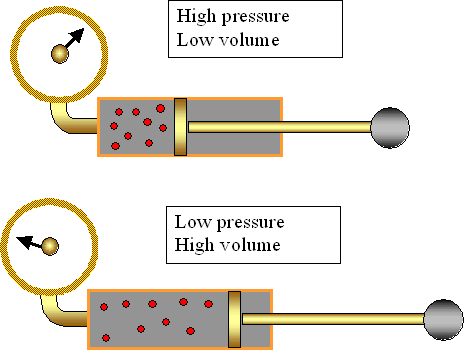
Boyle's law
The principle that at a constant temperature the volume of a confined ideal gas varies inversely with its pressure.
Diagram: Boyle's law
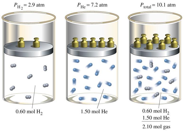
Dalton's Law
The principle that the pressure, i.e., the force applied uniformly against a container's surface, exerted by a mixture of gases equals the sum of the partial pressures of the gases in the mixture; the pressure of a gas in a mixture equals the pressure it would exert if it occupied the same volume alone at the same temperature.
Diagram Dalton's Law
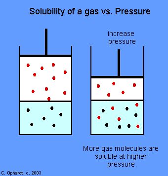
Henry's Law
The principle that when a mixture of gases comes into contact with a liquid, a gas will dissolve into the liquid in proportion to its partial pressure and its solubility coefficient, and will diffuse until equilibrium is achieved (gases diffuse into and out of liquids from high to low partial pressure).
Diagram: Henry's law
List: The gases, nitrogen (N2), oxygen (O2), and carbon dioxide (CO2), in order from largest to smallest solubility coefficient, i.e., from most to least soluble in water.
Largest Solubility Coefficient, i.e., Most Soluble In Water
carbon dioxide (CO2)
oxygen (O2)
nitrogen (N2)
Smallest Solubility Coefficient, i.e., Least Soluble In Water
List: The gases, nitrogen (N2), oxygen (O2), and carbon dioxide (CO2), in order from largest to smallest partial pressure in the atmosphere.
Largest Partial Pressure In The Atmosphere
nitrogen (N2)
oxygen (O2)
carbon dioxide (CO2)
Smallest Partial Pressure In The Atmosphere
Describe: The effects of atmospheric pressure on the partial pressures of the gases nitrogen (N2), oxygen (O2), and carbon dioxide (CO2), at:
(a) sea level, (b) at high altitudes, and (c) under water during deep diving.
The change in the effect of atmospheric pressure on the partial pressures of these three gases, nitrogen (N2), oxygen (O2), and carbon dioxide (CO2), is the same.
Sea Level: standard conditions (normal)
High Altitude: decreased
Under Water During Deep Diving: increased
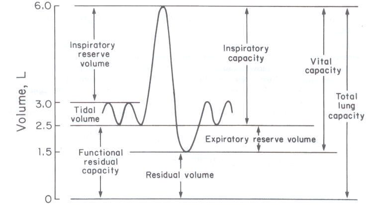
spirometer
An instrument for measuring the volume of air entering and leaving the lungs and recording the volumes moved during different types of breathing on a graph.
spirogram
A record of breathing made with a spirometer.
tidal volume
The amount of air moved in and out of the lungs with each relaxed or resting breath; usually about 0.5 L; ~70% of this volume reaches the respiratory section of the lungs, and ~30% remains in the conducting section of the lungs (the anatomic dead space).
anatomic dead space
The total volume of the conducting airways from the nose or mouth down to the level of the terminal bronchioles; normally it is ~0.15 L (~30% of a tidal volume); this air is "wasted" in the sense that it does not participate in gas exchange.
minute volume of respiration
The total volume of air ventilated over a period of one minute.
alveolar ventilation rate
The portion of the minute volume of respiration/ventilation which reaches those areas of the lung concerned with gas exchange; normally it is ~3.5 to 5.0 liters per minute; this measure of external respiratory function is, perhaps, the best criterion for effectiveness of breathing.
inspiratory reserve volume
The amount of air which can be taken into the lungs, beyond one's tidal volume, using a forced inspiration; normally it is ~3.0 L of air.
expiratory reserve volume
The amount of air which can be forced out of the lungs by contracting the chest and abdominal muscles beyond tidal volume ventiliation; normally it is ~1.5 L.
forced expiratory reserve volume
The volume of air which can be expelled from the lungs in a prescribed amount of time (FEV1 = for 1 second, FEV2 = for 2 seconds, FEV3 = for 3 seconds, etc.) with a maximal effort following a maximal inhalation; in cases of COPD, (Chronic Obstructive Pulmonary Disease), e.g., in cases of asthma and emphysema, the FEV is low, while the total lung capacity, functional residual capacity, and residual volumes are high; in restrictive pulmonary diseases, the vital capacity, total lung capacity, functional residual capacity and residual volume are low due to the inability of the lungs to expand.
residual volume
The amount of air remaining in lungs even after deepest possible expiration; normally it is ~1.0 L; (it cannot be directly measured by spirometry); this "dead air" decreases the efficiency of gas exchange by diluting the oxygen of the inspired air; however, this "dead air" keeps the alveoli from collapsing between breaths.
inspiratory capacity
The maximum volume of air which can be inspired into the lungs after the end of a tidal or resting exhalation; normally it is ~3.5 L; it is equivalent to the sum of the inspiratory reserve volume and the tidal volume.
functional residual capacity
The amount of air remaining in lungs after the end of a tidal or resting exhalation; normally it is ~1.5 L; it is equivalent to the sum of the expiratory reserve volume and the residual volume.
vital capacity
The maximum volume of air that can be moved in and out of the lungs during a single breath; normally it is ~5.0 L; it is equivalent to the sum of the tidal volume, the inspiratory reserve volume, and the expiratory reserve volume; it varies depending on body size, fitness level, and degree of health or disease.
total lung capacity
The total amount of air contained in the lungs after the deepest possible inhalation; normally it is ~6.0 L; it is equivalent to the sum of the tidal volume, the inspiratory reserve volume, the expiratory reserve volume, and the residual volume; it varies depending on body size, fitness level, and degree of health or disease.
3.6 L to 9.4 L in an adult male.
2.5 L to 6.9 Ls in an adult female.
The Volumes and Capacities Derived from Spirometry:

SPIROMETRY EQUATIONS FOR RESPIRATORY CAPACITIES:
Total Lung Capacity = Inspiratory Reserve Volume (IRV) + Tidal Volume (TV) + Expiratory Reserve Volume (ERV) + Residual Volume (RV)
Vital Capacity = Inspiratory Reserve Volume (IRV) + Tidal Volume (TV) + Expiratory Reserve Volume (ERV)
Inspiratory Capacity = Inspiratory Reserve Volume (IRV) + Tidal Volume (TV)
Functional Residual Capacity = Expiratory Reserve Volume (ERV) + Residual Volume (RV)
List:The lung volumes and capacities. Distinguish between a lung volume and a lung capacity.
Lung Volumes (measured):tidal volume, inspiratory reserve volume, expiratory reserve volume, residual volume
Lung Capacities (calculated):total lung capacity, vital capacity, inspiratory capacity, functional residual capacity
The lung volumes are measured variable using spirometry or other techniques while lung capacities are calculated variables obtained by summing the appropriate measured lung volumes.
Sketch and label: A spirogram.
oxygen = O
A nonmetallic element comprising 21% of the atmosphere by volume which occurs as a colorless odorless tasteless nonflammable diatomic gas, O2, and in many compounds such as water and iron ore; it combines with most elements, is essential for plant and animal respiration, and is required for nearly all combustion; atomic number 8; atomic weight 15.9994.
carbon monoxide = CO
A colorless, odorless, highly poisonous gas formed by the incomplete combustion of carbon or a carbonaceous material, such as gasoline; its toxicity is due to its greater affinity for hemoglobin than oxygen such that it greatly reduces effective oxygen transport in the blood.
carbon dioxide = CO2
A colorless, odorless, incombustible gas formed during respiration, combustion, and organic decomposition and used in food refrigeration, carbonated beverages, inert atmospheres, fire extinguishers, and aerosols; it combines spontaneously with water to form carbonic acid.
surface area
The extent of a 2-dimensional covering of a 3-dimensional object, or that portion of it enclosed within a boundary.
diffusion distance
The extent of space between two points, structures or surfaces over which intermingling molecules or particles in solution spread as a result of spontaneous random thermal agitation.
solubility
The relative amount of a substance which can be dissolved in a given amount of solvent; the quality, condition, or degree of a solute being soluble in a solvent, usually expressed as a decimal fraction.
oxygen transport
The general term for the set of physiological mechanisms used to carry oxygen from the atmospheric air to the mitochondria of every cell in the body; this involves bulk transport (ventilation, circulation) and diffusion (in alveoli, across the alveolar membrane into the erythrocytes and from the capillary beds through the interstitial fluid and into the cell cytoplasm and ultimately to the mitochondria); in a more restricted sense, the term implies the means by which oxygen is carried within the blood: 1.5% of the O2, is dissolved directly in the water of plasma while the remaining 98.5% is carried bound to hemoglobin within the erythrocytes.
List: The chemical forms in which oxygen and carbon dioxide are transported in the blood.
oxygen (1) gas molecules dissolved in the water of plasma and (2) non-covalently bound to hemoglobin (oxyhemoglobin)
carbon dioxide (1) gas molecules dissolved in the water of plasma and (2) non-covalently bound to hemoglobin (carbaminohemoglobin) and (3) chemically transformed into the bicarbonate ion (HCO3-)
List: The form of the substance, carbon dioxide (CO2), in order from largest to smallest fraction being transported in blood in its three transport forms.
Largest Fraction
bicarbonate ion, HCO3- dissolved in water (~70%)
carbaminohemoglobin (bound to hemoglobin: ~20-23%)
gaseous CO2 dissolved in water (7-10%)
Smallest Fraction
hypoxia
A clinically significant deficiency (below physiological levels) in the amount of oxygen reaching body tissues, usually despite adequate perfusion of the tissue by blood; the most common symptom of hypoxia is cyanosis, a bluish cast to the skin, lips and/or fingernails.
hypoxic hypoxia
A clinically significant deficiency (below physiological levels) in the amount of oxygen reaching body tissues due to low O2 in the atmosphere (altitude, smoke inhalation, etc.) or suffocation/strangulation.
anemic hypoxia
A clinically significant deficiency (below physiological levels) in the amount of oxygen reaching body tissues due to reduced numbers of, or defective erythrocytes, e.g., any anemia, other hemolytic diseases, cancers and cancer treatments, malnutrition, etc.
ischemic (stagnant) hypoxia
A clinically significant deficiency (below physiological levels) in the amount of oxygen reaching body tissues due to inadequate perfusion of the tissue by blood, e.g., from heart failure, vessel obstructions, e.g., blood clot or other embolus, etc.
histotoxic hypoxia
A clinically significant deficiency (below physiological levels) in the amount of oxygen reaching body tissues due to interference with O2 use in the mitochondria of the tissue cells, despite adequate perfusion of the tissue by blood; usually due to the presence of toxins or poisons, e.g., cyanide (cigarettes, chemicals), carbon monoxide (CO) (cigarettes, fires, automobile exhaust, etc.), botulinin toxin, etc.
List:The four clinical types of hypoxia and an example of each.
1.hypoxic hypoxia high altitude
smoke inhalation
suffocation/strangulation.
2.anemic hypoxia iron-deficiency anemia
pernicious anemia
hemorrhagic anemia
hemolytic anemia
aplastic anemia
sickle cell anemia
3.ischemic (stagnant) hypoxia heart failure
atherosclerotic vessel disease
vessel obstructions, e.g., blood clot
embolus
4.histotoxic hypoxia toxins or poisons, e.g., cyanide (cigarettes, chemicals), carbon monoxide (CO) (cigarettes, fires, automobile exhaust, etc.), botulinin toxin, etc.
ANS Control of Breathing
ANS Control of Breathing

respiratory center
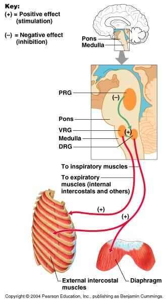
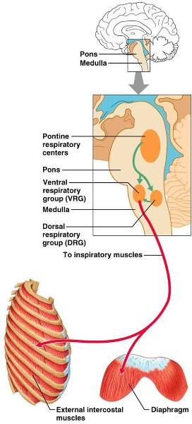
The series of paired and functionally related autonomic nuclei located bilaterally in the reticular formation of the brain stem; this control center consists of the medullary rhythmicity area (containing the dorsal respiratory group (DRG) (formerly the inspiratory area) and the ventral respiratory group (VRG) = (formerly the expiratory area) and the pontine respiratory center (formerly the pneumotaxic and the apneustic areas); these collections of neurons cooperate to regulate the rate and depth of breathing as an involuntary unconscious activity in response to the physiological needs of the body for O2 and CO2 exchange and for blood acid-base balance.
medullary rhythmicity area
A collection of neurons in the reticular formation within the medulla oblongata involved in establishing or modifying the pattern for breathing; within this area are two key components: (1) the ventral respiratory group (VRG) (formerly the inspiratory area) which autorhythmically stimulates spontaneous ventilation, resting or tidal breathing (eupnea), and (2) the dorsal respiratory group (DRG) (formerly the expiratory area) which responds to situations beyond those of the resting or tidal breathing (eupnea) to alter the pattern for ventilation in response to the physiological needs of the body for O2 and CO2 exchange and for blood acid-base balance.
dorsal respiratory group (DRG) (formerly the inspiratory area)
The collection of motor neurons forming nuclei within the dorsal portion of the medullary rhythmicity area of the reticular formation within the medulla oblongata which are involved in altering the pattern for ventilation in response to the physiological needs of the body for O2 and CO2 exchange and for blood acid-base balance; these neurons stimulate neurons in the ventral respiratory group (VRG) to achieve those effects; they are responsive to sensory information from chemoreceptors and mechanoreceptors.
ventral respiratory group (VRG) (formerly the expiratory area)
The collection of autorhythmic motor neurons forming nuclei within the ventral portion of the medullary rhythmicity area of the reticular formation within the medulla oblongata; this group contains both inspiratory and expiratory neurons; the inspiratory neurons stimulate the diaphragm and external intercostals for approximately 2 seconds to cause inspirations and then the antagonistic expiratory neurons fire for approximately 3 seconds to permit passive or stimulate active expirations; thereby inspiratory and expiratory neurons cooperate in a negative feedback control relationship, setting the basic rhythm of respiration (spontaneous ventilation, resting or tidal breathing (eupnea)); VRG neurons may be influenced by the dorsal respiratory group (DRG) for ventilations in situations other than eupnea.
pontine respiratory center (formerly pneumotaxic and apneustic areas)
A collection of neurons in the reticular formation within the pons which limit inspiratory duration by sending inhibitory signals to the medullary rhythmicity area reducing duration of inspiratory impulses causing shorter cycles which increases ventilation rate; these pontine respiratory neurons receive input from higher brain centers and peripheral receptors, and their output fine tunes the breathing rhythm during activities such as speaking, sleeping, or exercising.
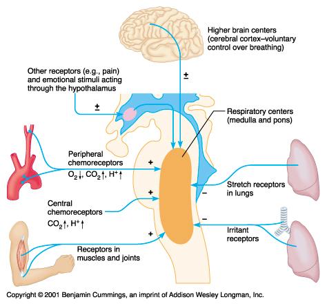
cortical influences
The action of higher, "conscious" centers in the cerebral cortex which permit voluntary control of ventilation by interacting with and over-riding the autonomic centers in the medullary rhythmicity area; examples include the control of ventilation during speech and singing, as well as deliberate forceful inspirations, expirations, or attempts at breath holding; pain and certain emotional states may also influence the rate and depth of ventilation in this fashion.
central chemoreceptors
A sensory neuron located within the CNS, usually in the brain stem or hypothalamus, which responds to chemical stimuli; a type of enteroreceptor sensitive to concentration changes of a variety of molecules in the blood or cerebrospinal fluid.
peripheral chemoreceptors
A sensory neuron located outside the CNS, usually in the wall of a blood vessel (e.g., the aortic body, the carotid body, the juxtaglomerular apparatus) which responds to chemical stimuli; a type of enteroreceptor sensitive to concentration changes of a variety of molecules in the blood or other body fluids.
carotid body
A group of peripheral chemoreceptors located near the bifurcations of the carotid arteries which monitor changes in the oxygen and CO2 content and pH of the blood and rely that sensory information to the hypothalmus and brain stem to help them control cardiovascular and respiratory functions; other cells in the carotid body respond to blood temperature and to certain chemicals, e.g., nicotine and cyanide.
aortic body
A group of peripheral chemoreceptors located in the arch of the aorta which monitor changes in the oxygen and CO2 content and pH of the blood and rely that sensory information to the hypothalmus and brain stem to help them control cardiovascular and respiratory functions.
baroreceptors
A type of mechanoreceptor; a specialized sensory end organ or sensory neuron which responds to mechanical stimuli such as tension (stretching) in the wall of a blood vessel or other tubular organ; important sensors in the regulation of blood pressure.
inflation reflex
A relatively rapid and predictable motor response by the skeletal muscles responsible for ventilation which occurs when stretch receptors in the visceral pleura, bronchioles, and alveoli are stimulated by being stretched; the motor response helps regulate the depth of breathing; over-stretching causes apnea, bronchodilation, increased heart rate and peripheral vasoconstriction.
Chemoreceptor Control of Breathing
Describe: The control of respiration (ventilation) by the respiratory center of the brain.
The ventral respiratory group (VRG) and the dorsal respiratory group (DRG) within the medullary rhythmicity area cooperate to establish the pattern for spontaneous ventilation and basal rate of ventilation which may be adjusted by impulses from related respiratory control centers in the pons; the ventral respiratory group (VRG) contains both inspiratory and expiratory neurons; the autorythmic inspiratory neurons stimulate the diaphragm and external intercostals for approximately 2 seconds to cause inspirations and then the antagonistic expiratory neurons fire for approximately 3 seconds to permit passive or stimulate active expirations; thereby inspiratory and expiratory neurons cooperate in a negative feedback control relationship, setting the basic rhythm of respiration (spontaneous ventilation, resting or tidal breathing (eupnea)); the dorsal respiratory group (DRG) neurons are involved in altering the pattern for ventilation in response to the physiological needs of the body for O2 and CO2 exchange and for blood acid-base balance; these neurons stimulate neurons in the ventral respiratory group (VRG) to achieve those effects; they are responsive to sensory information from chemoreceptors and mechanoreceptors.
DIagram: The control of respiration (ventilation) by the respiratory center of the brain.
Describe: The cortical, chemical, and neural influences on the respiratory center of the brain.
Cortical Influences voluntary controls: conscious decisions to change the rate and depth of breathing associated with speaking, singing, coughing, etc.
Chemical Influences changing levels of oxygen, carbon dioxide, hydrogen ion and bicarbonate ion in the blood are detected and this visceral sensory information is routed to the hypothalamus and medulla where it stimulates or inhibits the action of components of the respiratory center of the brain
Neural Influences (a) the medullary rhythmicity center with its authorythmic inspiratory and expiratory neurons sets the basal ventilation rate adjusted to the level of activity and metabolic demands of the body at any given moment
(b) the pontine respiratory center neurons limit inspiratory duration by sending inhibitory signals to the medullary rhythmicity area reducing duration of inspiratory impulses causing shorter cycles which increases ventilation rate; these pontine respiratory neurons receive input from higher brain centers and peripheral receptors, and their output fine tunes the breathing rhythm during activities such as speaking, sleeping, or exercising.
(c) proprioceptive stretch receptors in the lungs, pleura, and thoracic wall convey information about the degree of the filling of the lungs and overfilling will cause a reflex decrease in the strength of inspirations
Diagram:The cortical, chemical, and neural influences on the respiratory center of the brain.
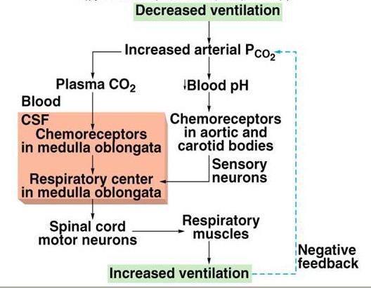
Sketch and label:The negative feedback pathway illustrating how the respiratory center of the brain that controls breathing by as a function of oxygen, carbon dioxide, and acidity levels of the blood.

Increased pO2, decreased pCO2, and a drop in [H+]* (alkalosis) all interact to discourage ventilation to retain more CO2 and, therefore, to restore normal CO2 levels.
Decreased pO2, increased pCO2, and a rise in [H+]* (acidosis) all interact to encourage ventilation to blow off more CO2 and, therefore, to restore normal CO2 levels.
[*Note: CO2 levels and acidity levels are directly proportional; when one rises the other rises, and vice versa.]
hypercapnia
A condition marked by an unusually high concentration of carbon dioxide in the blood as a result of hypoventilation; the additional CO2 may lead to respiratory acidosis.
hyperventilation
Abnormally fast or deep respiration, which results in the loss of carbon dioxide from the blood, thereby causing a fall in blood pressure, tingling of the extremities, and light-headedness, dizziness, and sometimes fainting and chest pain if continued; the loss of CO2 may lead to respiratory alkalosis.
hypocapnia
A condition marked by an unusually low concentration of carbon dioxide in the blood as a result of hyperventilation; the loss of CO2 may lead to respiratory alkalosis.
hypoventilation
Abnormally slow or shallow respiration, which results in the retention of carbon dioxide in the blood, the retention of CO2 may lead to respiratory acidosis.
asthma
A chronic respiratory disease (one of the COPDs), often arising from allergies, which is characterized by sudden recurring attacks of labored breathing, accompanied with a wheezing sound, a sense of chest constriction, spasmodic paradoxical contraction of the bronchi, and coughing and expectoration; changes in temperature or humidity, upper respiratory infections, exercise, stress or smoke (cigarette) can exacerbate the symptoms; treatments include bronchodilator drugs (oral and inhaled), and corticosteroids for more difficult cases.
pneumothorax
An accumulation of air or gas in the pleural cavity, occurring as a result of disease or injury, or sometimes induced to collapse the lung in the treatment of tuberculosis and other lung diseases; symptoms include shortness of breath, and severe one-sided (the affected side) chest pain on inhalation.
eupnea
Normal, unlabored breathing; the type observed in a normal individual under resting conditions; the normal resting adult respiratory rate is 12-20 breaths/minute.
apnea
A temporary absence or cessation of breathing.
dyspnea
Any difficulty in breathing, often associated with lung or heart disease and resulting in shortness of breath. nickname - air hunger
tachypnea
An abnormally rapid, usually shallow, respiratory rate; the clinically significant form of hyperventilation, which results in the loss of carbon dioxide from the blood, thereby causing a fall in blood pressure, tingling of the extremities, and light-headedness, dizziness, and sometimes fainting and chest pain if continued; the loss of CO2 may lead to respiratory alkalosis.
aspiration
(1) Expulsion of breath in speech. (2) The process of removing fluids or gases from the body with a suction device. (3) The act of inhaling fluid or a foreign body into the bronchi and lungs, often after vomiting.
atelectasis
The total or partial collapse of an expanded lung or a lobe of a lung, usually due to an obstruction of a bronchus by a mucus plug, infection, or cancer; also, the failure of the pulmonary alveoli to expand at birth; symptoms include low-grade fever, dry cough, chest pains and mild shortness of breath.
scuba
A portable apparatus containing compressed air and used for breathing under water; it is an acronym for self-contained underwater breathing apparatus.
nitrogen narcosis
A condition of confusion or stupor resulting from increased levels of dissolved nitrogen in the blood, potentially occurring in deep-sea divers breathing air under high pressure.
decompression sickness
A disorder, seen especially in deep-sea divers, caused by the formation of nitrogen bubbles (air embolism) in the blood and tissues following a sudden drop in the surrounding pressure, as when ascending rapidly from a dive, and characterized by severe pains in the joints and chest, skin irritation, cramps, and paralysis. aka - caisson disease; nickname - the bends
hyperbaric oxygenation
To treat, or infuse with oxygen at pressures higher than normal atmospheric pressure, e.g., in a hyperbaric chamber; it is a treatment for decompression sickness, carbon monoxide poisoning and for some burn patients receiving skin grafts.
bronchitis
Any chronic or acute inflammation of the mucous membrane of the bronchial tubes, usually secondary to infection (often viral), characterized by mucus-producing, i.e., "productive" cough.
emphysema
A common pathological condition of the lungs marked by an abnormal increase in the size of the air spaces, resulting in labored breathing and an increased susceptibility to infection; it can be caused by irreversible expansion of the alveoli or by the destruction of alveolar walls; it is classified among the chronic obstructive pulmonary diseases (COPDs).
pneumothorax
An accumulation of air or gas in the pleural cavity, due to an abrupt change in the intrapleural pressure occurring spontaneously or as a result of disease or injury, or sometimes induced to collapse the lung in the treatment of tuberculosis and other lung diseases; symptoms include shortness of breath and severe, one-sided (affected side) chest pain upon inhalation.
hemothorax
Any clinically significant bleeding into the pleural cavity; if severe, it may cause the lung to collapse; there may be tachycardia and hypotension due to the blood loss; the most common causes are chest injury, pneumothorax, pulmonary infarct, and as an undesirable effect of anticoagulant therapy; immediate chest drainage is the crucial therapy.
bronchogenic carcinoma
Squamous cell or "oat cell" "lung cancer" which arises in the mucosa of the large bronchi and produces a persistent, productive cough or the expectoration of blood or of blood-stained sputum; local growth causes bronchial obstruction and is observed radiologically as an enlarging lung mass; malignant tumor cells can be detected in the sputum, and they metastasize early to the thoracic lymph nodes and to the brain, adrenal glands, and other organs via the bloodstream; a highly malignant primary lung tumor which accounts for most cases (>90%) of lung cancer and has a very poor prognosis; cigarette smoking is the principal cause.
Picture: lung tumor
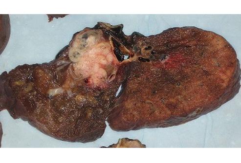
The tumor is the white mass within the lung shown.
pneumonia
An acute or chronic, potentially fatal, disease marked by inflammation of the lungs with congestion and consolidation (solidification into a firm dense mass due to the presence of cellular exudate (pus and other liquid) in the involved alveoli and resulting reduction in oxygenation of the blood; caused by viruses, bacteria, or other microorganisms and sometimes by physical and chemical irritants.
tuberculosis
An infectious disease caused by the tubercle bacillus (Mycobacterium tuberculi) and characterized by the formation of tubercles on the lungs and other tissues of the body, often developing long after the initial infection primary infection of the lungs is characterized by the coughing up of mucus and blood-stained sputum, fever, weight loss, and chest pain; it is transmitted from person to person by an aerosol of organisms suspended in tiny droplets which are inhaled; although deaths have decreased since the 1950s, it remains a major worldwide health problem and there has been some increase in incidence in recent years.
infant respiratory distress syndrome
A failure of the lungs of a newborn to inflate properly, as a result of lung immaturity and specifically to inadequate amounts of surfactant production in the alveoli; symptoms include rapid breathing, nasal flaring, a grunting noise with each breath and cyanosis around the lips and in the nail beds; more serious complications include sepsis, cranial hemorrhage, convulsions and shock; primarily a problem for premature babies; it is a significant (4%) cause of infant mortality.
coryza = rhinitis
an inflammation of the mucous membrane lining the nose (usually associated with nasal discharge); a symptom of the common cold and many other upper respiratory infections. nickname - runny nose
influenza
An acute contagious viral infection characterized by inflammation of the respiratory tract and by fever, chills, headache, muscular pain, and prostration; it may occur in isolated cases, in epidemics or in pandemics; involvement of the myocardium or the CNS occur infrequently; a necrotizing bronchitis and interstitial pneumonia develop in severe cases and predispose patients to secondary bacterial pneumonia.
pulmonary congestion = pulmonary edema
The clinical condition in which too much tissue fluid accumulates in the lungs; the excess fluid blocks the transport of oxygen from the lungs into the blood because it leaks into the air sacs and tissue of the lungs; emergency treatment with medicines and mechanical ventilation may be necessary.
pulmonary embolism
An obstruction of a pulmonary artery or one of its branches that is produced by foreign matter, most often by detached fragments of a blood clot = thromboembolus, usually originating in a vein of the leg or pelvis; clinically significant episodes are marked by labored breathing, chest pain, fainting, rapid heart rate, cyanosis, shock, and sometimes death; in other, rarer circumstances, material other than a blood clot is responsible; this may include fat or bone (usually in association with significant trauma), air (often when diving), clumped tumor cells, or amniotic fluid (during a difficult labor and delivery); treatment requires anticoagulant medication, such as heparin and warfarin, and rarely (in severe cases) thrombolysis or surgery.
cystic fibrosis (CF)
A hereditary disease (autosomal recessive) of the exocrine glands, usually developing during early childhood and affecting mainly the pancreas, respiratory system, and sweat glands (elevated chloride levels); it is characterized by the production of abnormally viscous mucus by the affected glands, usually resulting in chronic respiratory infections and impaired pancreatic and intestinal function.
pleurisy
Inflammation of the pleura, usually occurring as a complication of a disease such as pneumonia, accompanied by accumulation of fluid in the pleural cavity, chills, fever, and painful breathing and coughing.
Describe: The effect(s) of emphysema, pneumonia, pulmonary edema on the respiratory membrane.
A.emphysema (1) surface area of the alveolar walls dramatically reduced, (2) alveolar walls thickened, (3) character of alveolar wall degraded by scar tissue formation, (4) diffusion distance for respiratory gases within the alveolar walls increased
B.pneumonia (1) surface area of the alveolar walls somewhat reduced, (2) alveolar walls thickened and the fluid layer on the external surface of the alveolar walls thickened, (3) character of alveolar wall changed by inflammation, and possibly by the effects of the particular microbial pathogen at the site, (4) diffusion distance for respiratory gases within the alveolar walls dramatically increased
c.pulmonary edema (1) surface area of the alveolar walls somewhat reduced, (2) alveolar walls thickened and the fluid layer on the external surface of the alveolar walls thickened, (3) character of alveolar wall may be changed by fluid accumulation, (4) diffusion distance for respiratory gases within the alveolar walls dramatically increased