Artifact
anything that is not properly indicative of the structures or events
Benefits to artifacts
shadowing
enhancement
- tell the difference
between solid and fluid
- characterize masses
Negative attributes of artifacts
improper location
wrong size
wrong shape
wrong
acoustical properties
absent
present
absuring
Artifacts can be due to improper equipment settings
receiver gain
compensation settings
pre/post processing
range ambiguity
Artifacts can be due to ultrasound pysics
ring down
enhancement
edge shadowing
shadowing
reverberation
mirroring
Artifacts occur from assumptions
straight line
along the beam axis
amplitude related to
the object struck
speed of sound in soft tissue
Doppler artifacts
incorrect spectral flow
aliasing
range ambiguity
mirror imaging
speckle
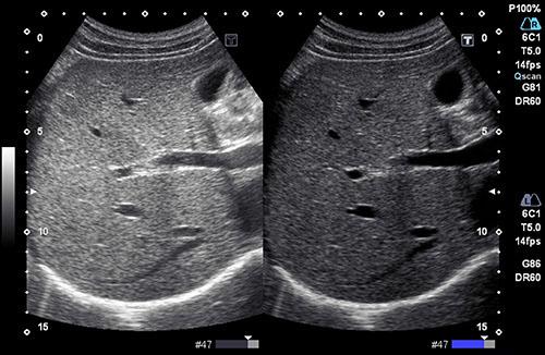
Slice thickness artifact
when 3D is flattened to convert to 2D external echoes show up in the image
Cause of slice thickness artifact
beam is not razor thin
AKA slice thickness artifact
section thickness
elevational resolution
partial volume artifact
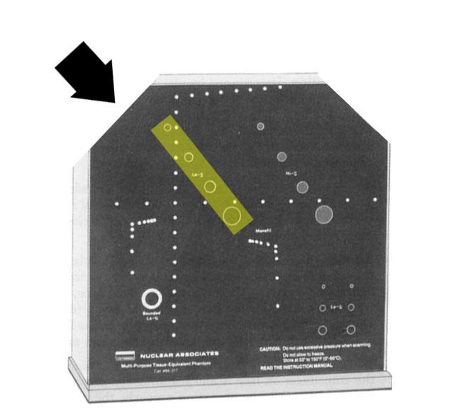
What transducers are prone to slice thickness artifacts?
linear array
due to poor elevational resolution
why are machines good at axial resolution?
axial resolution does not change with depth
what artifacts is similar to multipath?
mirror
What causes multipath?
returning echoes do not return in a direct line
Multipath
gives wrong depth
cause of acoustic speckle
small amplitudes of sound waves interfering with each other
Acoustic speckle
liver
specular reflections
incorrect texture
How to improve acoustic speckle?
THI
What are the problems associated with acoustic speckle?
if too bad you will not be able to see (fat encompassing)
liver - pathological degrees of fatty filtration
Reverberation
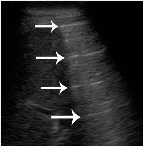
bouncy ball
multiple echos (strong reflectors) that
appear on the screen
multiple bounces between two or
more surfaces
always equal distance
normal
declining intensity
Ring down artifact
ring down occurs with gas filled loops of bowel
tends
to occur with dirty shadowing
Comet trails
squeezed out reverberation
reflectors close
mirror image
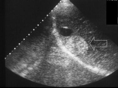
and improper location caused by a change in propagation speed
caused by an adjacent strong reflector
What frequently causes mirror image?
diaphragm
Spectral mirroring
when spectral doppler appears in both sides of the baseline
what is a common cause of spectral doppler?
receiver gain too high
How do you fix spectral mirroring?
change angle
turn down receiver gain
change to higher frequency
What type of transducer produces side lobes?
single element
What type of transducer produces grating lobes?
array transducers
What causes lobes?
weak beams that might otherwise be ignored hit a strong reflector.
the reflected echoes become misplaced.
How do you get rid of lobes?
subdicing the element
using dynamic apodization
apodization
weakening of the outside elements
listening is decreased on the side
How do speed errors occur?
system works on the assumption that sound will make a round trip through soft tissue in 13 μs/cm
Where is the object placed if the speed is faster than 13 μs/cm?
closer
Where is the object placed if the speed is slower than 13 μs/cm?
farther
Split off artifact
object in front of another object causes the sound to travel faster or slower only on a portion of the second object. Part of the object usually the diaphragm is placed incorrectly and the object appears cut
step off artifact
aka split off artifact
cut artifact
aka split off artifact
What causes Range ambiguity?
caused by deep echoes from a previous pulse
what is range ambiguity?
object is placed closer and near the scan plane
How can you correct range ambiguity?
Change frequency
change depth
change PRF
most systems will adjust PRF
What is shadowing
shadows distal to strong attenuator
What causes shadowing?
strong reflector bouncing all sound back imediately
strong absorber - no echoes left to return
What causes edge shadowing?
reflections bouncing away when hitting a curved object - none return
Dirty shadowing
usually occurs with bowels
shadowing when internal echoes are present
What is enhancement
hyperechoic areas distal to a a weak attenuator
cysts
What causes enhancement
an unexpected increase in amplitude
focal enhancement
increased enhancement at the focus
What can you do to correct focal enhancement?
spacial compounding
what is thru transmission?
aka enhancement
Aliasing
peaks cut off and place on bottom
speeding ticket
blood travels faster than the nyquist limit
What can cause aliasing?
insufficient spatial sampling
insufficient temporal sampling
What can correct aliasing?
raise PRF - chance of range ambiguity
increase doppler shift
shift baseline - cosmetic
switch to continuous wave
What is the nyquist limit?
1/2 PRF
PRF
pulses per second
Artifact cause
axial resolution
pulse length
Artifact cause
lateral resolution
pulse width
Artifact cause
section thickness
pulse width
Artifact cause
speckle
interference
Artifact cause
reverberation
multiple reflections
Artifact cause
refraction
refraction
Artifact cause
multipath
multiple reflections
Artifact cause
mirror image
multiple reflections
Artifact cause
side lobes
side lobes
Artifact cause
grating lobes
grating lobes
Artifact cause
comet trail
reverberation
Artifact cause
ring down
resonance
Artifact cause
speed error
speed error
Artifact cause
range ambiguity
high PRF
Artifact cause
shadowing
high attenuation
Artifact cause
enhancement
low attenuation
Artifact cause
edge shadowing
refraction
Artifact cause
focal enhancement
focusing
Artifact cause
aliasing
low PRF
Artifact cause
spectral mirroring
high doppler gain
Which testing is the most challenging?
Doppler
What part of the system is most likely to break down?
transducer
electric shock
Why do we perform performance testing?
to prevent degradation of image
What do acoustic output testers evaluate?
beam former and transducer acting together as a source of ultrasound
what do flow testers evaluate?
Doppler
What do detail testers evaluate?
lateral and axial resolution
What so output testers evaluate?
sound output
What is the goal of system testers?
detect gradual changes in system performance
Who does the responsibility of quality assurance rest?
sonographer
How often should equipment be tested?
one a month
Perfecting these methods is _____ but must be done & must be ______.
difficult
repeatable
What do AIUM test objects test for?
slice thickness
beam width
detail resolution\depth accuaracy
measurement accuracy
dead zone
What is the main problem with AIUM test objects?
no attenuation properties
What is the AIUM test object usually filled with?
water
but sometimes no water
What is the main advantage of the AIUM test object
price
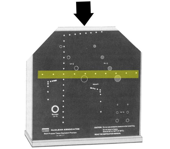
What does the tissue/cyst phantum test for?
detail resolution
dynamic range
time gain compensation
contrast resolution
What does the cyst phantom contain
columns of simulated cysts
How do you fix lateral smearing?
decrease depth (increases frame rate - improves temporal resolution)
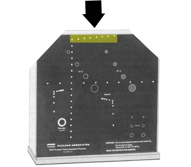
Describe the constructions of the tissue phantum
rubber face & sides
plexiglass base
cosists of cystic, nylon other materials
What is the test phantom filled with?
gel - 1.54
rubber - 1.45

How would you test the dead zone?

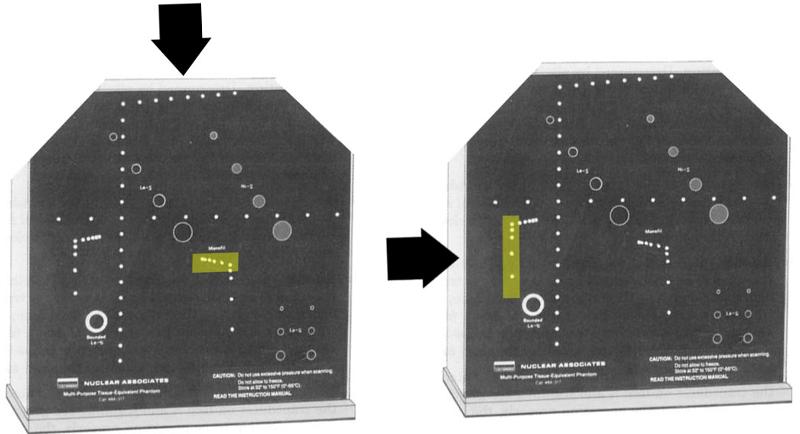
How do you evaluate cysts

What does the cyst evaluation show?
size and depth variation
How would you evaluate axial resolution?

What does this test for?
axial resolution
smallest distance two pins can be seen as two separate pins

How would you evaluate vertical registration?
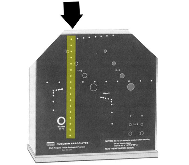

What does this test for?
vertical registration / range accuracy
ability to display echoes at the proper depth or top line

How would you evaluate Horizontal registration?
What does this test for?
Horizontal registration
the ability to position echoes in the their correct position along a line that is perpendicular to the ultrasound beam

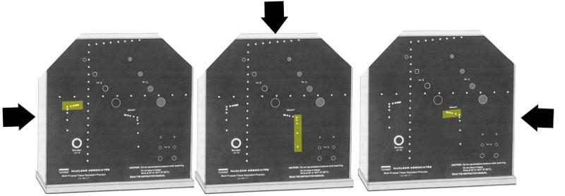
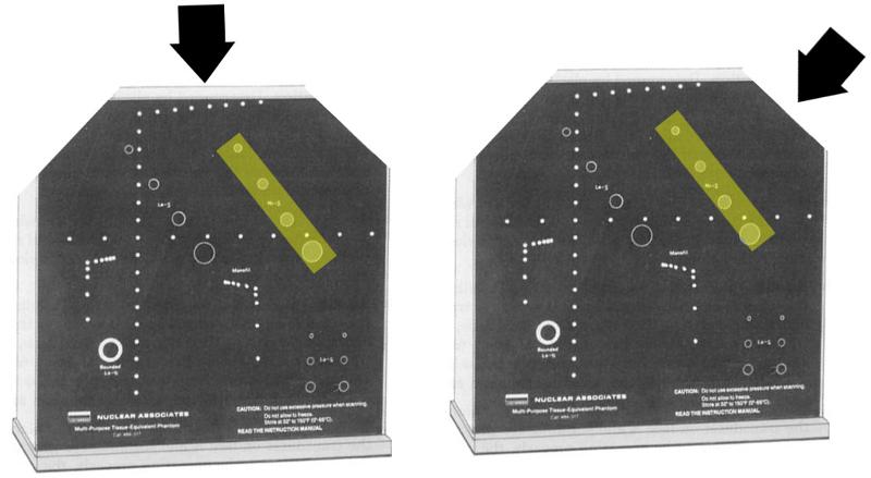
How would you evaluate lateral resolution?

What does this test for?
lateral resolution
minimum distance two pins can be seen as two separate pins at a specific depth

How would you evaluate grey scale?


What does this test for?
grayscale / contrast resolution
the ability to discriminate between 2 different objects that have different shades of grey
What is the difference between these two test for contrast resolution?
the first one has size variation
the second one has depth variation
Minimum sensitivity
start by making TGC flat then increase gain from minimum value
the point the echo appears on screen is minimum sensitivity
Normal sensitivity
is the point in which all the pins on an AIUM test object are displayed
Sensitivity
is the range that echoes are barely visible to fully sensitivity
lateral resolution
the minimum distance that two rods are displayed as two separate images at a specific depth
Focal zone
the depth at which the intensity is the highest and beam is the narrowest
this may be found using beam profiler or hydrophone
Dynamic range / greyscale
change in gain should result in a change in greyscale
vertical registration / range accuracy
the machines ability to display echoes at the proper depth
depth calibration
the accuracy of B mode, m mode and A mode on displaying the depth of reflectors
horisontal calibration
the machines ability to position echoes in the correct postiion perpendicular to the U/S beam
longitudinal resolution
smallest distance at which two pins are displayed as two separate echoes in their position parallel to the beam
What do the Doppler performance tools evaluate?
the effective position of the Doppler beam (penetration)
accuracy of measured flow
accuracy volume and flow speed
What do we use for Doppler testing?
Blood tissue phantom
Doppler testing object
What is a Blood tissue phantom
Doppler testing the mimicks blood
- sephadex in water
- polystyrene microspheres in water/glycerol
- water + machine cutting oil
- starch suspensions
What is a Doppler testing object?
Doppler test object uses controlled movement of strings
- moving solid object
- usually a string ultrasound
- pulsatile motions
- reverse motion
What are the disadvantages of the Doppler string phantom?
- over estimates peak velocities
- causes artifactual spectral broadening
What does a Blood tissue phantom contain?
an image face
medium
a flow conduit
pump
blood mimick
reservoir
How does a Blood tissue phantom work?
complex
tube connects to a pump
pumps an echogenic fluid through out a known velocity simulates a stenosis.
What is a microprobe?
small transducer on a hollow needle
What is a hydrophone?
large piezoelectric membrane with electrodes on both sides
What are microprobe and hydrophone used for?
Measure intensity
produce a waveform on an oscilloscope
What do microprobe and hydrophone use as a piezoelectric material?
PVDF
How do microprobe and hydrophone work?
receive sound from all directions measure pressure at a given point within the beam in response to the varying pressure
the hydrophone produces a varying voltage
What is the voltage produced by a hydrophone displayed on?
oscilloscope
What does an oscilloscope display?
bandwidth
What do hydrophones measure?
frequency
PRF
duty factor
pressure amplitude
wavelength
SPL
intensity
Which type of U/S produces the greatest acoustic output?
Pulsed Spectral Doppler
Color Doppler
M mode
B mode
Intensity and output indexes have already been copulated using a ______.
hydrophone
What is the #1 assumption of risk?
U/S is energy and any energy applied to human cells can cause change
What is the rule of U/S?
risk benefit ratio
any possible benefit must outweigh possible risks
- whenever possible
- increase benefit
- decrease risk
How do we increase benefit?
Better equipment
- choose best greatest depth higher resolution - output gain will not need to be increased
- harmonic imaging
- appropriate transducer
Better operator
- knowledgable
- formal
- informal learning
- experience
How do we decrease risk?
decrease output/exposure
- use only when indicated
- minimize length of exam
decrease bioeffects
- lower exposure intensity
- decrease length of exam
- use low output imaging methods
Do you need a Doctors orders for an ultrasound exam?
Yes
medical device regulates by the FDA
What are the two types of mechanical bioeffects?
cavitation
radiation
What is radiation?
the amount of force that a beam exerts on an absorber or reflector
What are the two types of bioeffects?
heating - attenuation of U/S is primary by heat
cavitation - motion of microbubbles
What is heating primarily due to?
absorption
Why does absorption cause heating?
absorption involves conversion of U/S to heat
U/S produce a temp. rise as it propagates through tissue
...
What happens to heat when intensity goes up?
rises
What happens to heat when frequency goes up?
rises
What can Rayleighs scattering cause?
Thermal injury
Where are temperature elevations more likely to occur?
tissue-bone interface
What area is of great concern to thermal injury?
soft tissue adjacent to bone in a fetus
What is SPTA associated with?
tissue heating
What exam can be used without fear?
any exam that causes elevation in temperature of less than 2 degrees Celcius
What exam may cause harm in a fetus?
an exam that causes an elevation of temperature of greater than 41 degrees Celcius
What does Rayleigh scattering cause?
RBC to scatter
What is Rayleigh scattering?
when the wavelength of the incident sound beam is smaller than the size of the RBC
Which causes higher Temperature elevation Pulsed wave or Continuous wave?
CW causes higher temperature elevation
Which causes higher Temperature elevation Focused or unfocused?
unfocused causes higher temperature elevation
* focused has a narrow beam and heat is easily dispersed
What is cavitation?
the production and behavior of bubbles in a liquid medium
Where would a sound wave may cause cavitation?
Nucleation sites
tissue with gas bubbles
What are the two types of cavitation?
stable cavitation
transient cavitation
What is stable cavitation?
expand and contract
bubbles that oscillate in diameter with the passing pressure of a sound wave
bubbles don't burst
causes shear stress - cutting force
What does shear force cause?
microstreaming
What is microstreaming?
rapid rotational flow that occurs in intact blood vessels
What is shear stress?
cutting force
What is transient cavitation?
bubbles expand and collapse violently
bubbles burst
What is another name for transient cavitation?
Collapse / inertial cavitation
What does transient cavitation cause?
destructive effects
shockwaves
light emissions
high temperature
What is Thermal index?
heat production index -
deals with bioeffects caused by heating
If a machine can exceed thermal or mechanical indexes what must be?
acoustic output displayed
What is Mechanical index
Motion production index -
deals with bioeffects caused by cavitation
What is the thermal index measured in?
degrees celcius
Which display is preferred SPTA or Thermal index?
Thermal index - more accurate
What should SPTA be below?
720 mw/cm2
TIS
Thermal index soft tissue
TIB
Thermal index bone
TIC
Thermal index cranial
TI of 2 or less =
expected heating of 2 degrees or less
Mechanical index formula
MI = derated peak rarefractional pressure / sqrt (U/S center frequency
What is threshold for MI?
.3 or less
What is normal body temperature?
37 degrees C
What is the max body temp rise?
39 degrees C
C to F
37°C x 9/5 + 32 = 98.6°F
F to C
(98.6°F - 32) x 1.8 = 37°C
(98.6°F - 32) x 5/9 = 37°C